

Tom Wunderling, M.Sc.
Research Interests
- Real-time representation and interactive manipulation of flexible, tubular structures in VR/AR
- Hand-tracked and multimodal interaction for medical XR applications
- Machine learning for processing 3D data as a basis for modeling and simulation
- Immersive training environments for medical education
Contacts: ResearchGate |
Google Scholar |
ORCID |
LinkedIn | 🌐 Personal website
Publications
2025
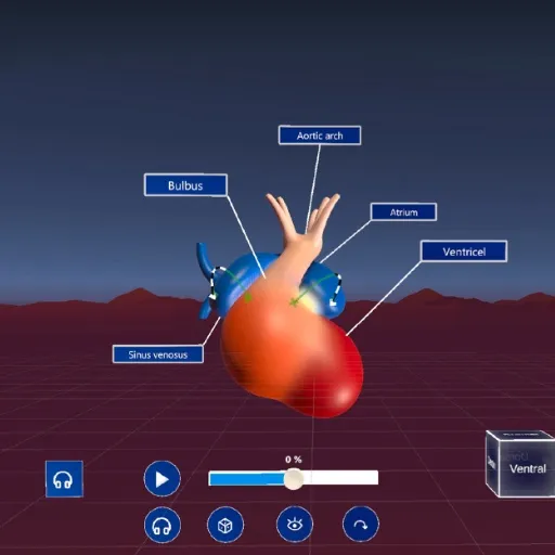
Kunz, M; Schott, D; Wunderling, T; Halloul, M; Hansen, C; Albrecht, A; Braun-Dullaeus, R
Embryonic Heart Development as an Immersive Experience: Unveiling Learning Effects and Influential Factors in Virtual Learning Environments Journal Article
In: Comput. Biol. Med., vol. 187, no. C, 2025.
@article{kunz_embryonic_2025,

title = {Embryonic Heart Development as an Immersive Experience: Unveiling Learning Effects and Influential Factors in Virtual Learning Environments},

author = {M Kunz and D Schott and T Wunderling and M Halloul and C Hansen and A Albrecht and R Braun-Dullaeus},

url = {https://doi.org/10.1016/j.compbiomed.2024.109638

https://pubmed.ncbi.nlm.nih.gov/39923588/, PubMed},

doi = {10.1016/j.compbiomed.2024.109638},

year  = {2025},

date = {2025-04-01},

urldate = {2025-04-01},

journal = {Comput. Biol. Med.},

volume = {187},

number = {C},

abstract = {As the quality and availability of Virtual Reality (VR) technologies improve, their potential applications in medical education, particularly VR Learning Environments (VRLEs), are increasingly explored. VRLEs offer a dynamic platform where educators and students can interact, access materials, and engage in learning beyond traditional classrooms. However, questions remain about their long-term learning effects and potential confounding factors. This study investigates these aspects through a VR application designed for teaching heart embryology. For this reason we conducted a user study with medical students in their early years of training (N = 143). Our findings reveal significant short-term and sustained learning outcomes two to four weeks following a single VR session. Importantly, these outcomes appear largely independent of users’ technical affinity and are minimally influenced by their immersion tendencies. Instead, the quality of the VRLE and its user experience emerge as critical factors. These results underscore the efficacy of well-designed VRLEs in higher education and highlight key areas for future development.},

keywords = {},

pubstate = {published},

tppubtype = {article}

}
2023
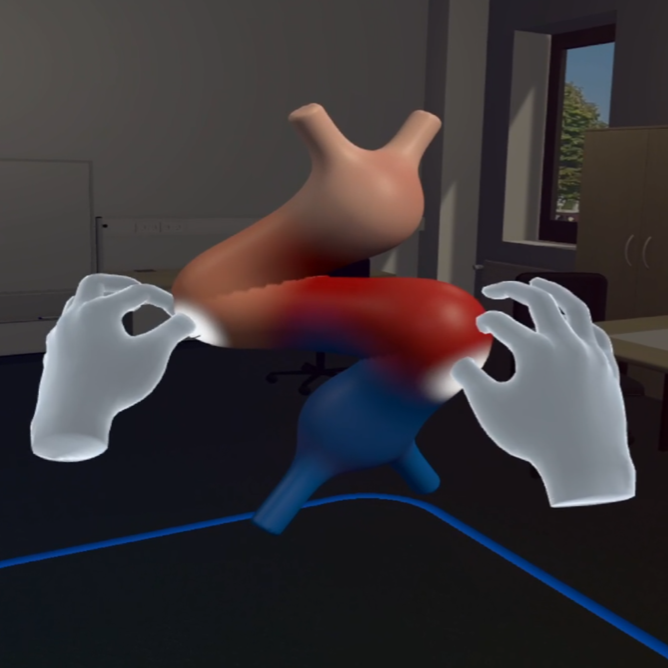
Schott, D; Kunz, M; Wunderling, T; Heinrich, F; Braun-Dullaeus, R; Hansen, C
CardioGenesis4D: Interactive Morphological Transitions of Embryonic Heart Development in a Virtual Learning Environment Journal Article
In: IEEE Transactions on Visualization and Computer Graphics, vol. 29, no. 5, pp. 2615–2625, 2023.
@article{schott_cardiogenesis4d_2023,

title = {CardioGenesis4D: Interactive Morphological Transitions of Embryonic Heart Development in a Virtual Learning Environment},

author = {D Schott and M Kunz and T Wunderling and F Heinrich and R Braun-Dullaeus and C Hansen},

url = {https://ieeexplore.ieee.org/document/10049681},

doi = {10.1109/TVCG.2023.3247110},

year  = {2023},

date = {2023-05-01},

urldate = {2023-05-01},

journal = {IEEE Transactions on Visualization and Computer Graphics},

volume = {29},

number = {5},

pages = {2615–2625},

abstract = {In the embryonic human heart, complex dynamic shape changes take place in a short period of time on a microscopic scale, making this development difficult to visualize. However, spatial understanding of these processes is essential for students and future cardiologists to properly diagnose and treat congenital heart defects. Following a user centered approach, the most crucial embryological stages were identified and translated into a virtual reality learning environment (VRLE) to enable the understanding of the morphological transitions of these stages through advanced interactions. To address individual learning types, we implemented different features and evaluated the application regarding usability, perceived task load, and sense of presence in a user study. We also assessed spatial awareness and knowledge gain, and finally obtained feedback from domain experts. Overall, students and professionals rated the application positively. To minimize distraction from interactive learning content, such VRLEs should consider features for different learning types, allow for gradual habituation, and at the same time provide enough playful stimuli. Our work previews how VR can be integrated into a cardiac embryology education curriculum.},

keywords = {},

pubstate = {published},

tppubtype = {article}

}
2018
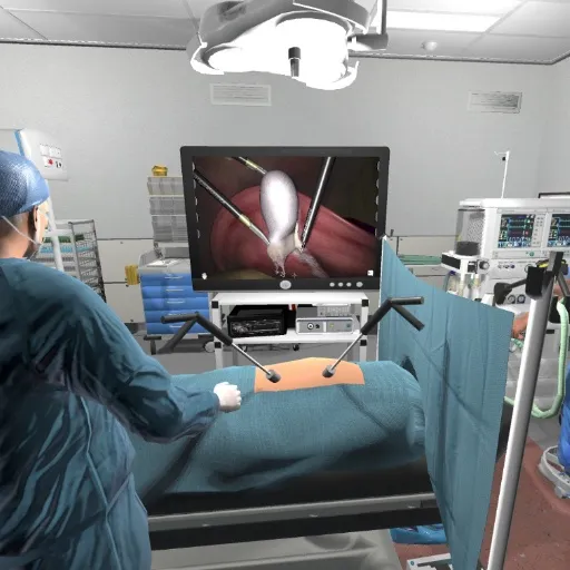
Huber, T; Wunderling, T; Paschold, M; Lang, H; Kneist, W; Hansen, C
Highly Immersive Virtual Reality Laparoscopy Simulation: Development and Future Aspects Journal Article
In: International Journal of Computer Assisted Radiology and Surgery, vol. 13, no. 2, pp. 281–290, 2018.
@article{huber_highly_2018,

title = {Highly Immersive Virtual Reality Laparoscopy Simulation: Development and Future Aspects},

author = {T Huber and T Wunderling and M Paschold and H Lang and W Kneist and C Hansen},

url = {http://link.springer.com/10.1007/s11548-017-1686-2

https://pubmed.ncbi.nlm.nih.gov/29151194/, PubMed},

doi = {10.1007/s11548-017-1686-2},

year  = {2018},

date = {2018-02-01},

urldate = {2018-02-01},

journal = {International Journal of Computer Assisted Radiology and Surgery},

volume = {13},

number = {2},

pages = {281–290},

abstract = {Purpose Virtual Reality (VR) applications with head-mounted displays (HMDs) have had an impact on information and multimedia technologies. The current work aimed to describe the process of developing a highly-immersive VR simulation for laparoscopic surgery.

Methods We combined a VR laparoscopy simulator (LapSim) and a VR HMD to create a user-friendly VR simulation scenario. Continuous clinical feedback was an essential aspect of the development process. We created an artificial VR (AVR) scenario by integrating the simulator video output with VR game components of figures and equipment in an operating room. We also created a highly-immersive VR surrounding (IVR) by integrating the simulator video output with a 360° video of a standard laparoscopy scenario in the department's operating room.

Results Clinical feedback led to optimization of the visualization, synchronization, and resolution of the virtual operating rooms (in both the IVR and the AVR). Preliminary testing results revealed that individuals experienced a high degreee of exhilaration and presence, with rare events of motion sickness. The technical performance showed no significant difference compared to that achieved with the standard LapSim.

Conclusion Our results provided a proof-of-concept for the technical feasibility of an custom highly immersive VR-HMD setup. Future technical research is needed to improve the visualization, immersion, and capability of interacting within the virtual scenario.},

keywords = {},

pubstate = {published},

tppubtype = {article}

}
Methods We combined a VR laparoscopy simulator (LapSim) and a VR HMD to create a user-friendly VR simulation scenario. Continuous clinical feedback was an essential aspect of the development process. We created an artificial VR (AVR) scenario by integrating the simulator video output with VR game components of figures and equipment in an operating room. We also created a highly-immersive VR surrounding (IVR) by integrating the simulator video output with a 360° video of a standard laparoscopy scenario in the department's operating room.
Results Clinical feedback led to optimization of the visualization, synchronization, and resolution of the virtual operating rooms (in both the IVR and the AVR). Preliminary testing results revealed that individuals experienced a high degreee of exhilaration and presence, with rare events of motion sickness. The technical performance showed no significant difference compared to that achieved with the standard LapSim.
Conclusion Our results provided a proof-of-concept for the technical feasibility of an custom highly immersive VR-HMD setup. Future technical research is needed to improve the visualization, immersion, and capability of interacting within the virtual scenario.
2017
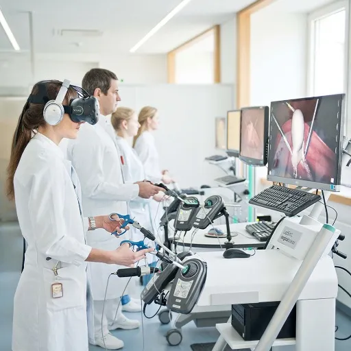
Huber, T; Paschold, M; Hansen, C; Wunderling, T; Lang, H; Kneist, W
New Dimensions in Surgical Training: Immersive Virtual Reality Laparoscopic Simulation Exhilarates Surgical Staff Journal Article
In: Surgical Endoscopy, vol. 31, no. 11, pp. 4472–4477, 2017.
@article{huber_new_2017,

title = {New Dimensions in Surgical Training: Immersive Virtual Reality Laparoscopic Simulation Exhilarates Surgical Staff},

author = {T Huber and M Paschold and C Hansen and T Wunderling and H Lang and W Kneist},

url = {https://doi.org/10.1007/s00464-017-5500-6

https://pubmed.ncbi.nlm.nih.gov/28378077/, PubMed},

doi = {10.1007/s00464-017-5500-6},

year  = {2017},

date = {2017-11-01},

urldate = {2017-11-01},

journal = {Surgical Endoscopy},

volume = {31},

number = {11},

pages = {4472–4477},

abstract = {Virtual reality (VR) and head mount displays (HMDs) have been advanced for multimedia and information technologies but have scarcely been used in surgical training. Motion sickness and individual psychological changes have been associated with VR. The goal was to observe first experiences and performance scores using a new combined highly immersive virtual reality (IVR) laparoscopy setup.},

keywords = {},

pubstate = {published},

tppubtype = {article}

}
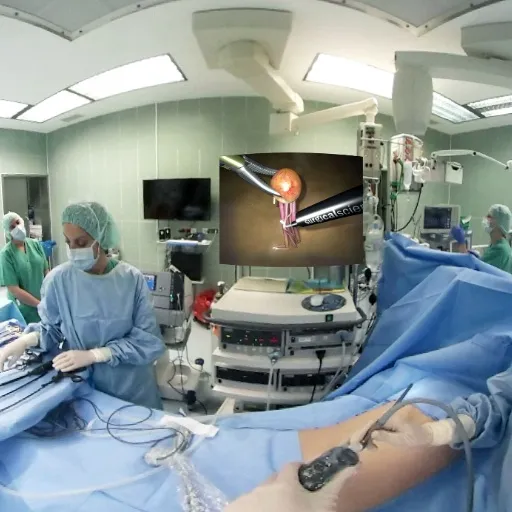
Huber, T; Paschold, M; Hansen, C; Wunderling, T; Lang, H; Kneist, W
Neue Dimensionen in der Laparoskopischen Simulation – Erste Erfahrungen mit der totalen, hoch-immersiven virtuellen Realität Proceedings Article
In: Zeitschrift für Gastroenterologie, pp. KV 533, Georg Thieme Verlag KG, 2017.
@inproceedings{Huber_2017,

title = {Neue Dimensionen in der Laparoskopischen Simulation – Erste Erfahrungen mit der totalen, hoch-immersiven virtuellen Realität},

author = {T Huber and M Paschold and C Hansen and T Wunderling and H Lang and W Kneist},

url = {https://www.thieme-connect.com/products/ejournals/html/10.1055/s-0037-1605273},

doi = {10.1055/s-0037-1605273},

year  = {2017},

date = {2017-08-01},

urldate = {2017-08-01},

booktitle = {Zeitschrift für Gastroenterologie},

volume = {55},

number = {08},

pages = {KV 533},

publisher = {Georg Thieme Verlag KG},

abstract = {Virtual Reality (VR) und sogenannte Head-Mount-Displays (HMD) haben die Informations- und Multimediatechnologie verändert. Vor dem Hintergrund einer niedrigen Nutzung laparoskopischer Trainingsmöglichkeiten wurde eine totale, hoch-immersive VR Simulation (TVRL) für laparoskopische Chirurgie entwickelt. In einer ersten Pilotuntersuchung sollten Leistungsparameter und Erfahrungen der Teilnehmer untersucht werden.

Die Entwicklung der TVRL wurde durch die Kombination eines HMD (HTC Vive®) mit einem VR Laparoskopie Simulator (LapSim®) realisiert. Die Simulationsübungen wurden in eine aufgenommene 360 ° Videoumgebung einer nachgestellten laparoskopischen Operation im Operationssaal projiziert. In einer ersten Pilotuntersuchung wurden 10 Probanden zur Immersion und Präsenz in der TVRL anhand validierter Fragebögen befragt, sowie die Leistung am Simulator anhand von drei Übungen (Ringtausch, Feine Dissektion, Cholezystektomie) analysiert.

Die Teilnehmer benötigten für die Feine Dissektion in der TVRL länger als in der herkömmlichen VR Simulation. Bei der Cholezystektomie wurden mehr Fehler gemacht. Die Teilnehmer waren von der TVRL begeistert, dachten selten an andere Personen im Raum und gaben ein hohes Maß an Präsenz in der computergenerierten Welt an.

In dieser ersten Pilotuntersuchung konnte die klinische und technische Machbarkeit der TVRL gezeigt werden. Teilnehmer gaben ein hohes Maß an Präsenz und Immersion an. Das neu entwickelte Trainingssetup eröffnet völlig neue Möglichkeiten für die laparoskopische Simulation.},

keywords = {},

pubstate = {published},

tppubtype = {inproceedings}

}
Die Entwicklung der TVRL wurde durch die Kombination eines HMD (HTC Vive®) mit einem VR Laparoskopie Simulator (LapSim®) realisiert. Die Simulationsübungen wurden in eine aufgenommene 360 ° Videoumgebung einer nachgestellten laparoskopischen Operation im Operationssaal projiziert. In einer ersten Pilotuntersuchung wurden 10 Probanden zur Immersion und Präsenz in der TVRL anhand validierter Fragebögen befragt, sowie die Leistung am Simulator anhand von drei Übungen (Ringtausch, Feine Dissektion, Cholezystektomie) analysiert.
Die Teilnehmer benötigten für die Feine Dissektion in der TVRL länger als in der herkömmlichen VR Simulation. Bei der Cholezystektomie wurden mehr Fehler gemacht. Die Teilnehmer waren von der TVRL begeistert, dachten selten an andere Personen im Raum und gaben ein hohes Maß an Präsenz in der computergenerierten Welt an.
In dieser ersten Pilotuntersuchung konnte die klinische und technische Machbarkeit der TVRL gezeigt werden. Teilnehmer gaben ein hohes Maß an Präsenz und Immersion an. Das neu entwickelte Trainingssetup eröffnet völlig neue Möglichkeiten für die laparoskopische Simulation.
Wunderling, T; Golla, B; Poudel, P; Arens, C; Friebe, M; Hansen, C
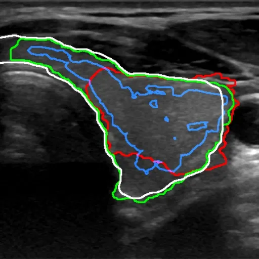
Comparison of Thyroid Segmentation Techniques for 3D Ultrasound Proceedings Article
In: Styner, M; Angelini, E (Ed.): Medical Imaging 2017: Image Processing, pp. 1013317, Orlando, Florida, United States, 2017.
@inproceedings{styner_comparison_2017,

title = {Comparison of Thyroid Segmentation Techniques for 3D Ultrasound},

author = {T Wunderling and B Golla and P Poudel and C Arens and M Friebe and C Hansen},

editor = {M Styner and E Angelini},

url = {http://proceedings.spiedigitallibrary.org/proceeding.aspx?doi=10.1117/12.2254234},

doi = {10.1117/12.2254234},

year  = {2017},

date = {2017-02-01},

urldate = {2017-02-01},

booktitle = {Medical Imaging 2017: Image Processing},

pages = {1013317},

address = {Orlando, Florida, United States},

abstract = {The segmentation of the thyroid in ultrasound images is a field of active research. The thyroid is a gland of the endocrine system and regulates several body functions. Measuring the volume of the thyroid is regular practice of diagnosing pathological changes. In this work, we compare three approaches for semi-automatic thyroid segmentation in freehand-tracked three-dimensional ultrasound images. The approaches are based on level set, graph cut and feature classification. For validation, sixteen 3D ultrasound records were created with ground truth segmentations, which we make publicly available. The properties analyzed are the Dice coefficient when compared against the ground truth reference and the effort of required interaction. Our results show that in terms of Dice coefficient, all algorithms perform similarly. For interaction, however, each algorithm has advantages over the other. The graph cut-based approach gives the practitioner direct influence on the final segmentation. Level set and feature classifier require less interaction, but offer less control over the result. All three compared methods show promising results for future work and provide several possible extensions.},

keywords = {},

pubstate = {published},

tppubtype = {inproceedings}

}
Wunderling, T; Huber, T; Paschold, M; Kneist, W; Hansen, C
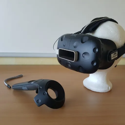
Immersives Laparoskopie-Training mit Hand-Tracking im virtuellen Operationssaal Proceedings Article
In: CURAC 2017 - Tagungsband, pp. 67–72, Hannover, 2017.
@inproceedings{wunderling_immersives_2017,

title = {Immersives Laparoskopie-Training mit Hand-Tracking im virtuellen Operationssaal},

author = {T Wunderling and T Huber and M Paschold and W Kneist and C Hansen},

url = {https://www.var.ovgu.de/pub/2017_CURAC_Wunderling.pdf},

year  = {2017},

date = {2017-01-01},

urldate = {2017-01-01},

booktitle = {CURAC 2017 - Tagungsband},

pages = {67–72},

address = {Hannover},

abstract = {In dieser Arbeit wird eine neue Methode des Laparoskopie-Trainings vorgestellt, dass die Vorteile etablierter Simulatoren mit der Immersion moderner Head-Mounted-Displays (HMDs) kombiniert. Die Komponenten dieses Systems und ihr Zusammenspiel werden beschrieben. Eine komplette, virtuelle Nachbildung eines Operationssaals wurde geschaffen, inklusive interaktiven laparoskopischen Instrumenten und Monitor. Mit dem Ziel, die Interaktion mit den Eingabegeräten in der virtuellen Realität (VR) zu verbessern, wurde im Zuge dieser Arbeit ein optisches Hand-Tracking mit dem Leap Motion Sensor realisiert. Das Hand-Tracking wurde im Rahmen einer Nutzerstudie mit 12 Teilnehmern evaluiert. Die Ergebnisse zeigen eine steile Lernkurve bei der Nutzung des Systems, die grundsätzliche Eignung als Hilfe für erfahrene Nutzer, aber auch Defizite bei der Verlässlichkeit und Usability des optischen Hand-Trackings.},

keywords = {},

pubstate = {published},

tppubtype = {inproceedings}

}