
Dr. Julian Alpers
Publications
2024
Schröer, S; Alpers, J; Gutberlet, M; Brüsch, I; Rumpel, R; Wacker, F; Hensen, B; Hansen, C
A probabilistic thermal dose model for the estimation of necrosis in MR-guided tumor ablations Journal Article
In: Medical Physics, vol. 51, no. 1, pp. 239–250, 2024, ISSN: 2473-4209, (_eprint: https://aapm.onlinelibrary.wiley.com/doi/pdf/10.1002/mp.16605).
@article{schroer_probabilistic_2024,
title = {A probabilistic thermal dose model for the estimation of necrosis in MR-guided tumor ablations},
author = {S Schröer and J Alpers and M Gutberlet and I Brüsch and R Rumpel and F Wacker and B Hensen and C Hansen},
url = {https://onlinelibrary.wiley.com/doi/abs/10.1002/mp.16605},
doi = {10.1002/mp.16605},
issn = {2473-4209},
year = {2024},
date = {2024-01-01},
urldate = {2024-01-01},
journal = {Medical Physics},
volume = {51},
number = {1},
pages = {239–250},
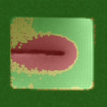
abstract = {Background Monitoring minimally invasive thermo ablation procedures using magnetic resonance (MR) thermometry allows therapy of tumors even close to critical anatomical structures. Unfortunately, intraoperative monitoring remains challenging due to the necessary accuracy and real-time capability. One reason for this is the statistical error introduced by MR measurement, which causes the prediction of ablation zones to become inaccurate. Purpose In this work, we derive a probabilistic model for the prediction of ablation zones during thermal ablation procedures based on the thermal damage model CEM43. By integrating the statistical error caused by MR measurement into the conventional prediction, we hope to reduce the amount of falsely classified voxels. Methods The probabilistic CEM43 model is empirically evaluated using a polyacrilamide gel phantom and three in-vivo pig livers. Results The results show a higher accuracy in three out of four data sets, with a relative difference in Sørensen–Dice coefficient from textbackslash-3.04%textbackslash to 3.97% compared to the conventional model. Furthermore, the ablation zones predicted by the probabilistic model show a false positive rate with a relative decrease of 11.89%–30.04% compared to the conventional model. Conclusion The presented probabilistic thermal dose model might help to prevent false classification of voxels within ablation zones. This could potentially result in an increased success rate for MR-guided thermal ablation procedures. Future work may address additional error sources and a follow-up study in a more realistic clinical context.},
note = {_eprint: https://aapm.onlinelibrary.wiley.com/doi/pdf/10.1002/mp.16605},
keywords = {},
pubstate = {published},
tppubtype = {article}
}
2022
Alpers, J; Rötzer, M; Gutberlet, M; Wacker, F; Hensen, B; Hansen, C
Adaptive simulation of 3D thermometry maps for interventional MR-guided tumor ablation using Pennes’ bioheat equation and isotherms Journal Article
In: Scientific Reports, vol. 12, no. 1, pp. 20356, 2022, ISSN: 2045-2322.
@article{alpers_adaptive_2022,
title = {Adaptive simulation of 3D thermometry maps for interventional MR-guided tumor ablation using Pennes’ bioheat equation and isotherms},
author = {J Alpers and M Rötzer and M Gutberlet and F Wacker and B Hensen and C Hansen},
url = {https://www.nature.com/articles/s41598-022-24911-1},
doi = {10.1038/s41598-022-24911-1},
issn = {2045-2322},
year = {2022},
date = {2022-11-01},
urldate = {2022-11-01},
journal = {Scientific Reports},
volume = {12},
number = {1},
pages = {20356},
abstract = {Abstract
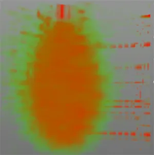
Minimally-invasive thermal ablation procedures have become clinically accepted treatment options for tumors and metastases. Continuous and reliable monitoring of volumetric heat distribution promises to be an important condition for successful outcomes. In this work, an adaptive bioheat transfer simulation of 3D thermometry maps is presented. Pennes’ equation model is updated according to temperature maps generated by uniformly distributed 2D MR phase images rotated around the main axis of the applicator. The volumetric heat diffusion and the resulting shape of the ablation zone can be modelled accurately without introducing a specific heat source term. Filtering the temperature maps by extracting isotherms reduces artefacts and noise, compresses information of the measured data and adds physical a priori knowledge. The inverse heat transfer for estimating values of the simulated tissue and heating parameters is done by reducing the sum squared error between these isotherms and the 3D simulation. The approach is evaluated on data sets consisting of 13 ex vivo bio protein phantoms, including six perfusion phantoms with simulated heat sink effects. Results show an overall average Dice score of 0.89 ± 0.04 (SEM < 0.01). The optimization of the parameters takes 1.05 ± 0.26 s for each acquired image. Future steps should consider the local optimization of the simulation parameters instead of a global one to better detect heat sinks without a priori knowledge. In addition, the use of a proper Kalman filter might increase robustness and accuracy if combined with our method.},
keywords = {},
pubstate = {published},
tppubtype = {article}
}
Minimally-invasive thermal ablation procedures have become clinically accepted treatment options for tumors and metastases. Continuous and reliable monitoring of volumetric heat distribution promises to be an important condition for successful outcomes. In this work, an adaptive bioheat transfer simulation of 3D thermometry maps is presented. Pennes’ equation model is updated according to temperature maps generated by uniformly distributed 2D MR phase images rotated around the main axis of the applicator. The volumetric heat diffusion and the resulting shape of the ablation zone can be modelled accurately without introducing a specific heat source term. Filtering the temperature maps by extracting isotherms reduces artefacts and noise, compresses information of the measured data and adds physical a priori knowledge. The inverse heat transfer for estimating values of the simulated tissue and heating parameters is done by reducing the sum squared error between these isotherms and the 3D simulation. The approach is evaluated on data sets consisting of 13 ex vivo bio protein phantoms, including six perfusion phantoms with simulated heat sink effects. Results show an overall average Dice score of 0.89 ± 0.04 (SEM < 0.01). The optimization of the parameters takes 1.05 ± 0.26 s for each acquired image. Future steps should consider the local optimization of the simulation parameters instead of a global one to better detect heat sinks without a priori knowledge. In addition, the use of a proper Kalman filter might increase robustness and accuracy if combined with our method.
Alpers, J; Hensen, B; Rötzer, M; Reimert, D; Gerlach, T; Vick, R; Gutberlet, M; Wacker, F; Hansen, C
Comparison study of reconstruction algorithms for volumetric necrosis maps from 2D multi-slice GRE thermometry images Journal Article
In: Scientific Reports, vol. 12, no. 1, pp. 11509, 2022, ISSN: 2045-2322.
@article{alpers_comparison_2022,

title = {Comparison study of reconstruction algorithms for volumetric necrosis maps from 2D multi-slice GRE thermometry images},

author = {J Alpers and B Hensen and M Rötzer and D Reimert and T Gerlach and R Vick and M Gutberlet and F Wacker and C Hansen},

url = {https://www.nature.com/articles/s41598-022-15712-7},

doi = {10.1038/s41598-022-15712-7},

issn = {2045-2322},

year  = {2022},

date = {2022-07-01},

urldate = {2022-07-01},

journal = {Scientific Reports},

volume = {12},

number = {1},

pages = {11509},

abstract = {Cancer is a disease which requires a significant amount of careful medical attention. For minimally‑

invasive thermal ablation procedures, the monitoring of heat distribution is one of the biggest

challenges. In this work, three approaches for volumetric heat map reconstruction (Delauney

triangulation, minimum volume enclosing ellipsoids (MVEE) and splines) are presented based on

uniformly distributed 2D MRI phase images rotated around the applicator’s main axis. We compare

them with our previous temperature interpolation method with respect to accuracy, robustness and

adaptability. All approaches are evaluated during MWA treatment on the same data sets consisting of

13 ex vivo bio protein phantoms, including six phantoms with simulated heat sink effects. Regarding

accuracy, the DSC similarity results show a strong trend towards the MVEE ( 0.80 ± 0.03) and the

splines (0.77 ± 0.04) method compared to the Delauney triangulation ( 0.75 ± 0.02) or the temperature

interpolation (0.73 ± 0.07). Robustness is increased for all three approaches and the adaptability

shows a significant trend towards the initial interpolation method and the splines. To overcome local

inhomogeneities in the acquired data, the use of adaptive simulations should be considered in the

future. In addition, the transfer to in vivo animal experiments should be considered to test for clinical

applicability.},

keywords = {},

pubstate = {published},

tppubtype = {article}

}
invasive thermal ablation procedures, the monitoring of heat distribution is one of the biggest
challenges. In this work, three approaches for volumetric heat map reconstruction (Delauney
triangulation, minimum volume enclosing ellipsoids (MVEE) and splines) are presented based on
uniformly distributed 2D MRI phase images rotated around the applicator’s main axis. We compare
them with our previous temperature interpolation method with respect to accuracy, robustness and
adaptability. All approaches are evaluated during MWA treatment on the same data sets consisting of
13 ex vivo bio protein phantoms, including six phantoms with simulated heat sink effects. Regarding
accuracy, the DSC similarity results show a strong trend towards the MVEE ( 0.80 ± 0.03) and the
splines (0.77 ± 0.04) method compared to the Delauney triangulation ( 0.75 ± 0.02) or the temperature
interpolation (0.73 ± 0.07). Robustness is increased for all three approaches and the adaptability
shows a significant trend towards the initial interpolation method and the splines. To overcome local
inhomogeneities in the acquired data, the use of adaptive simulations should be considered in the
future. In addition, the transfer to in vivo animal experiments should be considered to test for clinical
applicability.
2021
Wei, W; Haishan, X; Alpers, J; Rak, M; Hansen, C
A deep learning approach for 2D ultrasound and 3D CT/MR image registration in liver tumor ablation Journal Article
In: Computer Methods and Programs in Biomedicine, vol. 206, pp. 106117, 2021, ISSN: 0169-2607.
@article{wei_deep_2021,
title = {A deep learning approach for 2D ultrasound and 3D CT/MR image registration in liver tumor ablation},
author = {W Wei and X Haishan and J Alpers and M Rak and C Hansen},
url = {https://www.sciencedirect.com/science/article/pii/S0169260721001929},
doi = {10.1016/j.cmpb.2021.106117},
issn = {0169-2607},
year = {2021},
date = {2021-07-01},
urldate = {2021-07-01},
journal = {Computer Methods and Programs in Biomedicine},
volume = {206},
pages = {106117},
abstract = {Background and Objective
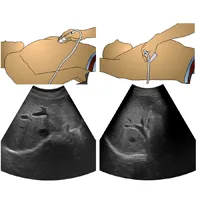
Liver tumor ablation is often guided by ultrasound (US). Due to poor image quality, intraoperative US is fused with preoperative computed tomography or magnetic tomography (CT/MR) images to provide visual guidance. As of today, the underlying 2D US to 3D CT/MR registration problem remains a very challenging task.
Methods
We propose a novel pipeline to address this registration problem. Contrary to previous work, we do not formulate the problem as a regression task, which - for the given registration problem - achieves a low performance regarding accuracy and robustness due to the limited US soft-tissue contrast and the inter-patient variability on liver vessels. Instead, we first estimate the US probe angle roughly by using a classification network. Given this coarse initialization, we then improve the registration by formulation of the problem as a segmentation task, estimating the US plane in the 3D CT/MR through segmentation.
Results
We benchmark our approach on 1035 clinical images from 52 patients, yielding average registration errors of 11.6° and 4.7 mm, which outperforms the state of the art SVR method[1].
Conclusion
Our results show the efficiency of the proposed registration pipeline, which has potential to improve the robustness and accuracy of intraoperative patient registration.},
keywords = {},
pubstate = {published},
tppubtype = {article}
}
Liver tumor ablation is often guided by ultrasound (US). Due to poor image quality, intraoperative US is fused with preoperative computed tomography or magnetic tomography (CT/MR) images to provide visual guidance. As of today, the underlying 2D US to 3D CT/MR registration problem remains a very challenging task.
Methods
We propose a novel pipeline to address this registration problem. Contrary to previous work, we do not formulate the problem as a regression task, which - for the given registration problem - achieves a low performance regarding accuracy and robustness due to the limited US soft-tissue contrast and the inter-patient variability on liver vessels. Instead, we first estimate the US probe angle roughly by using a classification network. Given this coarse initialization, we then improve the registration by formulation of the problem as a segmentation task, estimating the US plane in the 3D CT/MR through segmentation.
Results
We benchmark our approach on 1035 clinical images from 52 patients, yielding average registration errors of 11.6° and 4.7 mm, which outperforms the state of the art SVR method[1].
Conclusion
Our results show the efficiency of the proposed registration pipeline, which has potential to improve the robustness and accuracy of intraoperative patient registration.
Alpers, J; Reimert, D; Rötzer, M; Gerlach, T; Gutberlet, M; Wacker, F; Hensen, B; Hansen, C
2.5D Thermometry Maps for MRI-guided Tumor Ablation Book Section
In: vol. 12904, pp. 311–320, 2021, (arXiv:2108.05734 [eess]).
@incollection{alpers_25d_2021,
title = {2.5D Thermometry Maps for MRI-guided Tumor Ablation},
author = {J Alpers and D Reimert and M Rötzer and T Gerlach and M Gutberlet and F Wacker and B Hensen and C Hansen},
url = {http://arxiv.org/abs/2108.05734},
doi = {10.1007/978-3-030-87202-1_30},
year = {2021},
date = {2021-01-01},
urldate = {2021-01-01},
volume = {12904},
pages = {311–320},
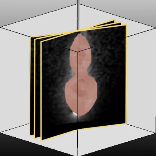
abstract = {Fast and reliable monitoring of volumetric heat distribution during MRI-guided tumor ablation is an urgent clinical need. In this work, we introduce a method for generating 2.5D thermometry maps from uniformly distributed 2D MRI phase images rotated around the applicator's main axis. The images can be fetched directly from the MR device, reducing the delay between image acquisition and visualization. For reconstruction, we use a weighted interpolation on a cylindric coordinate representation to calculate the heat value of voxels in a region of interest. A pilot study on 13 ex vivo bio protein phantoms with flexible tubes to simulate a heat sink effect was conducted to evaluate our method. After thermal ablation, we compared the measured coagulation zone extracted from the post-treatment MR data set with the output of the 2.5D thermometry map. The results show a mean Dice score of 0.75+-0.07, a sensitivity of 0.77+-0.03, and a reconstruction time within 18.02ms+-5.91ms. Future steps should address improving temporal resolution and accuracy, e.g., incorporating advanced bioheat transfer simulations.},
note = {arXiv:2108.05734 [eess]},
keywords = {},
pubstate = {published},
tppubtype = {incollection}
}
2020
Wei, W; Rak, M; Alpers, J; Hansen, C
Towards Fully Automatic 2D Us to 3D CT/MR Registration: A Novel Segmentation-Based Strategy Proceedings Article
In: 2020 IEEE 17th International Symposium on Biomedical Imaging (ISBI), pp. 433–437, IEEE, Iowa City, IA, USA, 2020, ISBN: 978-1-5386-9330-8.
@inproceedings{wei_towards_2020,

title = {Towards Fully Automatic 2D Us to 3D CT/MR Registration: A Novel Segmentation-Based Strategy},

author = {W Wei and M Rak and J Alpers and C Hansen},

url = {https://ieeexplore.ieee.org/document/9098379/},

doi = {10.1109/ISBI45749.2020.9098379},

isbn = {978-1-5386-9330-8},

year  = {2020},

date = {2020-04-01},

urldate = {2025-08-14},

booktitle = {2020 IEEE 17th International Symposium on Biomedical Imaging (ISBI)},

pages = {433–437},

publisher = {IEEE},

address = {Iowa City, IA, USA},

abstract = {2D-US to 3D-CT/MR registration is a crucial module during minimally invasive ultrasound-guided liver tumor ablations. Many modern registration methods still require manual or semi-automatic slice pose initialization due to insufficient robustness of automatic methods. The state-of-the-art regression networks do not work well for liver 2D US to 3D CT/MR registration because of the tremendous inter-patient variability of the liver anatomy. To address this unsolved problem, we propose a deep learning network pipeline which instead of a regression starts with a classification network to recognize the coarse ultrasound transducer pose followed by a segmentation network to detect the target plane of the US image in the CT/MR volume. The rigid registration result is derived using plane regression. In contrast to the state-of-the-art regression networks, we do not estimate registration parameters from multi-modal images directly, but rather focus on segmenting the target slice plane in the volume. The experiments reveal that this novel registration strategy can identify the initial slice phase in a 3D volume more reliably than the standard regression-based techniques. The proposed method was evaluated with 1035 US images from 52 patients. We achieved angle and distance errors of 12.7 ± 6.2 degrees and 4.9 ± 3.1 mm, clearly outperforming the state-of-the-art regression strategy which results in 37.0 ± 15.6 degrees angle error and 19.0 ± 11.6 mm distance error.},

keywords = {},

pubstate = {published},

tppubtype = {inproceedings}

}
2019
Wei, W; Xu, H; Alpers, J; Tianbao, Z; Wang, L; Rak, M; Hansen, C
Fast Registration for Liver Motion Compensation in Ultrasound-Guided Navigation Proceedings Article
In: 2019 IEEE 16th International Symposium on Biomedical Imaging (ISBI 2019), pp. 1132–1136, IEEE, Venice, Italy, 2019, ISBN: 978-1-5386-3641-1.
@inproceedings{wei_fast_2019,

title = {Fast Registration for Liver Motion Compensation in Ultrasound-Guided Navigation},

author = {W Wei and H Xu and J Alpers and Z Tianbao and L Wang and M Rak and C Hansen},

url = {https://ieeexplore.ieee.org/document/8759464/},

doi = {10.1109/ISBI.2019.8759464},

isbn = {978-1-5386-3641-1},

year  = {2019},

date = {2019-04-01},

urldate = {2025-08-14},

booktitle = {2019 IEEE 16th International Symposium on Biomedical Imaging (ISBI 2019)},

pages = {1132–1136},

publisher = {IEEE},

address = {Venice, Italy},

abstract = {In recent years, image-guided thermal ablations have become a considerable treatment method for cancer patients, including support through navigational systems. One of the most critical challenges in these systems is the registration between the intraoperative images and the preoperative volume. The motion secondary to inspiration makes registration even more difficult. In this work, we propose a coarse-fine fast patient registration technique to solve the problem of motion compensation. In contrast to other state-of-the-art methods, we focus on improving the convergence range of registration. To this end, we make use of a Deep Learning 2D UNet framework to extract the vessels and liver borders from intraoperative ultrasound images and employ the segmentation results as regions of interest in the registration. After an initial 3D-3D registration during breath hold, the following motion compensation is achieved using a 2D-3D registration. Our approach yields a convergence rate of over 70% with an accuracy of 1.97 ± 1.07 mm regarding the target registration error. The 2D-3D registration is GPU-accelerated with a time cost of less than 200 ms.},

keywords = {},

pubstate = {published},

tppubtype = {inproceedings}

}
Alpers, J; Hensen, B; Wacker, F; Rieder, C; Hansen, C
MRI-Guided Liver Tumor Ablation - A Workflow Design Prototype Journal Article
In: 2019.
@article{alpers_mri-guided_2019,

title = {MRI-Guided Liver Tumor Ablation - A Workflow Design Prototype},

author = {J Alpers and B Hensen and F Wacker and C Rieder and C Hansen},

year  = {2019},

date = {2019-01-01},

abstract = {Thermal ablation procedures such as radiofrequency ablation have become a clinically accepted treatment method for liver tumors. Using image guidance like magnetic resonance imaging (MRI), a needle-shaped instrument is navigated to the target position aiming at a complete destruction of the focal liver malignancies. Planning the intervention, navigating the instrument to the target position, and monitoring the ablation of the malignant tissue are currently three separated steps during MRI guided interventions. This is hampering the clinical workflow and results in an unnecessary amount of additional work for the clinicians due to data export and import or mental data transfer.},

keywords = {},

pubstate = {published},

tppubtype = {article}

}
Gabele, M; Thoms, A; Alpers, J; Hußlein, S; Hansen, C
Non-player character as a companion in cognitive rehabilitation for adults - Characteristics and representation Journal Article
In: 2019.
@article{gabele_non-player_2019,

title = {Non-player character as a companion in cognitive rehabilitation for adults - Characteristics and representation},

author = {M Gabele and A Thoms and J Alpers and S Hußlein and C Hansen},

year  = {2019},

date = {2019-01-01},

abstract = {A lack of social support reduces the chances of successful rehabilitation. However, the social environment changes considerably during this time. Therefore, fostering consistent social contacts is highly relevant. To address social relatedness in software-based cognitive therapy during rehabilitation we intend to use a non-player character as a companion. In this work we analyze possible forms of representation of the companion based on required characteristics, age and gender to achieve this goal. These were set in relation to age and gender of the user. Three female and three male companions in three age groups were created and subsequently tested in an explorative feasibility study with 40 participants. 50% of participants preferred a female middle-aged companion, 25% a younger male. Older companions were chosen only by women. Regarding the gender, 62.5% chose a female companion. We present an orientation for development of non-player characters as companion in software-based training for cognitive rehabilitation.},

keywords = {},

pubstate = {published},

tppubtype = {article}

}
Gabele, M; Thoms, A; Alpers, J; Hußlein, S; Hansen, C
Effects of interactive storytelling and quests in cognitive rehabilitation for adults Journal Article
In: 2019.
@article{gabele_effects_2019,

title = {Effects of interactive storytelling and quests in cognitive rehabilitation for adults},

author = {M Gabele and A Thoms and J Alpers and S Hußlein and C Hansen},

year  = {2019},

date = {2019-01-01},

abstract = {Software-based training in cognitive rehabilitation is often perceived as boring when used over a longer period. This may negatively affect adherence and therefore reduce therapy success. Regular use is essential and should be continued after clinical stay. In this work, medically approved therapy software for cognitive training of divided attention in clinical use is extended with suitable gamification elements. Based on interactive storytelling, we created a main quest for single-player about two training sections and demonstrate a method by a prototype to wrap it around the therapy. Based on an exploratory study in Germany with patients (n=4) and a control group with healthy participants (n=6), we found a subjectively perceived motivational tendency for patients to use the software without being exhausted or losing concentration. Patients stated interest in further use. This work lays a basis to influence motivation, for further clinical evaluations, and shows important fields for future research.},

keywords = {},

pubstate = {published},

tppubtype = {article}

}
2017
Alpers, J; Hansen, C; Ringe, K; Rieder, C
CT-Based Navigation Guidance for Liver Tumor Ablation Journal Article
In: 2017.
@article{alpers_ct-based_2017,

title = {CT-Based Navigation Guidance for Liver Tumor Ablation},

author = {J Alpers and C Hansen and K Ringe and C Rieder},

url = {https://www.var.ovgu.de/pub/2017_VCBM_Alpers.pdf},

year  = {2017},

date = {2017-01-01},

abstract = {Image-guided thermal ablation procedures such as microwave ablation (MWA) or radiofrequency ablation (RFA) have become clinically accepted treatment options for liver tumors. The goal of these minimally invasive procedures is the destruction of focal liver malignancies using mostly needle-shaped instruments. Computed tomography (CT) imaging may be used to navigate the applicator to the target position in order to achieve complete tumor ablation. Due to limited image quality and resolution, the treatment target and risk structures may be hardly visible in intra-interventional CT-images, hampering verification of the intended applicator position.},

keywords = {},

pubstate = {published},

tppubtype = {article}

}