
2026
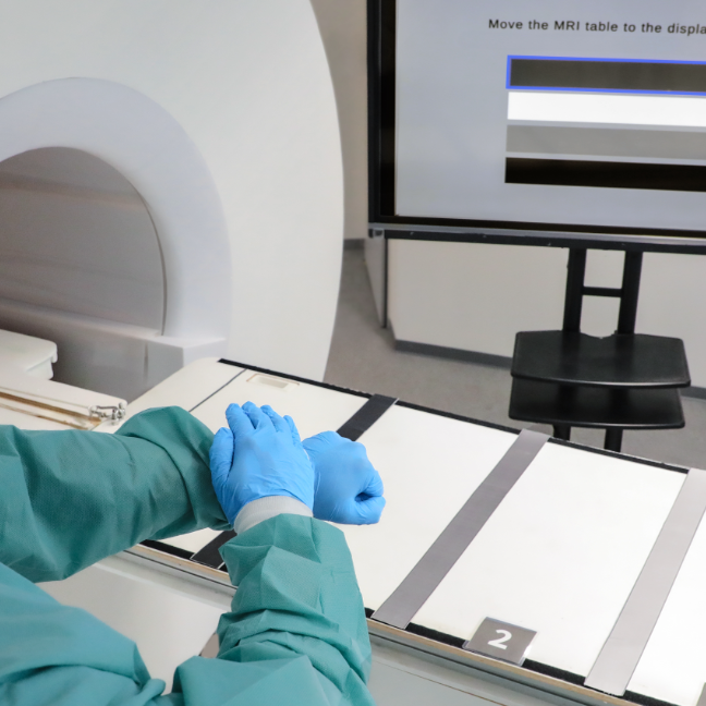
Klank, R; Heinrich, F; Schott, D; Hansen, C; Joeres, F
Testing before Building? Strengths and Weaknesses of VR-Based Usability Testing of a Gesture Interface Proceedings Article Forthcoming
In: ACM Symposium on Virtual Reality Software and Technology (VRST), Forthcoming.
@inproceedings{klank2026,

title = {Testing before Building? Strengths and Weaknesses of VR-Based Usability Testing of a Gesture Interface},

author = {R Klank and F Heinrich and D Schott and C Hansen and F Joeres },

year  = {2026},

date = {2026-11-16},

urldate = {2026-11-16},

booktitle = {ACM Symposium on Virtual Reality Software and Technology (VRST)},

keywords = {},

pubstate = {forthcoming},

tppubtype = {inproceedings}

}
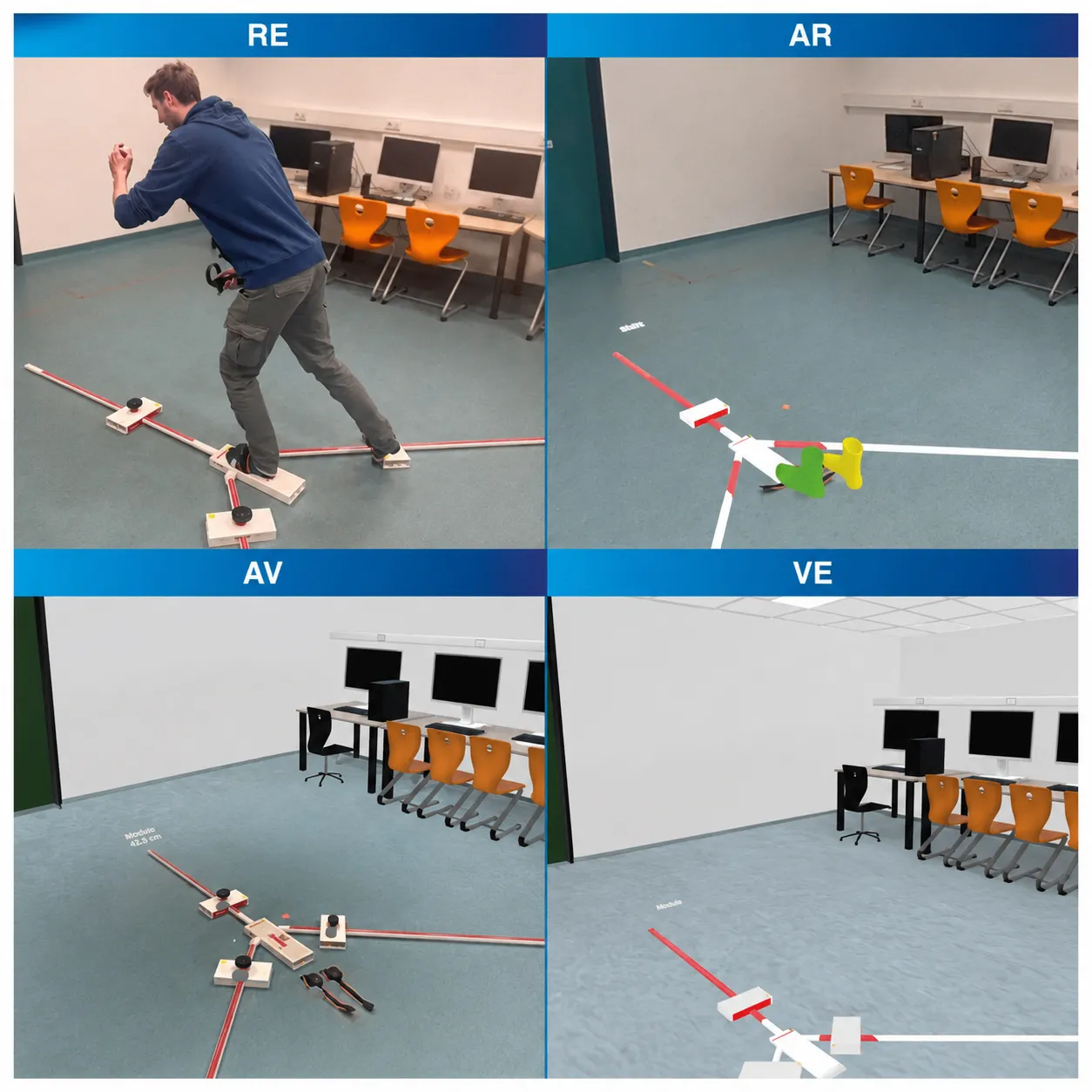
Schwadtke, A; Pastel, S; Kunz, M; Müller, P; Braun-Dullaeus, R; Hansen, C; Witte, K; Schott, D
Striving for Stability: Evaluating the Feasibility of Dynamic Balance Skills Across the Reality–Virtuality Continuum Proceedings Article Forthcoming
In: IEEE International Symposium on Mixed and Augmented Reality Adjunct (ISMAR-Adjunct), Forthcoming.
@inproceedings{schwadtke2026,

title = {Striving for Stability: Evaluating the Feasibility of Dynamic Balance Skills Across the Reality–Virtuality Continuum},

author = {A Schwadtke and S Pastel and M Kunz and P Müller and R Braun-Dullaeus and C Hansen and K Witte and D Schott},

year  = {2026},

date = {2026-10-07},

urldate = {2026-10-07},

booktitle = {IEEE International Symposium on Mixed and Augmented Reality Adjunct (ISMAR-Adjunct)},

journal = {ISMAR 2026 Posters},

abstract = {Dynamic balance assessment is important in sports and rehabilitation. Mixed Reality (MR) offers new opportunities for portable diagnostics. This study investigated whether the standardized YBalance Test Lower Quarter (YBT) can be administered in MR nvironments. Twenty-four participants performed the YBT in a real environment (RE), Augmented Reality (AR), Augmented Virtuality (AV), and Virtual Environment (VE) on two test days. Despite higher perceived demands, MR-based YBT outcomes were comparable to RE across all MR conditions (p > 0.05), with good to-moderate test-retest reliability. These findings support the feasibility of MR-based YBT assessment for diagnostics, warranting further validation.},

howpublished = {ISMAR Posters},

keywords = {},

pubstate = {forthcoming},

tppubtype = {inproceedings}

}
Klus, C; Bosch, J; Ludwig, C; Kunz, M; Hansen, C; Stoevesandt, D; Braun-Dullaeus, R; Schott, D
Curriculare Akzeptanz von XR in der medizinischen Ausbildung: Einfluss von XR-Erleben, Wissenszuwachs und Vorerfahrungen in einer Pilotstudie mit Medizinstudierenden Proceedings Article
In: Jahrestagung der Gesellschaft für Medizinische Ausbildung (GMA), Dresden, Germany, 2026, (DocV-0404).
@inproceedings{klus2026,

title = {Curriculare Akzeptanz von XR in der medizinischen Ausbildung: Einfluss von XR-Erleben, Wissenszuwachs und Vorerfahrungen in einer Pilotstudie mit Medizinstudierenden},

author = {C Klus and J Bosch and C Ludwig and M Kunz and C Hansen and D Stoevesandt and R Braun-Dullaeus and D Schott},

url = {https://series.publisso.de/en/conferences/gma2026/26gma022},

doi = {10.3205/26gma022},

year  = {2026},

date = {2026-09-17},

urldate = {2026-09-17},

booktitle = {Jahrestagung der Gesellschaft für Medizinische Ausbildung (GMA)},

address = {Dresden, Germany},

note = {DocV-0404},

keywords = {},

pubstate = {published},

tppubtype = {inproceedings}

}
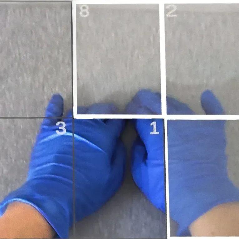
Schwenderling, L; Schotte, M; Joeres, F; Heinrich, F; Hansen, C
Augmented Reality Training of Attention Allocation in Medical Dual-Task Learning Applications Proceedings Article Forthcoming
In: ACM International Conference on Mobile Human-Computer Interaction 2026 (MobileHCI 2026), Forthcoming.
@inproceedings{schwenderling2026,

title = {Augmented Reality Training of Attention Allocation in Medical Dual-Task Learning Applications},

author = {L Schwenderling and M Schotte and F Joeres and F Heinrich and C Hansen},

year  = {2026},

date = {2026-08-31},

urldate = {2026-08-31},

booktitle = {ACM International Conference on Mobile Human-Computer Interaction 2026 (MobileHCI 2026)},

journal = {The ACM International Conference on Mobile Human-Computer Interaction 2026 (MobileHCI 2026)},

keywords = {},

pubstate = {forthcoming},

tppubtype = {inproceedings}

}
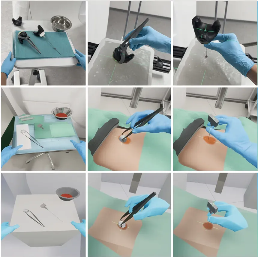
Heinrich, F; Kohpeiß, M; Hansen, C; Schott, D
Mixed Reality Simulation of Guided Needle Insertion: Effects of Interaction and Visual Fidelity Journal Article
In: International Journal of Computer Assisted Radiology and Surgery, 2026.
@article{heinrich2026a,

title = {Mixed Reality Simulation of Guided Needle Insertion: Effects of Interaction and Visual Fidelity},

author = {F Heinrich and M Kohpeiß and C Hansen and D Schott},

url = {https://link.springer.com/content/pdf/10.1007/s11548-026-03756-3.pdf},

doi = {10.1007/s11548-026-03756-3},

year  = {2026},

date = {2026-08-10},

urldate = {2026-08-10},

journal = {International Journal of Computer Assisted Radiology and Surgery},

keywords = {},

pubstate = {published},

tppubtype = {article}

}
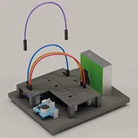
Batz, V; Cavkovski, P; Loebel, JM; Lameski, P; Trajkovik, V; Hansen, C; Herzog, MA
Usability Assessment of Low-cost Sensor Kits for Educational Air Quality Sensing Proceedings Article
In: HCI International (HCII), Lecture Notes in Computer Science, pp. 23-42, 2026.
@inproceedings{nokeyc,

title = {Usability Assessment of Low-cost Sensor Kits for Educational Air Quality Sensing},

author = {V Batz and P Cavkovski and JM Loebel and P Lameski and V Trajkovik and C Hansen and MA Herzog},

url = {https://link.springer.com/chapter/10.1007/978-3-032-30542-8_2},

doi = {10.1007/978-3-032-30542-8_2},

year  = {2026},

date = {2026-07-07},

urldate = {2026-07-07},

booktitle = {HCI International (HCII), Lecture Notes in Computer Science},

volume = {16731},

pages = {23-42},

abstract = {This study assesses the usability of the Pulse.Eco Combo Cube v3 particulate‑matter sensor kit and examines its suitability as an educational toolkit for citizen science in schools. The goal is to develop a low-cost DIY sensor kit that can be used for participatory data collection in school MAKER workshops, thereby promoting environmental awareness and environmentally conscious behavior among students. We further show that our design-thinking workflow generalizes to other educational sensing toolkits.},

keywords = {},

pubstate = {published},

tppubtype = {inproceedings}

}
Schrader, C; Diekmann, C; Kunz, M; Albrecht, A; Hansen, C; Schott, D
In: Computers & Education: X Reality, vol. 9, pp. 100172, 2026.
@article{schrader2026,

title = {Applying the Signaling Principle in Immersive Virtual Reality for Medical Education: Effects of Color Coding on Virtual Presence, Learning-Centered Emotions, Cognitive Load, and Learning Outcomes },

author = {C Schrader and C Diekmann and M Kunz and A Albrecht and C Hansen and D Schott},

url = {https://www.sciencedirect.com/science/article/pii/S2949678026000401},

doi = {10.1016/j.cexr.2026.100172},

year  = {2026},

date = {2026-07-01},

urldate = {2026-07-01},

journal = {Computers & Education: X Reality},

volume = {9},

pages = {100172},

keywords = {},

pubstate = {published},

tppubtype = {article}

}
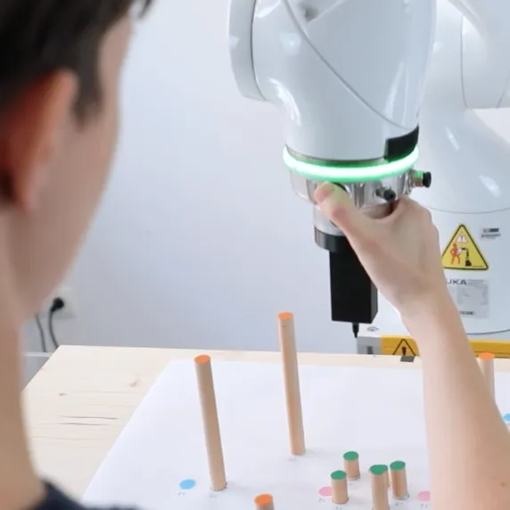
Schreiter, J; Pandi, P; Klank, R; Hansen, C; Heinrich, F
Touch-Based Interaction and Clutching Strategies in Collaborative Robotics across Task Types Proceedings Article Forthcoming
In: IEEE/RSJ International Conference on Intelligent Robots and Systems (IROS), Forthcoming.
@inproceedings{schreiter2026,

title = {Touch-Based Interaction and Clutching Strategies in Collaborative Robotics across Task Types},

author = {J Schreiter and P Pandi and R Klank and C Hansen and F Heinrich},

year  = {2026},

date = {2026-06-30},

urldate = {2026-06-30},

booktitle = {IEEE/RSJ International Conference on Intelligent Robots and Systems (IROS)},

keywords = {},

pubstate = {forthcoming},

tppubtype = {inproceedings}

}
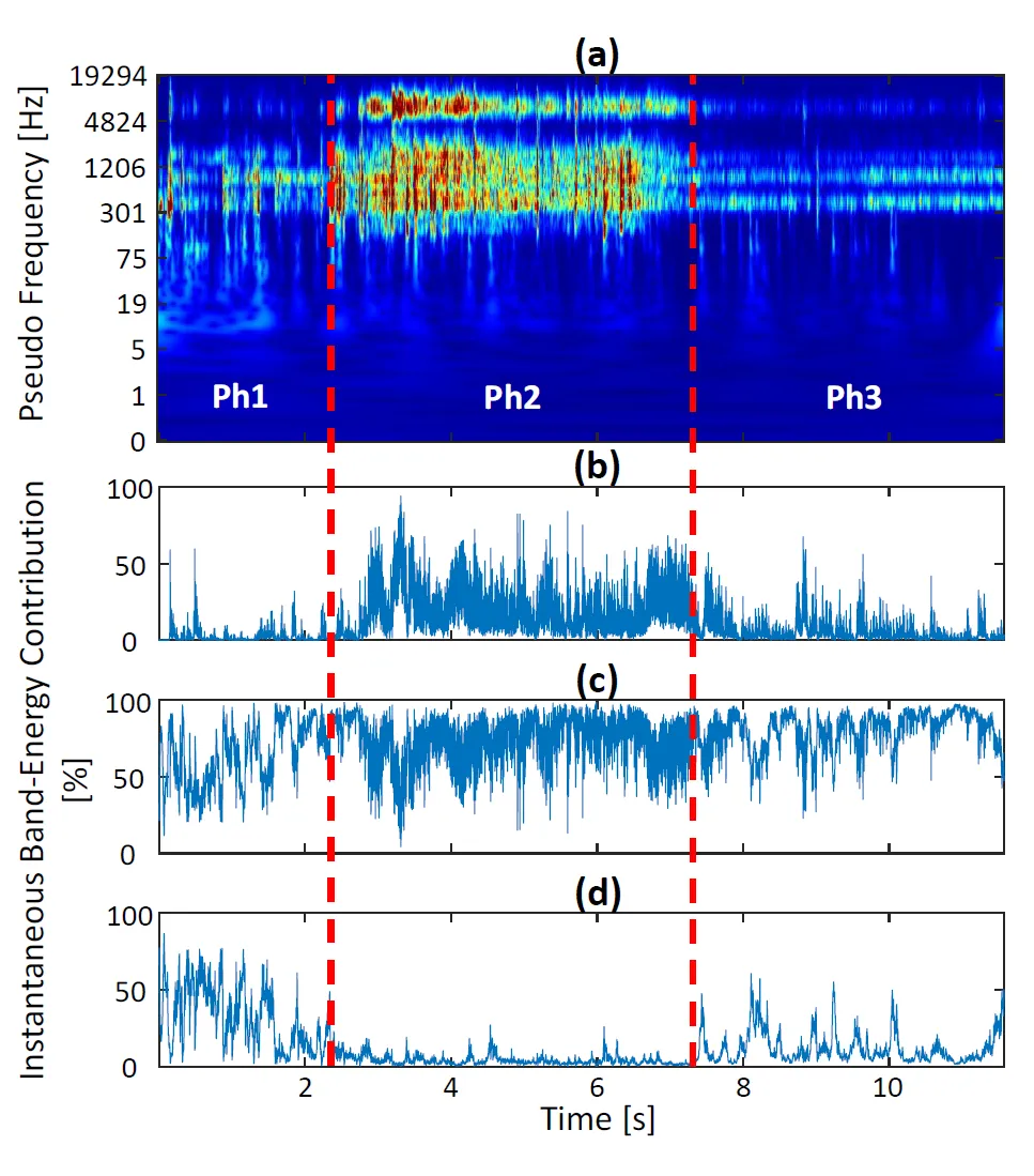
Esmaeili, N; Urrutia, R; Espejo, D; Boese, A; Horvath, P; Hansen, C; Hagenah, J; Illanes, A
When Tissue Speaks: Vibroacoustic Monitoring of Tissue Response during Electrosurgery Proceedings Article Forthcoming
In: Medical Image Computing and Computer Assisted Intervention (MICCAI), Forthcoming.
@inproceedings{esmaeili2026,

title = {When Tissue Speaks: Vibroacoustic Monitoring of Tissue Response during Electrosurgery},

author = {N Esmaeili and R Urrutia and D Espejo and A Boese and P Horvath and C Hansen and J Hagenah and A Illanes},

year  = {2026},

date = {2026-06-19},

urldate = {2026-06-19},

publisher = {Medical Image Computing and Computer Assisted Intervention (MICCAI)},

keywords = {},

pubstate = {forthcoming},

tppubtype = {inproceedings}

}
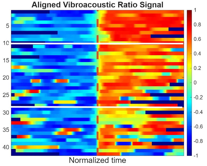
Urrutia, R; Esmaeili, N; Matinfar, S; Boese, A; Davaris, N; Arens, C; Hansen, C; Illanes, A
Where Is the Needle Tip? Vibroacoustic Sensing Reveals Hidden Needle-Tissue Transitions During Ultrasound Guidance Proceedings Article Forthcoming
In: Medical Image Computing and Computer Assisted Intervention (MICCAI), Forthcoming.
@inproceedings{urrutia2026,

title = {Where Is the Needle Tip? Vibroacoustic Sensing Reveals Hidden Needle-Tissue Transitions During Ultrasound Guidance},

author = {R Urrutia and N Esmaeili and S Matinfar and A Boese and N Davaris and C Arens and C Hansen and A Illanes},

year  = {2026},

date = {2026-06-18},

urldate = {2026-06-18},

issuetitle = {Medical Image Computing and Computer Assisted Intervention (MICCAI)},

publisher = {Medical Image Computing and Computer Assisted Intervention (MICCAI)},

keywords = {},

pubstate = {forthcoming},

tppubtype = {inproceedings}

}
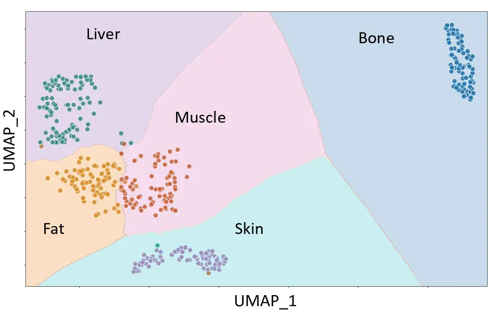
Illanes, A; Esmaeili, N; Urrutia, R; Espejo, D; Hagenah, J; Hansen, C; Boese, A
Ex Vivo Tissue Differentiation Using Vibroacoustic Sensing at the Proximal End of Laparoscopic Instruments Proceedings Article Forthcoming
In: IEEE Engineering in Medicine and Biology Society (EMBC), Forthcoming.
@inproceedings{illanes2026,

title = {Ex Vivo Tissue Differentiation Using Vibroacoustic Sensing at the Proximal End of Laparoscopic Instruments},

author = {A Illanes and N Esmaeili and R Urrutia and D Espejo and J Hagenah and C Hansen and A Boese},

year  = {2026},

date = {2026-06-17},

urldate = {2026-06-17},

booktitle = { IEEE Engineering in Medicine and Biology Society (EMBC)},

keywords = {},

pubstate = {forthcoming},

tppubtype = {inproceedings}

}
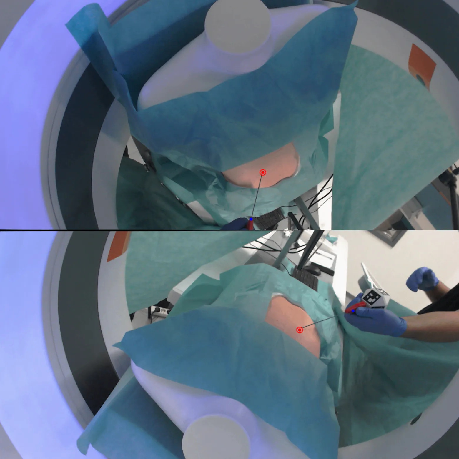
Steiger, M; Rezapourian, M; Rak, M; Hansen, C
Dynamic Uncertainty-level Assessment Framework for Real-time Needle tracking in CT-guided Surgical Environments Journal Article
In: International Journal of Computer Assisted Radiology and Surgery, 2026.
@article{steiger2026a,

title = {Dynamic Uncertainty-level Assessment Framework for Real-time Needle tracking in CT-guided Surgical Environments},

author = {M Steiger and M Rezapourian and M Rak and C Hansen},

url = {https://link.springer.com/article/10.1007/s11548-026-03725-w},

doi = {10.1007/s11548-026-03725-w},

year  = {2026},

date = {2026-06-05},

urldate = {2026-06-05},

journal = {International Journal of Computer Assisted Radiology and Surgery},

abstract = {Abstract Purpose: Accurate needle tracking is critical for the success of computed tomography (CT)-guided interventions, where even minor deviations may compromise procedural safety and clinical outcomes. However, existing image-guided tracking systems typically lack mechanisms to quantify and communicate the reliability of their predictions in real time, leaving clinicians to act on guidance of uncertain trustworthiness.


Methods: We propose a Dynamic Uncertainty Level Assessment Framework that provides a quantitative, real-time estimate of tracking reliability by linearly linking a predicted uncertainty score to spatial tracking error. The framework consists of three different approaches: (1) a classic method based on dynamically weighted, interpretable reliability metrics; (2) a lightweight convolutional neural network (CNN) that predicts uncertainty directly from multi-view image data; and (3) a hybrid CNN that adaptively optimizes metric weights while preserving interpretability. The uncertainty level is defined on a fixed scale, with 0% corresponding to ideal tracking (error of 0 mm) and 100% to a tracking error of 10 mm.


Results: Experimental validation on clinical and clinically realistic laboratory datasets of ∼30,000 frames demonstrates a strong positive correlation between uncertainty and error (Pearson r > 0.82) and achieves a stable tracking error estimation (error difference < 0.6 mm) with real-time performance (5 ms per frame). Conclusion: By enabling an intuitive uncertainty-to-error mapping, the proposed framework supports more informed intraprocedural decision-making, enhances operator trust in guidance data, and establishes a practical basis for integration into uncertainty-aware CT-guided intervention systems.},

keywords = {},

pubstate = {published},

tppubtype = {article}

}
Methods: We propose a Dynamic Uncertainty Level Assessment Framework that provides a quantitative, real-time estimate of tracking reliability by linearly linking a predicted uncertainty score to spatial tracking error. The framework consists of three different approaches: (1) a classic method based on dynamically weighted, interpretable reliability metrics; (2) a lightweight convolutional neural network (CNN) that predicts uncertainty directly from multi-view image data; and (3) a hybrid CNN that adaptively optimizes metric weights while preserving interpretability. The uncertainty level is defined on a fixed scale, with 0% corresponding to ideal tracking (error of 0 mm) and 100% to a tracking error of 10 mm.
Results: Experimental validation on clinical and clinically realistic laboratory datasets of ∼30,000 frames demonstrates a strong positive correlation between uncertainty and error (Pearson r > 0.82) and achieves a stable tracking error estimation (error difference < 0.6 mm) with real-time performance (5 ms per frame). Conclusion: By enabling an intuitive uncertainty-to-error mapping, the proposed framework supports more informed intraprocedural decision-making, enhances operator trust in guidance data, and establishes a practical basis for integration into uncertainty-aware CT-guided intervention systems.
Steeg, K; Urrutia, R; Illanes, A; Fuentealba, P; Gawron, J; Strama, K; Hansen, C; Scherberich, J; Windfelder, A; Krombach, G; Friebe, M
Multimodal Alignment of MicroCT Imaging to Vibroacoustic Signals to Validate Soft Tissue Needle Transitions in Manduca sexta Journal Article
In: 2026.
@article{Steeg_2026,

title = {Multimodal Alignment of MicroCT Imaging to Vibroacoustic Signals to Validate Soft Tissue Needle Transitions in \textit{Manduca sexta}},

author = {K Steeg and R Urrutia and A Illanes and P Fuentealba and J Gawron and K Strama and C Hansen and J Scherberich and A Windfelder and G Krombach and M Friebe},

url = {http://dx.doi.org/10.64898/2026.06.07.730726},

doi = {10.64898/2026.06.07.730726},

year  = {2026},

date = {2026-06-01},

urldate = {2026-06-01},

publisher = {openRxiv},

keywords = {},

pubstate = {published},

tppubtype = {article}

}
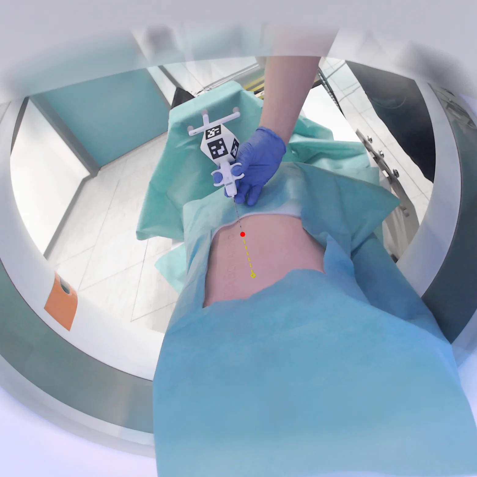
Steiger, M; Mielke, T; Bashkanov, O; Großer, OS; Pech, M; Hansen, C; Rak, M
Real-time Marker-less Needle Tracking for CT-guided Interventions using Multiple RGB Cameras Journal Article
In: International Journal of Computer Assisted Radiology and Surgery, 2026, (in print).
@article{steiger2026b,

title = {Real-time Marker-less Needle Tracking for CT-guided Interventions using Multiple RGB Cameras},

author = {M Steiger and T Mielke and O Bashkanov and OS Großer and M Pech and C Hansen and M Rak},

url = {https://link.springer.com/article/10.1007/s11548-026-03622-2

https://pubmed.ncbi.nlm.nih.gov/42033583/, PubMed},

doi = {10.1007/s11548-026-03622-2},

year  = {2026},

date = {2026-05-10},

urldate = {2026-05-10},

journal = {International Journal of Computer Assisted Radiology and Surgery},

abstract = {Purpose: In CT-guided interventions, instrument tracking reduces patient risk while improving the overall outcome. Though useful, marker-based tracking systems suffer from occlusion, are complex to set up, and fail when needles deflect. A markerless tracking system could overcome these challenges, enabling streamlined integration.

Methods: We developed and evaluated a proof-of-concept system combining standard RGB cameras with a U-Net-ConvNeXt architecture for needle detection and tracking. We compiled a fully annotated dataset of 35,000 frames, spanning multiple needle types and scenarios. We evaluated tracking accuracy against state-of-the-art marker-based systems, utilizing an IR-based and an image-based system on static positioning, dynamic movement, and needle-deflection scenarios. Moreover, our marker-less system was evaluated using both known and unknown needle types.

Results: Our proof-of-concept system demonstrated a 3D tracking error of 2.70 ± 1.07mm at 25FPS with 3 cameras, outperforming ArUco marker-based tracking (5.29 ± 1.85mm). Tracking accuracy remained consistent for unknown needle types not included in the training data, suggesting strong generalization. The system maintained a 99.4% needle-tip detection rate across all frames with robust performance during needle deflection, while marker-based systems failed under identical conditions. With only 2 cameras, the tracking error of our system increased modestly (4.81 ± 2.38mm).

Conclusion: Our proof-of-concept demonstrates the feasibility of marker-less needle tracking, suggesting potential for clinically relevant accuracy, reliability, and reduced complexity while exhibiting robustness to needle deflection and varying needle appearances. These preliminary results indicate promising directions for future clinical development.},

note = {in print},

keywords = {},

pubstate = {published},

tppubtype = {article}

}
Methods: We developed and evaluated a proof-of-concept system combining standard RGB cameras with a U-Net-ConvNeXt architecture for needle detection and tracking. We compiled a fully annotated dataset of 35,000 frames, spanning multiple needle types and scenarios. We evaluated tracking accuracy against state-of-the-art marker-based systems, utilizing an IR-based and an image-based system on static positioning, dynamic movement, and needle-deflection scenarios. Moreover, our marker-less system was evaluated using both known and unknown needle types.
Results: Our proof-of-concept system demonstrated a 3D tracking error of 2.70 ± 1.07mm at 25FPS with 3 cameras, outperforming ArUco marker-based tracking (5.29 ± 1.85mm). Tracking accuracy remained consistent for unknown needle types not included in the training data, suggesting strong generalization. The system maintained a 99.4% needle-tip detection rate across all frames with robust performance during needle deflection, while marker-based systems failed under identical conditions. With only 2 cameras, the tracking error of our system increased modestly (4.81 ± 2.38mm).
Conclusion: Our proof-of-concept demonstrates the feasibility of marker-less needle tracking, suggesting potential for clinically relevant accuracy, reliability, and reduced complexity while exhibiting robustness to needle deflection and varying needle appearances. These preliminary results indicate promising directions for future clinical development.
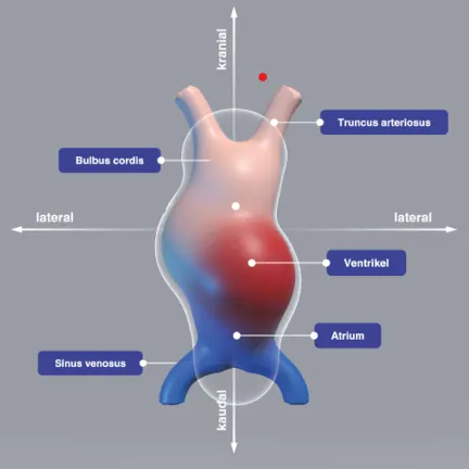
Schott, D; Kunz, M; Schrader, C; Ringler, E; Schwadtke, A; Mandel, J; Müller, P; Kleinbeck, C; Roth, D; Albrecht, A; Hansen, C
The Influence of Environmental Fidelity on Virtual Presence, Intrinsic Motivation, Cognitive Load and Learning Outcomes in Medical VR Journal Article
In: IEEE Transactions on Visualization and Computer Graphics, vol. 32, no. 5, pp. 4302-4311, 2026.
@article{nokeyb,

title = {The Influence of Environmental Fidelity on Virtual Presence, Intrinsic Motivation, Cognitive Load and Learning Outcomes in Medical VR},

author = {D Schott and M Kunz and C Schrader and E Ringler and A Schwadtke and J Mandel and P Müller and C Kleinbeck and D Roth and A Albrecht and C Hansen},

url = {https://ieeexplore.ieee.org/document/11475220/},

doi = {10.1109/TVCG.2026.3680702},

year  = {2026},

date = {2026-05-01},

urldate = {2026-05-01},

journal = {IEEE Transactions on Visualization and Computer Graphics},

volume = {32},

number = {5},

pages = {4302-4311},

howpublished = {IEEE Conference on Virtual Reality and 3D User Interfaces (VR)},

keywords = {},

pubstate = {published},

tppubtype = {article}

}
Weiss, S; Hanke, L; Schröder, E; Weinberg, M; Dicks, T; Castell, C; Wessels, M; Hecht, H; Dietz, P; Huber, T; Hansen, C
Bridging the Reality Gap: Immersive VR Simulation for Enhancing Stress Resilience in Medical Students Proceedings Article
In: IEEE Conference on Virtual Reality and 3D User Interfaces Abstracts and Workshops (VRW), pp. 814-818, 2026.
@inproceedings{weiss2026,

title = {Bridging the Reality Gap: Immersive VR Simulation for Enhancing Stress Resilience in Medical Students},

author = {S Weiss and L Hanke and E Schröder and M Weinberg and T Dicks and C Castell and M Wessels and H Hecht and P Dietz and T Huber and C Hansen},

doi = {10.1109/VRW70859.2026.00156},

year  = {2026},

date = {2026-05-01},

urldate = {2026-05-01},

booktitle = {IEEE Conference on Virtual Reality and 3D User Interfaces Abstracts and Workshops (VRW)},

journal = {2026 IEEE Conference on Virtual Reality and 3D User Interfaces Abstracts and Workshops (VRW)},

pages = { 814-818},

abstract = {Medical students often face a “reality gap” between theory and clinical practice, leading to burnout. We present a system to induce stress as first step for later stress resilience trainings through immersive simulations of clinical conflicts. Based on a survey of 374 professionals, we developed four scenarios targeting stressors like “conflict with superiors” and “time pressure,” utilizing realistic locations and NPCs. Our framework uses a Meta Quest 3 and a custom Android app for real-time experimenter control. To overcome static training limitations, we explore local Large Language Models (LLMs) for generative NPC behavior. We detail a work-in-progress evaluation protocol using physiological measures (HR, HRV, BF) and questionnaires, providing a scalable framework for medical stress inoculation training.},

howpublished = { IEEE Conference on Virtual Reality and 3D User Interfaces Abstracts and Workshops},

keywords = {},

pubstate = {published},

tppubtype = {inproceedings}

}
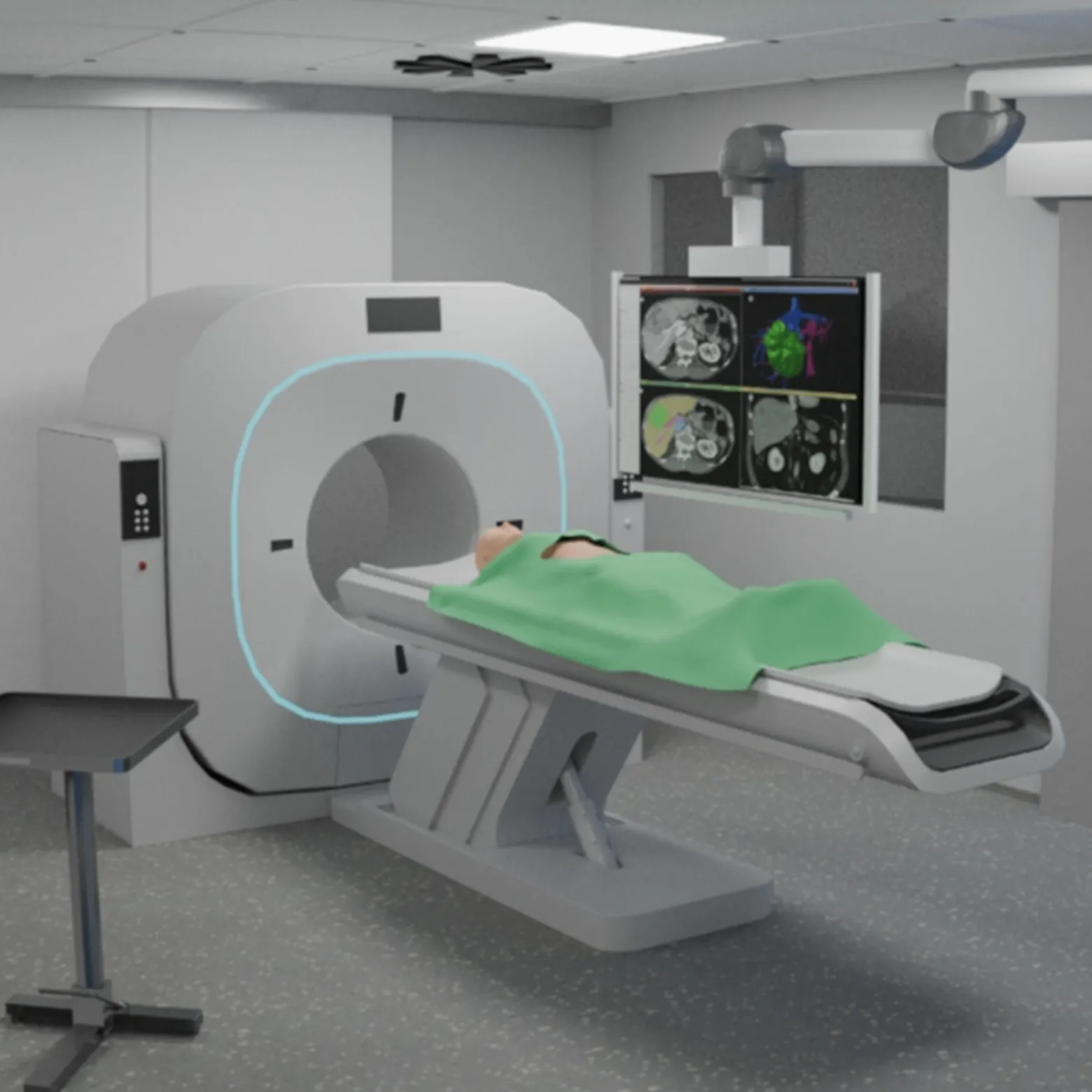
Heinrich, F; Schülke, L; Kohpeiss, M; Schott, D; Hansen, C
Towards Simulating Projector-Based Augmented Reality in Virtual Reality for Interventional Evaluation Proceedings Article
In: IEEE Conference on Virtual Reality and 3D User Interfaces Abstracts and Workshops (VRW), pp. 758-763, IEEE, 2026.
@inproceedings{heinrich2026b,

title = {Towards Simulating Projector-Based Augmented Reality in Virtual Reality for Interventional Evaluation},

author = {F Heinrich and L Schülke and M Kohpeiss and D Schott and C Hansen},

doi = {10.1109/VRW70859.2026.00145},

year  = {2026},

date = {2026-05-01},

urldate = {2026-05-01},

booktitle = {IEEE Conference on Virtual Reality and 3D User Interfaces Abstracts and Workshops (VRW)},

pages = {758-763},

publisher = {IEEE},

abstract = {Evaluating projector-based augmented reality (AR) systems for interventional guidance in clinical environments is costly, timeconsuming, and subject to strict safety constraints. We present a virtual reality (VR)–based simulation approach that enables earlystage evaluation of projector-based AR guidance concepts without requiring physical hardware. A virtual interventional room was created based on a real CT suite and combined with hand-tracked needle interaction and a simulated projector-based guidance visualization. We evaluated the system in a virtual needle insertion task using performance metrics as well as established presence and user experience questionnaires. Results indicate high perceived presence and user experience, while revealing limitations in absolute insertion accuracy. Our initial findings suggest that VR-based simulation is a promising tool for early interventional evaluation of projector-based AR concepts, particularly for usability and user experience studies.},

howpublished = { IEEE Conference on Virtual Reality and 3D User Interfaces Abstracts and Workshops},

keywords = {},

pubstate = {published},

tppubtype = {inproceedings}

}
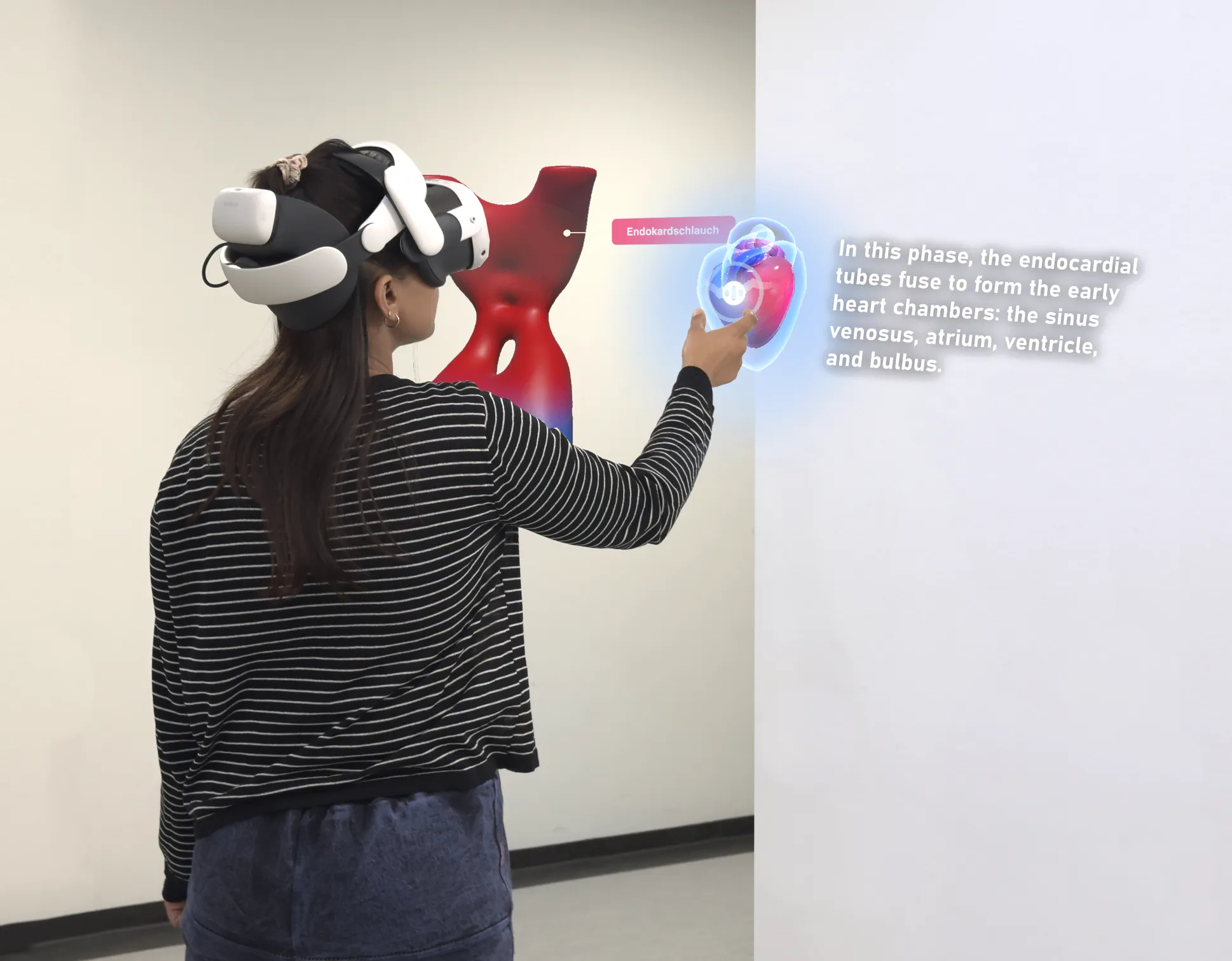
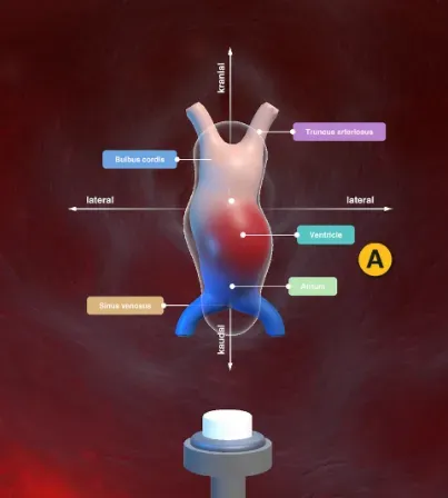
Sabir, A; Ringler, E; Mandel, J; Hansen, C; Schott, D
Evaluation of a State-Aware conversational AI agent for Immersive Anatomy Education Proceedings Article
In: IEEE Conference on Virtual Reality and 3D User Interfaces (VRW), pp. 790-794, 2026.
@inproceedings{Sabir2026,

title = {Evaluation of a State-Aware conversational AI agent for Immersive Anatomy Education},

author = {A Sabir and E Ringler and J Mandel and C Hansen and D Schott},

url = {https://ieeexplore.ieee.org/document/11489889},

doi = {10.1109/VRW70859.2026.00151},

year  = {2026},

date = {2026-04-30},

urldate = {2026-04-30},

booktitle = {IEEE Conference on Virtual Reality and 3D User Interfaces (VRW)},

pages = {790-794},

abstract = {The dynamic 3D morphology and temporal complexity of embryonic heart development make it difficult to comprehend. We introduce Reinheart, a conversational AI tutor for anatomy education that is state-aware and integrated into a mixed-reality learning environment. The AI tutor uses OpenAI services to provide voice-based tutoring in real-time with interactive 3D visualization. Using a mixed-method analysis, ten anatomy experts assessed pedagogical performance, usability, and trust. The results indicated high ease-of-use and positive social presence, while qualitative feedback revealed limited uncertainty in communication and instability in challenge tasks. Experts supported the tutor’s potential for conceptual reinforcement and guided exploration. To improve contextual reasoning and instructional reliability in MR anatomy learning, future work should extend this framework with domain-specific, uncertainty-aware AI.},

keywords = {},

pubstate = {published},

tppubtype = {inproceedings}

}
Schreiter, J; Tan, M; Klank, R; Hansen, C; Joeres, F
Exploring Augmented Reality Visualizations for Communicating Robot Intent in Robotic Ultrasound Proceedings Article
In: IEEE Conference on Virtual Reality and 3D User Interfaces (VRW), pp. 739-744, 2026.
@inproceedings{Schreiter2026,

title = {Exploring Augmented Reality Visualizations for Communicating Robot Intent in Robotic Ultrasound},

author = {J Schreiter and M Tan and R Klank and C Hansen and F Joeres},

doi = {10.1109/VRW70859.2026.00142},

year  = {2026},

date = {2026-04-30},

urldate = {2026-04-30},

booktitle = {IEEE Conference on Virtual Reality and 3D User Interfaces (VRW)},

pages = {739-744},

abstract = {Working safely in close proximity to robots, such as during robot-assisted medical procedures, requires communication of robot intent. We investigated how augmented reality (AR) visualizations can convey robot poses, trajectories, and motion progress by comparing several designs in a laboratory study with six participants. Results showed that arrow-based visualizations are preferred for representing robot poses and trajectories. Explicit progress displays were perceived as clearer and safer, but trajectory-based cues were preferred overall due to reduced distraction. Overall, visualizations that conveyed essential information while minimizing occlusion were rated most valuable. This work contributes an initial perspective towards structuring the AR design space for human-robot interaction with a robotic arm, illustrated through the example of robotic ultrasound},

howpublished = { IEEE Conference on Virtual Reality and 3D User Interfaces Abstracts and Workshops (VRW)},

keywords = {},

pubstate = {published},

tppubtype = {inproceedings}

}
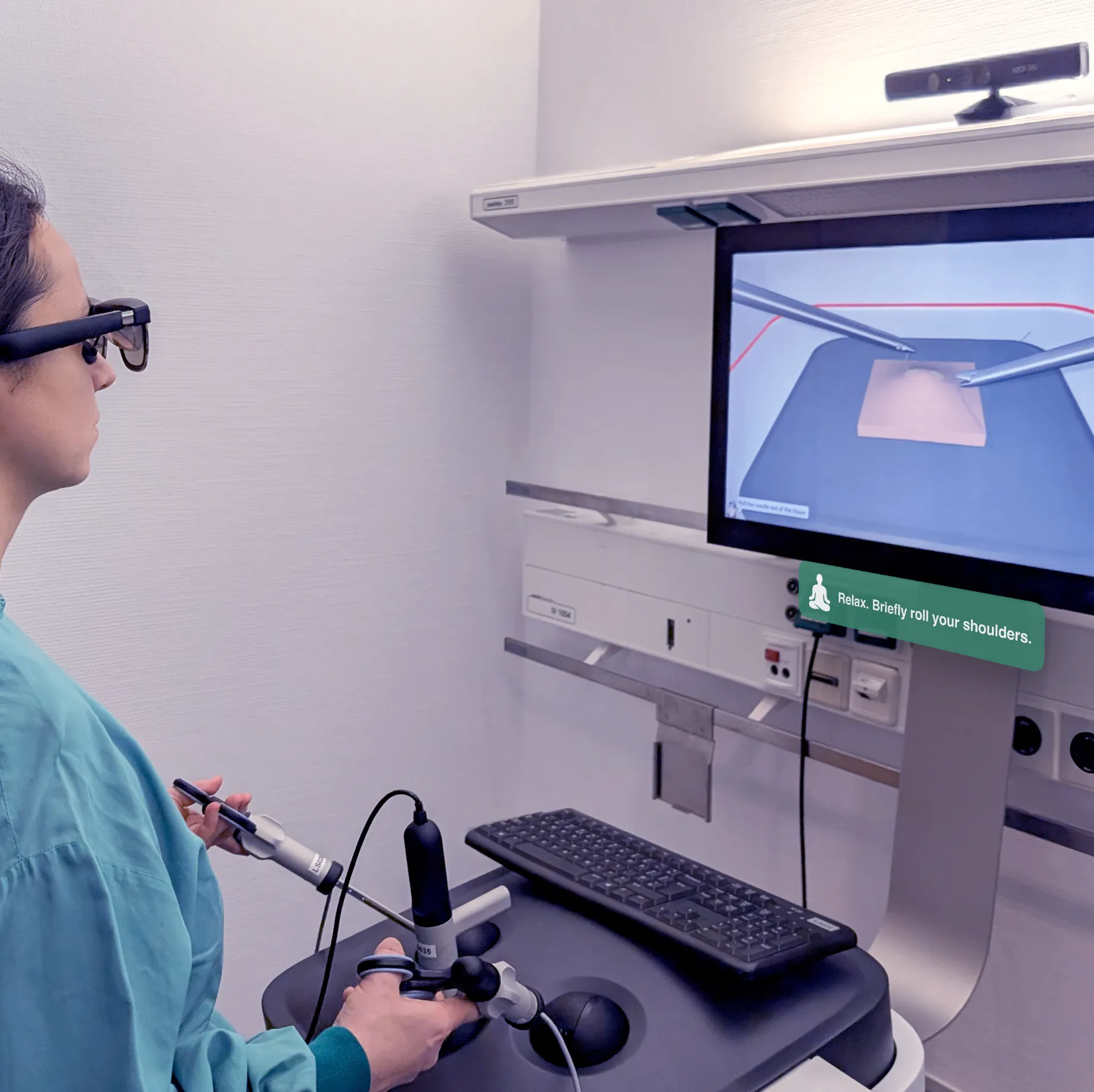
Heinrich, F; Mielke, T; Klank, R; Ortega, F; Hansen, C; Huber, T; Huettl, F
HoloNote: Exploring Augmented Reality Notifications for Ergonomic Feedback in Laparoscopic Training Proceedings Article
In: Extended Abstracts of the CHI Conference on Human Factors in Computing Systems (CHI EA), pp. 1-6, Association for Computing Machinery, 2026.
@inproceedings{holonote,

title = {HoloNote: Exploring Augmented Reality Notifications for Ergonomic Feedback in Laparoscopic Training},

author = {F Heinrich and T Mielke and R Klank and F Ortega and C Hansen and T Huber and F Huettl},

url = {https://dl.acm.org/doi/10.1145/3772363.3799086},

doi = {10.1145/3772363.3799086},

year  = {2026},

date = {2026-04-13},

urldate = {2026-03-01},

booktitle = {Extended Abstracts of the CHI Conference on Human Factors in Computing Systems (CHI EA)},

number = {368},

pages = {1-6},

publisher = {Association for Computing Machinery},

abstract = {Laparoscopic surgery is a physically demanding task that can lead to ergonomic strain. While notification-based approaches have been used to support ergonomics in other domains, their application in laparoscopic surgery remains limited.

Therefore, an augmented reality (AR) notification concept was implemented using lightweight optical see-through AR glasses, examining their suitability for laparoscopic training.

A pilot study with ten surgeons was conducted using a laparoscopic simulator, where participants performed a stitching and knot-tying task with and without AR-based notifications.

All participants were able to complete the tasks while wearing the AR glasses. While task completion times were longer in the AR condition, no conclusive differences in perceived workload were observed. User experience and wearing comfort were rated positively, whereas the notifications were perceived as useful but not consistently effective. The findings suggest that lightweight AR glasses are a feasible platform for ergonomic feedback in laparoscopic training and motivate further investigation.},

keywords = {},

pubstate = {published},

tppubtype = {inproceedings}

}
Therefore, an augmented reality (AR) notification concept was implemented using lightweight optical see-through AR glasses, examining their suitability for laparoscopic training.
A pilot study with ten surgeons was conducted using a laparoscopic simulator, where participants performed a stitching and knot-tying task with and without AR-based notifications.
All participants were able to complete the tasks while wearing the AR glasses. While task completion times were longer in the AR condition, no conclusive differences in perceived workload were observed. User experience and wearing comfort were rated positively, whereas the notifications were perceived as useful but not consistently effective. The findings suggest that lightweight AR glasses are a feasible platform for ergonomic feedback in laparoscopic training and motivate further investigation.
Mielke, T; Georgiades, M; Grosser, O; Pech, M; Hansen, C; Heinrich, F
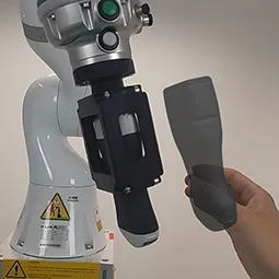
Enhancing Gesture-Based Human–Robot Interaction: Investigating the Role of Force Automation Proceedings Article
In: ACM/IEEE International Conference on Human-Robot Interaction (HRI), pp. 619-628, 2026.
@inproceedings{nokeyd,

title = {Enhancing Gesture-Based Human–Robot Interaction: Investigating the Role of Force Automation},

author = {T Mielke and M Georgiades and O Grosser and M Pech and C Hansen and F Heinrich},

doi = {10.1145/3757279.3785568},

year  = {2026},

date = {2026-03-16},

urldate = {2026-03-16},

booktitle = {ACM/IEEE International Conference on Human-Robot Interaction (HRI)},

pages = {619-628},

abstract = {Directly controlling robot motion through human–robot interaction enables the integration of human expertise into robot control. However, this requires efficient interaction methods. While previous work has shown that hand gestures offer potential for natural control, the lack of haptic feedback remains a key challenge. One solution is partial automation, in which the contact force is autonomously controlled. We implemented force automation enhanced gesture control, as well as the state-of-the-art baseline of hand-guiding, to systematically investigate hand gesture interaction for robot control. A user study (n=28) was conducted to investigate the efficiency and workload of these interaction concepts. As the interaction method efficiency may depend on task demands, two tasks with different precision requirements were evaluated in the context of robotic ultrasound. The results indicate that force automation significantly reduces perceived workload and task duration when using hand gestures. Moreover, gesture-based interaction can match the efficiency of hand-guiding for broad tasks and even outperform it in precise tasks. These findings highlight the potential of gesture-based interaction for robot control, particularly when the absence of haptic feedback is compensated by partial automation.},

keywords = {},

pubstate = {published},

tppubtype = {inproceedings}

}
2025
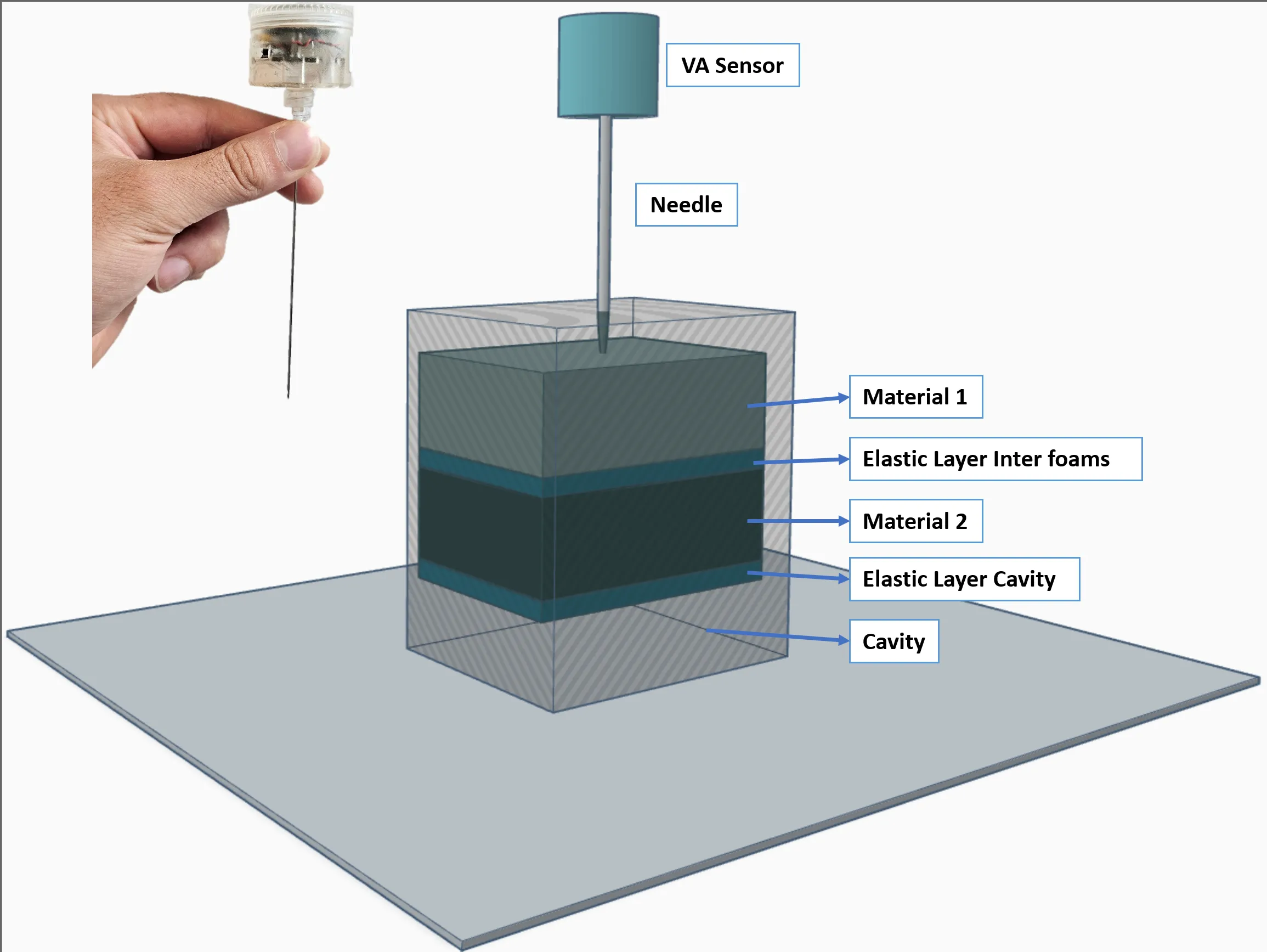
Urrutia, R; Ayman, F; Boese, A; Hansen, C; Illanes, A
Needle Puncture Detection Using Vibroacoustic Sensing in Layered Phantoms Journal Article
In: Current Directions in Biomedical Engineering, vol. 11, iss. 2, pp. 112-115, 2025.
@article{urrutia2025,

title = {Needle Puncture Detection Using Vibroacoustic Sensing in Layered Phantoms},

author = {R Urrutia and F Ayman and A Boese and C Hansen and A Illanes},

doi = {10.1515/cdbme-2025-0329},

year  = {2025},

date = {2025-12-01},

urldate = {2025-12-01},

journal = {Current Directions in Biomedical Engineering},

volume = {11},

issue = {2},

pages = {112-115},

abstract = {This study evaluates the use of vibroacoustic (VA)sensing to detect material transitions during needle insertions into layered synthetic phantoms. Experiments were conducted using foams with different pore densities and configurations including internal cavities. Time-domain and wavelet analyses showed that denser foams produced more frequent vibrational events,  while  less  dense  foams  resulted  in  sparser  signals. In setups with elastic membranes and cavities, differences in time–frequency patterns were observed: high-frequency activity appeared after membrane rupture but was absent in cavity insertions. These results support the use of VA sensing to identify mechanical changes during minimally invasive procedures.},

keywords = {},

pubstate = {published},

tppubtype = {article}

}
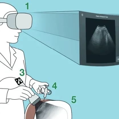
Schreiter, J; Pandi, P; Klank, R; Großer, OS; Hansen, C; Heinrich, F
Mixed Reality Assistance for Needle Insertion under Ultrasound Imaging: A Pilot Study Journal Article
In: Current Directions in Biomedical Engineering, vol. 11, no. 2, pp. 104–107, 2025, ISSN: 2364-5504.
@article{Schreiter2025,

title = {Mixed Reality Assistance for Needle Insertion under Ultrasound Imaging: A Pilot Study},

author = {J Schreiter and P Pandi and R Klank and OS Großer and C Hansen and F Heinrich},

url = {https://www.researchgate.net/publication/399914083_Mixed_Reality_Assistance_for_Needle_Insertion_under_Ultrasound_Imaging_A_Pilot_Study},

doi = {10.1515/cdbme-2025-0327},

issn = {2364-5504},

year  = {2025},

date = {2025-12-01},

urldate = {2025-12-01},

journal = {Current Directions in Biomedical Engineering},

volume = {11},

number = {2},

pages = {104–107},

publisher = {Walter de Gruyter GmbH},

abstract = {Ultrasound (US)-guided needle interventions require precise coordination between imaging and instrument handling, which can be ergonomically challenging with conventional displays. This work presents a mixed reality (MR) prototype that integrates live US and pre-interventional CT data into a video passthrough head-mounted display (HMD), enabling a spatially registered in-situ view and interactive virtual displays. Unlike prior work relying on optical see-through HMDs, our system combines immersive guidance and interactive tools to support image-guided needle insertion. A preliminary user study with six radiologists assessed usability and clinical potential. Results indicate slightly above-average usability, with participants valuing field-of-view integration. User feedback highlighted the need for features such as interactive planning and extended visualizations, e.g. patientspecific anatomy.With these refinements, the prototype shows strong potential to improve ergonomics and spatial awareness during US-guided procedures.

},

keywords = {},

pubstate = {published},

tppubtype = {article}

}
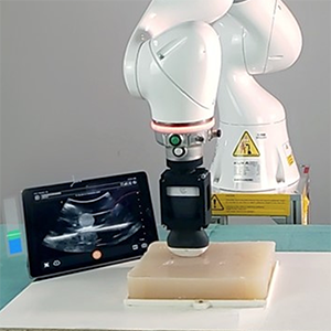
Mielke, T; Allgaier, M; Hansen, C; Heinrich, F
Extended Reality Check: Evaluating XR Prototyping for Human-Robot Interaction in Contact-Intensive Tasks Journal Article
In: IEEE Transactions on Visualization and Computer Graphics, vol. 31, iss. 11, pp. 10035 – 10044, 2025.
@article{mielke_extended_2025,

title = {Extended Reality Check: Evaluating XR Prototyping for Human-Robot Interaction in Contact-Intensive Tasks},

author = {T Mielke and M Allgaier and C Hansen and F Heinrich},

doi = {10.1109/TVCG.2025.3616753},

year  = {2025},

date = {2025-10-02},

urldate = {2025-10-02},

journal = {IEEE Transactions on Visualization and Computer Graphics},

volume = {31},

issue = {11},

pages = {10035 - 10044},

abstract = {Extended Reality (XR) has the potential to improve efficiency and safety in the user-centered development of human-robot interaction. However, the validity of using XR prototyping for user studies for contact-intensive robotic tasks remains underexplored. These in-contact tasks are particularly relevant due to challenges arising from indirect force perception in robot control. Therefore, in this work, we investigate a representative example of such a task: robotic ultrasound. A user study was conducted to assess the transferability of results from a simulated user study to real-world conditions, comparing two force-assistance approaches. The XR simulation replicates the physical study set-up employing a virtual robotic arm, its control interface, ultrasound imaging, and two force-assistance methods: automation and force visualization. Our results indicate that while differences in force deviation, perceived workload, and trust emerge between real and simulated setups, the overall findings remain consistent. Specifically, partial automation of robot control improves performance and trust while reducing workload, and visual feedback decreases force deviation in both real and simulated conditions. These findings highlight the potential of XR for comparative studies, even in complex robotic tasks.},

keywords = {},

pubstate = {published},

tppubtype = {article}

}
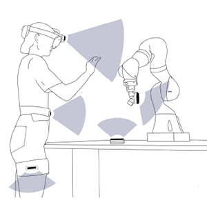
Mielke, T; Heinrich, F; Hansen, C
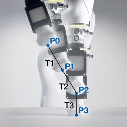
Gesturing Towards Efficient Robot Control: Exploring Sensor Placement and Control Modes for Mid-Air Human-Robot Interaction Proceedings Article
In: 2025 IEEE International Conference on Robotics and Automation (ICRA), IEEE, 2025.
@inproceedings{mielke_gesturing_2025,

title = {Gesturing Towards Efficient Robot Control: Exploring Sensor Placement and Control Modes for Mid-Air Human-Robot Interaction},

author = {T Mielke and F Heinrich and C Hansen},

doi = {10.1109/ICRA55743.2025.11127519},

year  = {2025},

date = {2025-09-02},

urldate = {2025-09-02},

booktitle = {2025 IEEE International Conference on Robotics and Automation (ICRA)},

publisher = {IEEE},

abstract = {While collaborative robots effectively combine robotic precision with human capabilities, traditional control methods such as button presses or hand guidance can be slow and physically demanding. This has led to an increasing interest in natural user interfaces that integrate hand gesturebased interactions for more intuitive and flexible robot control. Therefore, this paper systematically explores mid-air robot control by comparing position and rate control modes with different state-of-the-art and novel sensor placements. A user study was conducted to evaluate each combination in terms of accuracy, task duration, perceived workload, and physical exertion. Our results indicate that position control is more efficient than rate control. Traditional desk-mounted sensors can provide a good balance between accuracy and comfort. However, robot-mounted sensors are a viable alternative for short-term, accurate control with less spatial requirements. Legmounted sensors, while comfortable, pose challenges to handeye coordination. Based on these findings, we provide design implications for improving the usability and comfort of midair human-robot interaction. Future research should extend this evaluation to a wider range of tasks and environments.},

keywords = {},

pubstate = {published},

tppubtype = {inproceedings}

}
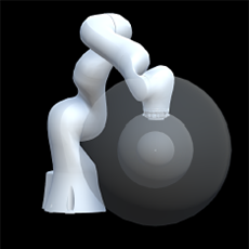
Mielke, T; Heinrich, F; Hansen, C
Enhancing AR-to-Robot Registration Accuracy: A Comparative Study of Marker Detection Algorithms and Registration Parameters Proceedings Article
In: 2025 IEEE International Conference on Robotics and Automation (ICRA), IEEE, 2025.
@inproceedings{mielke_enhancing_2025,

title = {Enhancing AR-to-Robot Registration Accuracy: A Comparative Study of Marker Detection Algorithms and Registration Parameters},

author = {T Mielke and F Heinrich and C Hansen},

doi = {10.1109/ICRA55743.2025.11128039},

year  = {2025},

date = {2025-09-02},

urldate = {2025-09-02},

booktitle = {2025 IEEE International Conference on Robotics and Automation (ICRA)},

publisher = {IEEE},

abstract = {Augmented Reality (AR) offers potential for enhancing human-robot collaboration by enabling intuitive interaction and real-time feedback. A crucial aspect of AR-robot integration is accurate spatial registration to align virtual content with the physical robotic workspace. This paper systematically investigates the effects of different tracking techniques and registration parameters on AR-to-robot registration accuracy, focusing on paired-point methods. We evaluate four marker detection algorithms - ARToolkit, Vuforia, ArUco, and retroreflective tracking - analyzing the influence of viewing distance, angle, marker size, point distance, distribution, and quantity. Our results show that ARToolkit provides the highest registration accuracy. While larger markers and positioning registration point centroids close to target locations consistently improved accuracy, other factors such as point distance and quantity were highly dependent on the tracking techniques used. Additionally, we propose an effective refinement method using point cloud registration, significantly improving accuracy by integrating data from points recorded between registration locations. These findings offer practical guidelines for enhancing AR-robot registration, with future work needed to assess the transferability to other AR devices and robots.},

keywords = {},

pubstate = {published},

tppubtype = {inproceedings}

}
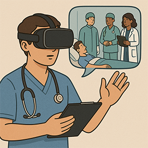
Hanke, L; Schwoerer, P; Huettl, F; Vradelis, L; Strelow, K; Boedecker, C; Saalfeld, P; Chheang, V; Buggenhagen, H; Lang, H; Hansen, C; Huber, T
Use of an Immersive Virtual Reality Application to Educate Medical Students in Patient Handover: Pilot Study Journal Article
In: JMIR Serious Games, vol. 13, pp. e73907, 2025.
@article{hanke_use_2025,

title = {Use of an Immersive Virtual Reality Application to Educate Medical Students in Patient Handover: Pilot Study},

author = {L Hanke and P Schwoerer and F Huettl and L Vradelis and K Strelow and C Boedecker and P Saalfeld and V Chheang and H Buggenhagen and H Lang and C Hansen and T Huber},

url = {https://games.jmir.org/2025/1/e73907

https://pubmed.ncbi.nlm.nih.gov/40864895/, PubMed},

doi = {10.2196/73907},

year  = {2025},

date = {2025-08-27},

urldate = {2025-08-27},

journal = {JMIR Serious Games},

volume = {13},

pages = {e73907},

keywords = {},

pubstate = {published},

tppubtype = {article}

}
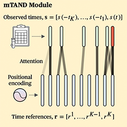
Bashkanov, O; Engelage, L; Behnel, N; Ehrlich, P; Hansen, C; Rak, M
Multimodal Data Fusion with Irregular PSA Kinetics for Automated Prostate Cancer Grading Journal Article
In: Computerized Medical Imaging and Graphics, vol. 124, pp. 102625, 2025.
@article{bashkanov_multimodal_2025,

title = {Multimodal Data Fusion with Irregular PSA Kinetics for Automated Prostate Cancer Grading},

author = {O Bashkanov and L Engelage and N Behnel and P Ehrlich and C Hansen and M Rak},

url = {https://www.sciencedirect.com/science/article/pii/S089561112500134X

https://pubmed.ncbi.nlm.nih.gov/40819603/, PubMed},

doi = {10.1016/j.compmedimag.2025.102625},

year  = {2025},

date = {2025-08-11},

urldate = {2025-08-11},

journal = {Computerized Medical Imaging and Graphics},

volume = {124},

pages = {102625},

keywords = {},

pubstate = {published},

tppubtype = {article}

}
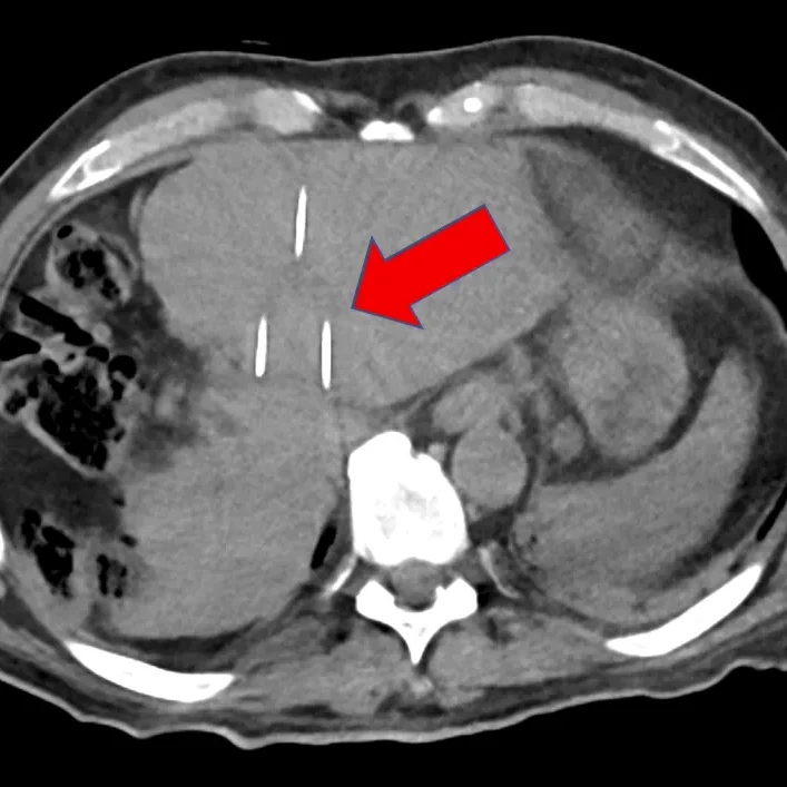
Joeres, F; Paetz, T; Hansen, C; Schenk, A
The necessity of parallel needle placement for tumor ablation using irreversible electroporation: a myth? Proceedings Article
In: CARS 2025—Computer Assisted Radiology and Surgery Proceedings of the 39th International Congress and Exhibition Berlin, Germany, June 17–20, 2025 , pp. 90–91, 2025.
@inproceedings{Joeres.2025,

title = {The necessity of parallel needle placement for tumor ablation using irreversible electroporation: a myth?},

author = {F Joeres and T Paetz and C Hansen and A Schenk},

url = {https://link.springer.com/article/10.1007/s11548-025-03373-6},

doi = {10.1007/s11548-025-03373-6},

year  = {2025},

date = {2025-05-29},

urldate = {2025-05-29},

booktitle = {CARS 2025—Computer Assisted Radiology and Surgery Proceedings of the 39th International Congress and Exhibition Berlin, Germany, June 17–20, 2025

},

pages = {90–91},

keywords = {},

pubstate = {published},

tppubtype = {inproceedings}

}
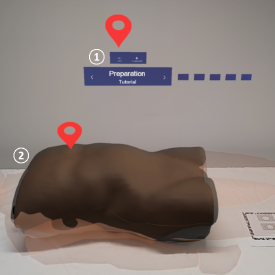
Schwenderling, L; Hanke, L; Holst, U; Huettl, F; Joeres, F; Huber, T; Hansen, C
Toward structured abdominal examination training using augmented reality Journal Article
In: International Journal of Computer Assisted Radiology and Surgery, vol. 20, no. 5, pp. 949–958, 2025, ISSN: 1861-6429.
@article{schwenderling_toward_2025,

title = {Toward structured abdominal examination training using augmented reality},

author = {L Schwenderling and L Hanke and U Holst and F Huettl and F Joeres and T Huber and C Hansen},

url = {https://doi.org/10.1007/s11548-024-03311-y

https://pubmed.ncbi.nlm.nih.gov/39755835/, PubMed},

doi = {10.1007/s11548-024-03311-y},

issn = {1861-6429},

year  = {2025},

date = {2025-05-01},

urldate = {2025-05-01},

journal = {International Journal of Computer Assisted Radiology and Surgery},

volume = {20},

number = {5},

pages = {949–958},

abstract = {Structured abdominal examination is an essential part of the medical curriculum and surgical training, requiring a blend of theory and practice from trainees. Current training methods, however, often do not provide adequate engagement, fail to address individual learning needs or do not cover rare diseases.},

keywords = {},

pubstate = {published},

tppubtype = {article}

}
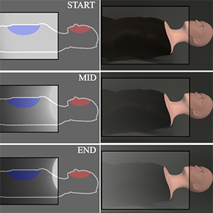
Schwenderling, L; Schotte, M; Joeres, F; Heinrich, F; Hanke, L; Huettl, F; Huber, T; Hansen, C
Teach Me Where to Look: Dual-task Attention Training in Augmented Reality Proceedings Article
In: Proceedings of the Extended Abstracts of the CHI Conference on Human Factors in Computing Systems, pp. 1–8, ACM, Yokohama Japan, 2025, ISBN: 979-8-4007-1395-8.
@inproceedings{schwenderling_teach_2025,

title = {Teach Me Where to Look: Dual-task Attention Training in Augmented Reality},

author = {L Schwenderling and M Schotte and F Joeres and F Heinrich and L Hanke and F Huettl and T Huber and C Hansen},

url = {https://dl.acm.org/doi/10.1145/3706599.3720198},

doi = {10.1145/3706599.3720198},

isbn = {979-8-4007-1395-8},

year  = {2025},

date = {2025-04-01},

urldate = {2025-04-01},

booktitle = {Proceedings of the Extended Abstracts of the CHI Conference on Human Factors in Computing Systems},

pages = {1–8},

publisher = {ACM},

address = {Yokohama Japan},

abstract = {Regular eye contact is essential in medicine to recognize signs of pain. However, it is difficult to remember this during training as attention is tied up in learning. While augmented reality (AR) has shown promising results for medical education, there is no training for attention allocation yet. Therefore, three auditory and three visual attention guidance tools in AR are evaluated for their use in medical dual-task training settings. In expert reviews with six participants in human-computer interaction and medical didactics, advantages, disadvantages, and refinements for the cues were developed. For visual cues, an overt but less occluding cue was preferred for constant visibility of the primary task. A more diegetic cue design was proposed for the auditory cues to use a patient simulation as a reminder of the regular face glance. In general, several cues were found to be suitable for gaze guidance training, requiring only minor changes for improvement.},

keywords = {},

pubstate = {published},

tppubtype = {inproceedings}

}
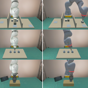
Mielke, T; Allgaier, M; Schott, D; Hansen, C; Heinrich, F
Virtual Studies, Real Results? Assessing the Impact of Virtualization on Human-Robot Interaction Proceedings Article
In: Proceedings of the Extended Abstracts of the CHI Conference on Human Factors in Computing Systems, pp. 1–8, ACM, Yokohama Japan, 2025, ISBN: 979-8-4007-1395-8.
@inproceedings{mielke_virtual_2025,

title = {Virtual Studies, Real Results? Assessing the Impact of Virtualization on Human-Robot Interaction},

author = {T Mielke and M Allgaier and D Schott and C Hansen and F Heinrich},

url = {https://dl.acm.org/doi/10.1145/3706599.3719724},

doi = {10.1145/3706599.3719724},

isbn = {979-8-4007-1395-8},

year  = {2025},

date = {2025-04-01},

urldate = {2025-04-01},

booktitle = {Proceedings of the Extended Abstracts of the CHI Conference on Human Factors in Computing Systems},

pages = {1–8},

publisher = {ACM},

address = {Yokohama Japan},

abstract = {Extended Reality (XR) shows potential for human-centered evaluation of real-world scenarios and could improve efficiency and safety in robotic research. However, the validity of XR Human-Robot Interaction (HRI) studies remains underexplored. This paper investigates the transferability of HRI studies across virtualization levels for three tasks. Our results indicate XR study validity is task-specific, with task virtualization as a key influencing factor. Partially virtualized settings with virtual tasks and a real robot, as well as fully virtualized setups with a simulated robot, yielded results comparable to real setups for pick-and-place and robotic ultrasound. However, for precision-dependent peg-in-hole, differences were observed between real and virtualized conditions regarding completion time, perceived workload, and ease. Demonstrating the task dependency of XR transferability and comparing virtualization levels, our work takes an important step in assessing XR study validity. Future work should isolate factors affecting transferability and assess relative validity in the absence of absolute validity.},

keywords = {},

pubstate = {published},

tppubtype = {inproceedings}

}
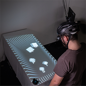
Heinrich, F; Schott, D; Schwenderling, L; Hansen, C
Do You See What I See? Evaluating Relative Depth Judgments Between Real and Virtual Projections Proceedings Article
In: Proceedings of the Extended Abstracts of the CHI Conference on Human Factors in Computing Systems, pp. 1–8, Association for Computing Machinery, New York, NY, USA, 2025, ISBN: 979-8-4007-1395-8.
@inproceedings{heinrich_you_2025,

title = {Do You See What I See? Evaluating Relative Depth Judgments Between Real and Virtual Projections},

author = {F Heinrich and D Schott and L Schwenderling and C Hansen},

url = {https://doi.org/10.1145/3706599.3720157},

doi = {10.1145/3706599.3720157},

isbn = {979-8-4007-1395-8},

year  = {2025},

date = {2025-04-01},

urldate = {2025-04-01},

booktitle = {Proceedings of the Extended Abstracts of the CHI Conference on Human Factors in Computing Systems},

pages = {1–8},

publisher = {Association for Computing Machinery},

address = {New York, NY, USA},

series = {CHI EA '25},

abstract = {Projector-based augmented reality (AR) is promising in different domains with less issues in discomfort or shortage of space. However, due to limitations like high costs and cumbersome calibration, this AR modality remains underused. To address this problem, a stereoscopic projector-based AR simulation was implemented for a cost-effective video see-through AR headset. To evaluate the validity of this simulation, a relative depth judgment experiment was conducted to compare this method with a physical projection system. Consistent results suggest that a known interaction effect between visualization and disparity mode could be successfully reproduced using both the physical projection and the virtual simulation. In addition, first findings indicate that there are no significant differences between these projection modalities. The results indicate that other perception-related effects observed for projector-based AR may also be applicable to virtual projection simulations and that future findings determined using only these simulations may also be applicable to real projections.},

keywords = {},

pubstate = {published},

tppubtype = {inproceedings}

}
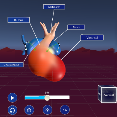
Kunz, M; Schott, D; Wunderling, T; Halloul, M; Hansen, C; Albrecht, A; Braun-Dullaeus, R
Embryonic heart development as an immersive experience: Unveiling learning effects and influential factors in virtual learning environments Journal Article
In: Comput. Biol. Med., vol. 187, no. C, 2025, ISSN: 0010-4825.
@article{kunz_embryonic_2025,

title = {Embryonic heart development as an immersive experience: Unveiling learning effects and influential factors in virtual learning environments},

author = {M Kunz and D Schott and T Wunderling and M Halloul and C Hansen and A Albrecht and R Braun-Dullaeus},

url = {https://doi.org/10.1016/j.compbiomed.2024.109638

https://pubmed.ncbi.nlm.nih.gov/39923588/, PubMed},

doi = {10.1016/j.compbiomed.2024.109638},

issn = {0010-4825},

year  = {2025},

date = {2025-04-01},

urldate = {2025-04-01},

journal = {Comput. Biol. Med.},

volume = {187},

number = {C},

abstract = {As the quality and availability of Virtual Reality (VR) technologies improve, their potential applications in medical education, particularly VR Learning Environments (VRLEs), are increasingly explored. VRLEs offer a dynamic platform where educators and students can interact, access materials, and engage in learning beyond traditional classrooms. However, questions remain about their long-term learning effects and potential confounding factors. This study investigates these aspects through a VR application designed for teaching heart embryology. For this reason we conducted a user study with medical students in their early years of training (N = 143). Our findings reveal significant short-term and sustained learning outcomes two to four weeks following a single VR session. Importantly, these outcomes appear largely independent of users’ technical affinity and are minimally influenced by their immersion tendencies. Instead, the quality of the VRLE and its user experience emerge as critical factors. These results underscore the efficacy of well-designed VRLEs in higher education and highlight key areas for future development.},

keywords = {},

pubstate = {published},

tppubtype = {article}

}
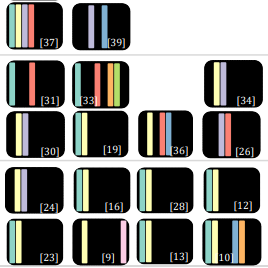
Schreiter, J; Heinrich, F; Hatscher, B; Schott, D; Hansen, C
Multimodal human–computer interaction in interventional radiology and surgery: a systematic literature review Journal Article
In: International Journal of Computer Assisted Radiology and Surgery, vol. 20, no. 4, pp. 807–816, 2025, ISSN: 1861-6429.
@article{schreiter_multimodal_2025,

title = {Multimodal human–computer interaction in interventional radiology and surgery: a systematic literature review},

author = {J Schreiter and F Heinrich and B Hatscher and D Schott and C Hansen},

url = {https://doi.org/10.1007/s11548-024-03263-3

https://pubmed.ncbi.nlm.nih.gov/39467893/, PubMed},

doi = {10.1007/s11548-024-03263-3},

issn = {1861-6429},

year  = {2025},

date = {2025-04-01},

urldate = {2025-04-01},

journal = {International Journal of Computer Assisted Radiology and Surgery},

volume = {20},

number = {4},

pages = {807–816},

abstract = {As technology advances, more research dedicated to medical interactive systems emphasizes the integration of touchless and multimodal interaction (MMI). Particularly in surgical and interventional settings, this approach is advantageous because it maintains sterility and promotes a natural interaction. Past reviews have focused on investigating MMI in terms of technology and interaction with robots. However, none has put particular emphasis on analyzing these kind of interactions for surgical and interventional scenarios.},

keywords = {},

pubstate = {published},

tppubtype = {article}

}
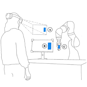
Mielke, T; Heinrich, F; Hansen, C
SensARy Substitution: Augmented Reality Techniques to Enhance Force Perception in Touchless Robot Control Journal Article
In: IEEE Transactions on Visualization and Computer Graphics, vol. 31, no. 5, pp. 3235–3244, 2025, ISSN: 1941-0506.
@article{mielke_sensary_2025,

title = {SensARy Substitution: Augmented Reality Techniques to Enhance Force Perception in Touchless Robot Control},

author = {T Mielke and F Heinrich and C Hansen},

url = {https://ieeexplore.ieee.org/document/10926846},

doi = {10.1109/TVCG.2025.3549856},

issn = {1941-0506},

year  = {2025},

date = {2025-03-14},

urldate = {2025-05-01},

journal = {IEEE Transactions on Visualization and Computer Graphics},

volume = {31},

number = {5},

pages = {3235–3244},

abstract = {The lack of haptic feedback in touchless human-robot interaction is critical in applications such as robotic ultrasound, where force perception is crucial to ensure image quality. Augmented reality (AR) is a promising tool to address this limitation by providing sensory substitution through visual or vibrotactile feedback. The implementation of visual force feedback requires consideration not only of feedback design but also of positioning. Therefore, we implemented two different visualization types at three different positions and investigated the effects of vibrotactile feedback on these approaches. Furthermore, we examined the effects of multimodal feedback compared to visual or vibrotactile output alone. Our results indicate that sensory substitution eases the interaction in contrast to a feedback-less baseline condition, with the presence of visual support reducing average force errors and being subjectively preferred by the participants. However, the more feedback was provided, the longer users needed to complete their tasks. Regarding visualization design, a 2D bar visualization reduced force errors compared to a 3D arrow concept. Additionally, the visualizations being displayed directly on the ultrasound screen were subjectively preferred. With findings regarding feedback modality and visualization design our work represents an important step toward sensory substitution for touchless human-robot interaction.},

keywords = {},

pubstate = {published},

tppubtype = {article}

}
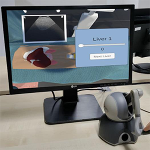
Allgaier, M; Dangszat, E; Huettl, F; Hanke, L; Huber, T; Preim, B; Hansen, C
Impact of Input and Output Devices on a Virtual Ultrasound Training Proceedings Article
In: 2025 IEEE Conference on Virtual Reality and 3D User Interfaces Abstracts and Workshops (VRW), pp. 937–941, IEEE, Saint Malo, France, 2025, ISBN: 979-8-3315-1484-6.
@inproceedings{allgaier_impact_2025,

title = {Impact of Input and Output Devices on a Virtual Ultrasound Training},

author = {M Allgaier and E Dangszat and F Huettl and L Hanke and T Huber and B Preim and C Hansen},

url = {https://ieeexplore.ieee.org/document/10972939/},

doi = {10.1109/VRW66409.2025.00191},

isbn = {979-8-3315-1484-6},

year  = {2025},

date = {2025-03-01},

urldate = {2025-03-01},

booktitle = {2025 IEEE Conference on Virtual Reality and 3D User Interfaces Abstracts and Workshops (VRW)},

pages = {937–941},

publisher = {IEEE},

address = {Saint Malo, France},

abstract = {Performing ultrasound requires mental skills that have to be trained hands on. Virtual simulations can be employed to provide novice surgeons with a safe training environment prior to performing ultrasound on the patient. For both input and output there is a wide range of devices that are used in existing ultrasound simulations. Because the devices have their limitations and benefits regarding realism, costs, and access, we compared three technical setups: desktop with mouse interaction, desktop with a haptic device, and virtual reality with a haptic device. In a user study with 19 participants we investigated the usability and assessed qualitative user feedback in a semi-structured interview. Significant differences regarding usability and training time were found between both versions with the haptic device and the desktop with mouse version. Based on the feedback, using a haptic device seems to be more relevant than the two output devices in this training case.},

keywords = {},

pubstate = {published},

tppubtype = {inproceedings}

}
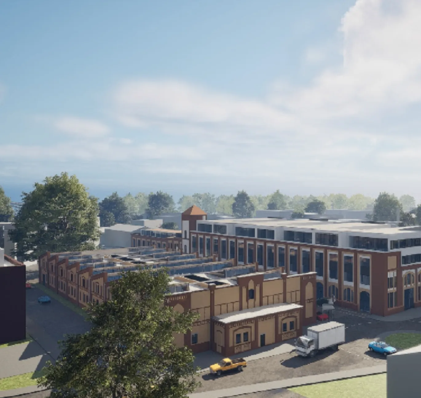
Herbrich, W; Zittlau, P; Joeres, F; Hansen, C
Prototype development of a cross-reality digital twin ecosystem: the web, open source and open data Proceedings Article
In: 2025 IEEE Conference on Virtual Reality and 3D User Interfaces Abstracts and Workshops (VRW), pp. 459–462, 2025.
@inproceedings{herbrich_prototype_2025,

title = {Prototype development of a cross-reality digital twin ecosystem: the web, open source and open data},

author = {W Herbrich and P Zittlau and F Joeres and C Hansen},

url = {https://ieeexplore.ieee.org/abstract/document/10972895},

doi = {10.1109/VRW66409.2025.00100},

year  = {2025},

date = {2025-03-01},

urldate = {2025-03-01},

booktitle = {2025 IEEE Conference on Virtual Reality and 3D User Interfaces Abstracts and Workshops (VRW)},

pages = {459–462},

abstract = {This work contributes to a broader initiative aimed at transforming a former industrial port area into a dynamic Knowledge Transfer Space (KTS). To support this transformation, we explore the development of a cross-reality (CR) digital twin of the port area, which integrates user interfaces with varying degrees of virtuality. We evaluate different web technologies, focusing on the balance between accessibility, immersion, scalability, and performance. By comparing client-side rendering with pixel streaming approaches, we aim to identify suitable solutions for prototyping a CR digital twin ecosystem. The development of a prototype is ongoing, based on a client-side rendering approach. The outcomes contribute to developing an open and transferable CR digital twin applicable to similar urban projects in other cities.},

keywords = {},

pubstate = {published},

tppubtype = {inproceedings}

}
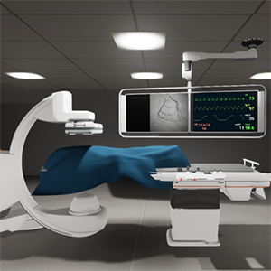
Razavizadeh, S; Kofler, M; Kunz, M; Kempfert, J; Braun-Dullaeus, R; Weidling, J; Preim, B; Hansen, C
A virtual patient authoring tool for transcatheter aortic valve replacement Journal Article
In: International Journal of Computer Assisted Radiology and Surgery, vol. 20, no. 2, pp. 379–389, 2025, ISSN: 1861-6429.
@article{razavizadeh_virtual_2025,

title = {A virtual patient authoring tool for transcatheter aortic valve replacement},

author = {S Razavizadeh and M Kofler and M Kunz and J Kempfert and R Braun-Dullaeus and J Weidling and B Preim and C Hansen},

url = {https://doi.org/10.1007/s11548-024-03293-x},

doi = {10.1007/s11548-024-03293-x},

issn = {1861-6429},

year  = {2025},

date = {2025-02-01},

urldate = {2025-02-01},

journal = {International Journal of Computer Assisted Radiology and Surgery},

volume = {20},

number = {2},

pages = {379–389},

abstract = {Computer-based medical training scenarios, derived from patient’s records, often lack variability, modifiability, and availability. Furthermore, generating image datasets and creating scenarios is resource-intensive. Therefore, patient authoring tools for rapid dataset-independent creation of virtual patients (VPs) is a pressing need.},

keywords = {},

pubstate = {published},

tppubtype = {article}

}
Schott, D; Kunz, M; Albrecht, A; Braun-Dullaeus, R; Hansen, C
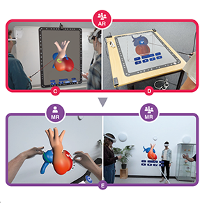
Too Heart to Handle? Exploring Self-Directed And Collaborative Virtual Learning Environments in Anatomy Education. Journal Article
In: EuroVis 2025 – Dirk Bartz Prize, 2025, (Artwork Size: 5 pages ISBN: 9783038682813 Publisher: The Eurographics Association).
@article{schott_too_2025,

title = {Too Heart to Handle? Exploring Self-Directed And Collaborative Virtual Learning Environments in Anatomy Education.},

author = {D Schott and M Kunz and A Albrecht and R Braun-Dullaeus and C Hansen},

url = {https://diglib.eg.org/handle/10.2312/evm20251973},

doi = {10.2312/EVM.20251973},

year  = {2025},

date = {2025-01-01},

urldate = {2025-01-01},

journal = {EuroVis 2025 - Dirk Bartz Prize},

abstract = {The integration of Extended Reality (XR) into medical education represents a transformative shift, particularly in anatomy training, where immersive simulations enhance cognitive engagement and knowledge retention. The developing heart is characterized by rapid morphological changes within a short time frame, which poses a significant pedagogical challenge. Conventional 2D imaging and static models often fail to convey these processes, limiting learners' ability to conceptualize critical spatial relationships-a barrier in understanding congenital anomalies. To address these limitations, this work leverages XRdriven visualization and interaction paradigms to create virtual learning environments. Based on this, we propose methods for designing XR educational modules that adapt to both collaborative and self-directed learning contexts, using embryonic cardiogenesis as an illustrating case study. We present findings from mixed-methods user studies involving a total of 264 students, along with feedback from lecturers, highlighting the importance of an iterative, user-centered design approach.},

note = {Artwork Size: 5 pages

ISBN: 9783038682813

Publisher: The Eurographics Association},

keywords = {},

pubstate = {published},

tppubtype = {article}

}
Schreiter, J; Mielke, T; Georgiades, M; Pech, M; Hansen, C; Heinrich, F
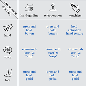
Exploring Interaction Concepts for the Manipulation of a Collaborative Robot: A Comparative Study Proceedings Article
In: ACM/IEEE International Conference on Human-Robot Interaction, pp. 55–64, IEEE Press, Melbourne, Australia, 2025.
@inproceedings{schreiter_exploring_2025,

title = {Exploring Interaction Concepts for the Manipulation of a Collaborative Robot: A Comparative Study},

author = {J Schreiter and T Mielke and M Georgiades and M Pech and C Hansen and F Heinrich},

url = {https://ieeexplore.ieee.org/document/10974113},

year  = {2025},

date = {2025-01-01},

urldate = {2025-01-01},

booktitle = {ACM/IEEE International Conference on Human-Robot Interaction},

pages = {55–64},

publisher = {IEEE Press},

address = {Melbourne, Australia},

series = {HRI '25},

abstract = {Robotic systems have the potential to enhance a wide range of domains, such as medical workflows, by automating individual steps of complex processes. However, human-robot interaction (HRI) is of critical importance, as effective collaboration between humans and robots is essential even in highly automated environments. Recent research has predominantly focused on the development of interaction methods rather than systematically comparing existing approaches. Therefore, we conducted a user study (n=20) to compare different HRI concepts for end effector manipulation combined with clutching mechanisms for manipulation activation in an alignment task using the example of robotic ultrasound (US). Manipulation methods included hand-guiding, teleoperation, and touchless interaction, while clutching mechanisms were realized through hand, voice, and foot interaction. The results indicate advantages of hand-guiding for manipulation. While no significant differences were observed between clutching mechanisms, strong evidence suggests comparable performance across these modalities. Notably, significant interaction effects on perceived workload reveal that the optimal clutching mechanism depends on the selected manipulation technique. This work underscores the critical importance of selecting appropriate HRI concepts and understanding the dependencies of manipulation techniques with clutching mechanisms. While our study included the usage of a robotic US, the insights gained are broadly transferable across various domains involving robotic manipulation tasks in human-robot collaboration.},

keywords = {},

pubstate = {published},

tppubtype = {inproceedings}

}
Bashkanov, O; Rak, M; Engelage, L; Hansen, C
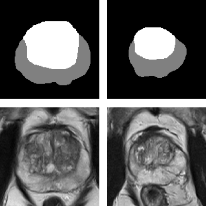
Augmenting Prostate MRI Dataset with Synthetic Volumetric Images from Zone-Conditioned Diffusion Generative Model Proceedings Article
In: Mukhopadhyay, A; Oksuz, I; Engelhardt, S; Merhof, D; Yuan, Y (Ed.): Deep Generative Models, pp. 160–168, Springer Nature Switzerland, Cham, 2025, ISBN: 978-3-031-72744-3.
@inproceedings{bashkanov_augmenting_2025,

title = {Augmenting Prostate MRI Dataset with Synthetic Volumetric Images from Zone-Conditioned Diffusion Generative Model},

author = {O Bashkanov and M Rak and L Engelage and C Hansen},

editor = {A Mukhopadhyay and I Oksuz and S Engelhardt and D Merhof and Y Yuan},

doi = {10.1007/978-3-031-72744-3_16},

isbn = {978-3-031-72744-3},

year  = {2025},

date = {2025-01-01},

urldate = {2025-01-01},

booktitle = {Deep Generative Models},

pages = {160–168},

publisher = {Springer Nature Switzerland},

address = {Cham},

abstract = {The need for artificial intelligence (AI)-driven computer-assist ed diagnosis (CAD) tools drives up the demand for large high-quality datasets in medical imaging. However, collecting the necessary amount of data is often impractical due to patient privacy concerns or restricted time for medical annotation. Recent advances in generative models in medical imaging with a focus on diffusion-based techniques could provide realistic-looking synthetic samples as a supplement for real data. In this work, we study whether synthetic volumetric MRIs generated by the diffusion model can be used to train downstream models, e.g., semantic segmentation. We can create an arbitrarily large dataset with ground truth by conditioning the diffusion model with a segmentation mask. Thus, the additional synthetic data can be used to control the dataset diversity. Experiments revealed that downstream tasks profit from additional synthetic data. However, the effect will eventually diminish when sufficient real samples are available. We showcase the strength of the synthetic data and provide practical recommendations for using the generated data in zonal prostate segmentation.},

keywords = {},

pubstate = {published},

tppubtype = {inproceedings}

}
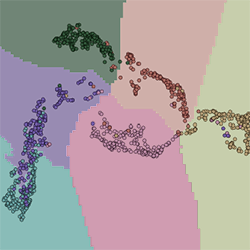
Urrutia, R; Espejo, D; Guerra, M; Vio, K; Sühn, T; Esmaeili, N; Boese, A; Fuentealba, P; Illanes, A; Hansen, C; Poblete, V
Exploring Deep Clustering Methods in Vibro-Acoustic Sensing for Enhancing Biological Tissue Characterization Journal Article
In: IEEE Access, vol. 13, pp. 80395–80406, 2025, ISSN: 2169-3536.
@article{urrutia_exploring_2025,

title = {Exploring Deep Clustering Methods in Vibro-Acoustic Sensing for Enhancing Biological Tissue Characterization},

author = {R Urrutia and D Espejo and M Guerra and K Vio and T Sühn and N Esmaeili and A Boese and P Fuentealba and A Illanes and C Hansen and V Poblete},

url = {https://ieeexplore.ieee.org/document/10981752/},

doi = {10.1109/ACCESS.2025.3566280},

issn = {2169-3536},

year  = {2025},

date = {2025-01-01},

urldate = {2025-01-01},

journal = {IEEE Access},

volume = {13},

pages = {80395–80406},

abstract = {Nonlinear dimensionality reduction techniques, often referred to as manifold learning, are increasingly valuable for data visualization and unsupervised clustering. In the context of surgery and medicine, these methods facilitate the analysis of complex datasets, enabling pattern recognition in surgical data. This study explores the characterization of six tissue types through manifold learning and unsupervised clustering, utilizing vibro-acoustic (VA) signals collected from manual palpation experiments. A wireless sensor mounted at the tip of a surgical instrument was used to acquire 1,680 VA signals, which were processed using Fourier transform and cepstral analysis for feature extraction. We assessed the performance of two dimensionality reduction techniques: uniform manifold approximation and projection (UMAP) and variational autoencoder (VAE). Results indicate that cepstral features combined with UMAP yield superior clustering performance compared to VAE, achieving higher classification accuracy ( 92 % vs. 87 % ) and better-defined clusters with greater compactness. The observed differences in performance are linked to the intrinsic properties of the tissues, particularly surface characteristics such as friction and moisture, which affect signal consistency. Additionally, we compared our approach with previous works, including a study utilizing the same dataset, where our methodology demonstrated improved accuracy. Future research will focus on refining the VAE model, increasing the diversity of tissue samples, and validating the proposed approach in real surgical settings to enhance its applicability in minimally invasive surgery.},

keywords = {},

pubstate = {published},

tppubtype = {article}

}
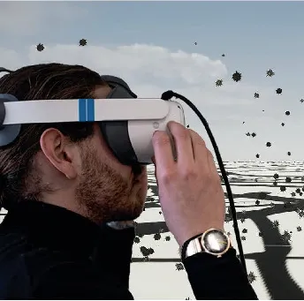
Batz, V; Liedtke, VB; Lameski, P; Trajkovik, V; Hußlein, S; Hansen, C; Herzog, MA
Enhancing Public Awareness of Air Quality: Evaluating Communication Strategies and Design Prototypes Using a Design-Based Implementation Research Approach Proceedings Article
In: International Conference on Human-Computer Interaction, pp. 3–25, Springer 2025.
@inproceedings{batz2025enhancing,

title = {Enhancing Public Awareness of Air Quality: Evaluating Communication Strategies and Design Prototypes Using a Design-Based Implementation Research Approach},

author = {V Batz and VB Liedtke and P Lameski and V Trajkovik and S Hußlein and C Hansen and MA Herzog},

url = {https://link.springer.com/chapter/10.1007/978-3-031-93221-2_1},

year  = {2025},

date = {2025-01-01},

urldate = {2025-01-01},

booktitle = {International Conference on Human-Computer Interaction},

pages = {3–25},

organization = {Springer},

abstract = {Despite the health risks associated with air pollution and the availability of data, public awareness of air quality remains limited. This paper examines communication strategies to enhance awareness in Germany and North Macedonia. Using the Design-Based Implementation Research approach, literature reviews, stakeholder interviews and surveys were conducted with 307 participants to identify knowledge gaps and effective methods.


Three citizen workshops led to the development and evaluation of five design prototypes, including an interactive installation (“Pollution Booth”), a VR application (“Visible Particulate Matter”), and a gamified learning experience (“End Game”). These prototypes addressed information accessibility, data representation, and environmentally conscious behavior. The results show that region-specific communication, interactive designs, and passive information methods in public spaces significantly improve the perception and understanding of air pollution data. Gamified approaches, such as the “End Game” prototype, were particularly effective in educating children about air pollution.


Thirty-two requirements for effective educational strategies were identified, including simplified data visualization, reduced content, and personalized information. The findings highlight the importance of local and personalized approaches, as well as methods for collective knowledge sharing, in promoting awareness and environmentally friendly behavior. Future work should further explore the long-term engagement and sustainable impact of such communication strategies.},

keywords = {},

pubstate = {published},

tppubtype = {inproceedings}

}
Three citizen workshops led to the development and evaluation of five design prototypes, including an interactive installation (“Pollution Booth”), a VR application (“Visible Particulate Matter”), and a gamified learning experience (“End Game”). These prototypes addressed information accessibility, data representation, and environmentally conscious behavior. The results show that region-specific communication, interactive designs, and passive information methods in public spaces significantly improve the perception and understanding of air pollution data. Gamified approaches, such as the “End Game” prototype, were particularly effective in educating children about air pollution.
Thirty-two requirements for effective educational strategies were identified, including simplified data visualization, reduced content, and personalized information. The findings highlight the importance of local and personalized approaches, as well as methods for collective knowledge sharing, in promoting awareness and environmentally friendly behavior. Future work should further explore the long-term engagement and sustainable impact of such communication strategies.
2024
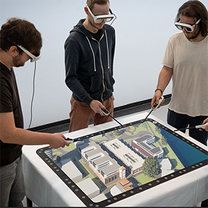
Joeres, F; Zittlau, P; Herbrich, W; Heinrich, F; Rose, G; Hansen, C
Concept development of a cross-reality ecosystem for urban knowledge transfer spaces Proceedings Article
In: 2024 IEEE International Symposium on Mixed and Augmented Reality Adjunct (ISMAR-Adjunct), pp. 166–169, 2024, (ISSN: 2771-1110).
@inproceedings{joeres_concept_2024,

title = {Concept development of a cross-reality ecosystem for urban knowledge transfer spaces},

author = {F Joeres and P Zittlau and W Herbrich and F Heinrich and G Rose and C Hansen},

url = {https://ieeexplore.ieee.org/abstract/document/10765174},

doi = {10.1109/ISMAR-Adjunct64951.2024.00043},

year  = {2024},

date = {2024-10-01},

urldate = {2024-10-01},

booktitle = {2024 IEEE International Symposium on Mixed and Augmented Reality Adjunct (ISMAR-Adjunct)},

pages = {166–169},

abstract = {This paper presents the development of a cross-reality (CR) ecosystem designed for an urban knowledge transfer space (KTS) in a post-industrial urban environment. The project is part of a larger initiative aimed at transforming a former industrial river port into a dynamic KTS, facilitating interactions between scientific, commercial, residential, and cultural stakeholders. Our research explores the potential of multimodal mixed reality (XR) technologies to enhance engagement with the content and stakeholders of the KTS. Through a three-phase process, we identified key stakeholders and their target audiences, selected appropriate XR technologies, and developed initial use cases that integrate web applications, mobile augmented reality (AR), and XR head-mounted displays. The preliminary findings indicate that these technologies can effectively cater to diverse user groups, providing different levels of virtuality and interaction. However, challenges remain, particularly in stakeholder engagement and the evolving nature of the KTS initiative. Ongoing work includes the development of a Web-XR-based prototype, which will be iteratively refined to better meet user needs and adapt to future technological advancements. This research contributes to the understanding of how CR technologies can be employed in urban transformation processes, offering insights into the design of flexible and scalable CR ecosystems.},

note = {ISSN: 2771-1110},

keywords = {},

pubstate = {published},

tppubtype = {inproceedings}

}
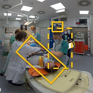
Hanke, L; Vradelis, L; Boedecker, C; Griesinger, J; Demare, T; Lindemann, N; Huettl, F; Chheang, V; Saalfeld, P; Wachter, N; Wollstädter, J; Spranz, M; Lang, H; Hansen, C; Huber, T
Immersive virtual reality for interdisciplinary trauma management – initial evaluation of a training tool prototype Journal Article
In: BMC Medical Education, vol. 24, no. 1, pp. 769, 2024, ISSN: 1472-6920.
@article{hanke_immersive_2024,

title = {Immersive virtual reality for interdisciplinary trauma management – initial evaluation of a training tool prototype},

author = {L Hanke and L Vradelis and C Boedecker and J Griesinger and T Demare and N Lindemann and F Huettl and V Chheang and P Saalfeld and N Wachter and J Wollstädter and M Spranz and H Lang and C Hansen and T Huber},

url = {https://doi.org/10.1186/s12909-024-05764-w

https://pubmed.ncbi.nlm.nih.gov/39026193/, PubMed},

doi = {10.1186/s12909-024-05764-w},

issn = {1472-6920},

year  = {2024},

date = {2024-07-01},

urldate = {2024-07-01},

journal = {BMC Medical Education},

volume = {24},

number = {1},

pages = {769},

abstract = {Emergency care of critically ill patients in the trauma room is an integral part of interdisciplinary work in hospitals. Live threatening injuries require swift diagnosis, prioritization, and treatment; thus, different medical specialties need to work together closely for optimal patient care. Training is essential to facilitate smooth performance. This study presents a training tool for familiarization with trauma room algorithms in immersive virtual reality (VR), and a first qualitative assessment.},

keywords = {},

pubstate = {published},

tppubtype = {article}

}
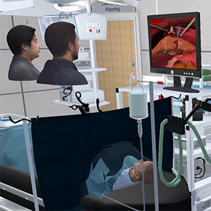
Chheang, V; Schott, D; Saalfeld, P; Vradelis, L; Huber, T; Huettl, F; Lang, H; Preim, B; Hansen, C
Advanced liver surgery training in collaborative VR environments Journal Article
In: Computers & Graphics, vol. 119, pp. 103879, 2024, ISSN: 0097-8493.
@article{chheang_advanced_2024,

title = {Advanced liver surgery training in collaborative VR environments},

author = {V Chheang and D Schott and P Saalfeld and L Vradelis and T Huber and F Huettl and H Lang and B Preim and C Hansen},

url = {https://www.sciencedirect.com/science/article/pii/S0097849324000050},

doi = {10.1016/j.cag.2024.01.006},

issn = {0097-8493},

year  = {2024},

date = {2024-04-01},

urldate = {2024-04-01},

journal = {Computers & Graphics},

volume = {119},

pages = {103879},

abstract = {Virtual surgical training systems are crucial for enabling mental preparation, supporting decision-making, and improving surgical skills. Many virtual surgical training environments focus only on training for a specific medical skill and take place in a single virtual room. However, surgical education and training include the planning of procedures as well as interventions in the operating room context. Moreover, collaboration among surgeons and other medical professionals is only applicable to a limited extent. This work presents a collaborative VR environment similar to a virtual teaching hospital to support surgical training and interprofessional collaboration in a co-located or remote environment. The environment supports photo-realistic avatars and scenarios ranging from planning to training procedures in the virtual operating room. It includes a lobby, a virtual surgical planning room with four surgical planning stations, laparoscopic liver surgery training with the integration of laparoscopic surgical instruments, and medical training scenarios for interprofessional team training in a virtual operating room. Each component was evaluated by domain experts as well as in a series of user studies, providing insights on usability, usefulness, and potential research directions. The proposed environment may serve as a foundation for future medical training simulators.},

keywords = {},

pubstate = {published},

tppubtype = {article}

}
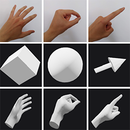
Schwenderling, L; Herbrich, W; Joeres, F; Hansen, C
A Novel Framework for Hand Visualization in Web-Based Collaborative XR Proceedings Article
In: 2024 IEEE Conference on Virtual Reality and 3D User Interfaces Abstracts and Workshops (VRW), pp. 18–23, IEEE, Orlando, FL, USA, 2024, ISBN: 979-8-3503-7449-0.
@inproceedings{schwenderling_novel_2024,

title = {A Novel Framework for Hand Visualization in Web-Based Collaborative XR},

author = {L Schwenderling and W Herbrich and F Joeres and C Hansen},

url = {https://ieeexplore.ieee.org/document/10536317/},

doi = {10.1109/VRW62533.2024.00010},

isbn = {979-8-3503-7449-0},

year  = {2024},

date = {2024-03-01},

urldate = {2024-03-01},

booktitle = {2024 IEEE Conference on Virtual Reality and 3D User Interfaces Abstracts and Workshops (VRW)},

pages = {18–23},

publisher = {IEEE},

address = {Orlando, FL, USA},

abstract = {Many extended reality (XR) applications are platform-specific, making accessibility and cross-platform collaboration difficult. Web-based collaborative XR can enhance adoption of XR technologies, using the browser as a platform-independent interface. However, challenges arise from the browser environment, such as performance limitations. To this end, we present a WebXR-based framework for hand interaction in cross-platform collaboration in XR. A network structure and methods for collaborative and individual object manipulation complement the integrated hand tracking. Three different fidelity levels to represent the hands of remote users were implemented to accommodate different performance capabilities. Concepts ranged from virtual hands to discrete poses with abstract objects. A sample application was implemented with a puzzle task. Two users collaborated in the browsers of the Microsoft HoloLens 2 and the Meta Quest 2. Qualitative and quantitative data on user performance (n=9), and frame rate recordings (n=1) were collected. All users were able to solve the puzzle together quickly and intu-itively. The Quest environment was preferred, as there were more performance issues with the HoloLens. Hand interaction was well-received and proved to be sufficient as the only form of communication. Simpler representations of the hands lead to a higher frame rate, whereby the effects were device-dependent. The impact on task performance was low. Hand interaction enables an intuitive exchange of objects and basic communication in cross-platform collaboration via browsers. Depending on the XR environment, however, device-specific performance limitations must be taken into account by modulating the amount of data and rendering effort.},

keywords = {},

pubstate = {published},

tppubtype = {inproceedings}

}
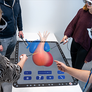
Schott, D; Kunz, M; Mandel, J; Schwenderling, L; Braun-Dullaeus, R; Hansen, C
An AR-Based Multi-User Learning Environment for Anatomy Seminars Proceedings Article
In: 2024 IEEE Conference on Virtual Reality and 3D User Interfaces Abstracts and Workshops (VRW), pp. 949–950, IEEE, Orlando, FL, USA, 2024, ISBN: 979-8-3503-7449-0.
@inproceedings{schott_ar-based_2024,

title = {An AR-Based Multi-User Learning Environment for Anatomy Seminars},

author = {D Schott and M Kunz and J Mandel and L Schwenderling and R Braun-Dullaeus and C Hansen},

url = {https://ieeexplore.ieee.org/document/10536568/},

doi = {10.1109/VRW62533.2024.00271},

isbn = {979-8-3503-7449-0},

year  = {2024},

date = {2024-03-01},

urldate = {2024-03-01},

booktitle = {2024 IEEE Conference on Virtual Reality and 3D User Interfaces Abstracts and Workshops (VRW)},

pages = {949–950},

publisher = {IEEE},

address = {Orlando, FL, USA},

abstract = {Understanding the intricate and rapid changes in shape during em-bryonic formation is vital for medical students. Using the example of embryonic human heart development, we introduce an AR-based multi-user approach to enhance understanding and foster a participatory learning environment. Through a user-centered approach, we created a prototype accommodating two player roles and enabling multi-modal inputs to encourage dynamic group discussions. We in-vited four anatomy experts to evaluate three system configurations in an interdisciplinary workshop to assess the feasibility of integration into anatomy seminars. The gathered data and feedback indicate the potential of our collaborative concept for integration into the medical curriculum.},

keywords = {},

pubstate = {published},

tppubtype = {inproceedings}

}
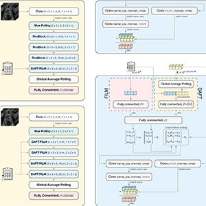
Bashkanov, O; Rak, M; Engelage, L; Hansen, C
Automatic Patient-level Diagnosis of Prostate Disease with Fused 3D MRI and Tabular Clinical Data Proceedings Article
In: Medical Imaging with Deep Learning, pp. 1225–1238, PMLR, 2024, (ISSN: 2640-3498).
@inproceedings{bashkanov_automatic_2024,

title = {Automatic Patient-level Diagnosis of Prostate Disease with Fused 3D MRI and Tabular Clinical Data},

author = {O Bashkanov and M Rak and L Engelage and C Hansen},

url = {https://proceedings.mlr.press/v227/bashkanov24a.html},

year  = {2024},

date = {2024-01-01},

urldate = {2024-01-01},

booktitle = {Medical Imaging with Deep Learning},

pages = {1225–1238},

publisher = {PMLR},

abstract = {Computer-aided diagnosis systems for automatic prostate cancer diagnosis can provide radiologists with decision support during image reading. However, in this case, patient-relevant information often remains unexploited due to the greater focus on the image recognition side, with various imaging devices and modalities, while omitting other potentially valuable clinical data. Therefore, our work investigates the performance of recent methods for the fusion of rich image data and heterogeneous tabular data. Those data may include patient demographics as well as laboratory data, e.g., prostate-specific antigen (PSA). Experiments on the large dataset (3800 subjects) indicated that when using the fusion method with demographic data in clinically significant prostate cancer (csPCa) detection tasks, the mean area under the receiver operating characteristic curve (ROC AUC) has improved significantly from 0.736 to 0.765. We also observed that the naïve concatenation performs similarly or even better than the textbackslashmboxstate-of-the-art fusion modules. We also achieved better prediction quality in grading prostate disease by including more samples from longitudinal PSA profiles in the tabular feature set. Thus, by including the three last PSA samples per patient, the best-performing model has reached AUC of 0.794 and a quadratic weighted kappa score (QWK) of 0.464, which constitutes a significant improvement compared with the image-only method, with ROC AUC of 0.736 and QWK of 0.342.},

note = {ISSN: 2640-3498},

keywords = {},

pubstate = {published},

tppubtype = {inproceedings}

}
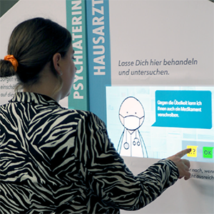
Wagnerberger, D; Schott, D; Schwenderling, L; Hansen, C; Schumacher, D
Empowering Patients: Improve Gender-Sensitive Medical Knowledge Through Interactive Edutainment Proceedings Article
In: Proceedings of the 13th Nordic Conference on Human-Computer Interaction, pp. 1–12, Association for Computing Machinery, New York, NY, USA, 2024, ISBN: 979-8-4007-0966-1.
@inproceedings{wagnerberger_empowering_2024,

title = {Empowering Patients: Improve Gender-Sensitive Medical Knowledge Through Interactive Edutainment},

author = {D Wagnerberger and D Schott and L Schwenderling and C Hansen and D Schumacher},

url = {https://dl.acm.org/doi/10.1145/3679318.3685500},

doi = {10.1145/3679318.3685500},

isbn = {979-8-4007-0966-1},

year  = {2024},

date = {2024-01-01},

urldate = {2024-01-01},

booktitle = {Proceedings of the 13th Nordic Conference on Human-Computer Interaction},

pages = {1–12},

publisher = {Association for Computing Machinery},

address = {New York, NY, USA},

series = {NordiCHI '24},

abstract = {Disregarding crucial gender-specific differences and potential risks in medicine leads to widespread gender inequalities. This paper introduces interactive edutainment concepts developed through a user-centered design approach to raise awareness of gender medicine. An interactive exhibition course and an accompanying deck of cards provide an engaging and sensitizing experience of medical gender inequalities. Qualitative feedback, self-assessment, and user experience and behavior were evaluated during a public display of the concepts (n=14). The results highlight the potential of our playful approach to raising awareness among the public as well as health-related professionals, paving new ways for communication and empowerment of patients of all genders. We believe these insights have broader applicability across various domains, supporting efforts to address all forms of inequality.},

keywords = {},

pubstate = {published},

tppubtype = {inproceedings}

}
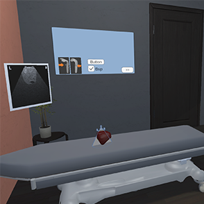
Allgaier, M; Huettl, F; Hanke, L; Huber, T; Preim, B; Saalfeld, S; Hansen, C
Gamification Concepts for a VR-based Visuospatial Training for Intraoperative Liver Ultrasound Proceedings Article
In: Extended Abstracts of the CHI Conference on Human Factors in Computing Systems, pp. 1–8, Association for Computing Machinery, New York, NY, USA, 2024, ISBN: 979-8-4007-0331-7.
@inproceedings{allgaier_gamification_2024,

title = {Gamification Concepts for a VR-based Visuospatial Training for Intraoperative Liver Ultrasound},

author = {M Allgaier and F Huettl and L Hanke and T Huber and B Preim and S Saalfeld and C Hansen},

url = {https://doi.org/10.1145/3613905.3650736},

doi = {10.1145/3613905.3650736},

isbn = {979-8-4007-0331-7},

year  = {2024},

date = {2024-01-01},

urldate = {2024-01-01},

booktitle = {Extended Abstracts of the CHI Conference on Human Factors in Computing Systems},

pages = {1–8},

publisher = {Association for Computing Machinery},

address = {New York, NY, USA},

series = {CHI EA '24},

abstract = {Gamification is widely used due to its positive influence on learning by adding emotions and steering behavior. In medical VR training applications, the use of gamification is rare, and when it is implemented, it often lacks thoughtful design decisions and empirical evaluation. Using a VR-based training for intraoperative ultrasound for liver surgery, we analyzed game elements regarding their suitability and examined two in more detail: difficulty levels and a kit, where the user has to assemble a virtual liver using US. In a broad audience study, levels achieved significantly better results regarding enjoyment. Qualitative feedback from medical students directly comparing the elements revealed that they prefer the kit as well as levels for training. Our studies indicate that levels and the more interactive kit improve the learning experience, which could also be taken as a basis for similar VR-based medical training applications.},

keywords = {},

pubstate = {published},

tppubtype = {inproceedings}

}
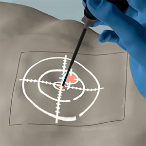
Polenz, L; Joeres, F; Hansen, C; Heinrich, F
Simulating projective Augmented Reality Visualizations in Virtual Reality: Is VR a feasible Environment for medical AR Evaluations? Proceedings Article
In: Extended Abstracts of the CHI Conference on Human Factors in Computing Systems, pp. 1–8, Association for Computing Machinery, New York, NY, USA, 2024, ISBN: 979-8-4007-0331-7.
@inproceedings{polenz_simulating_2024,

title = {Simulating projective Augmented Reality Visualizations in Virtual Reality: Is VR a feasible Environment for medical AR Evaluations?},

author = {L Polenz and F Joeres and C Hansen and F Heinrich},

url = {https://doi.org/10.1145/3613905.3650843},

doi = {10.1145/3613905.3650843},

isbn = {979-8-4007-0331-7},

year  = {2024},

date = {2024-01-01},

urldate = {2024-01-01},

booktitle = {Extended Abstracts of the CHI Conference on Human Factors in Computing Systems},

pages = {1–8},

publisher = {Association for Computing Machinery},

address = {New York, NY, USA},

series = {CHI EA '24},

abstract = {Augmented Reality (AR) has demonstrated potential in medical applications, such as enhancing surgical navigation. However, evaluating medical AR visualizations entails high costs and effort to provide suitable hardware solutions. This is particularly crucial in projective AR, as these systems require several error-prone calibration and registration steps. This work investigates the suitability of Virtual Reality (VR) as a cost-effective and controlled study environment for evaluating projective AR visualizations. A virtual twin of a real laboratory environment was created, and a user study comparing two needle navigation visualizations was conducted. The study simulated identical experiments in both AR and VR to assess if similar results would emerge. Our findings indicate that both AR and VR experiments exhibited comparable effects in terms of performance and workload of both needle insertion visualizations. This study serves as a preliminary step in demonstrating the feasibility of using VR as an evaluation environment for projective AR visualizations.},

keywords = {},

pubstate = {published},

tppubtype = {inproceedings}

}
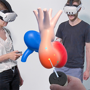
Schott, D; Kunz, M; Heinrich, F; Mandel, J; Albrecht, A; Braun-Dullaeus, R; Hansen, C
Stand Alone or Stay Together: An In-situ Experiment of Mixed-Reality Applications in Embryonic Anatomy Education Proceedings Article
In: Proceedings of the 30th ACM Symposium on Virtual Reality Software and Technology, pp. 1–11, Association for Computing Machinery, New York, NY, USA, 2024, ISBN: 979-8-4007-0535-9.
@inproceedings{schott_stand_2024,

title = {Stand Alone or Stay Together: An In-situ Experiment of Mixed-Reality Applications in Embryonic Anatomy Education},

author = {D Schott and M Kunz and F Heinrich and J Mandel and A Albrecht and R Braun-Dullaeus and C Hansen},

url = {https://dl.acm.org/doi/10.1145/3641825.3687706},

doi = {10.1145/3641825.3687706},

isbn = {979-8-4007-0535-9},

year  = {2024},

date = {2024-01-01},

urldate = {2024-01-01},

booktitle = {Proceedings of the 30th ACM Symposium on Virtual Reality Software and Technology},

pages = {1–11},

publisher = {Association for Computing Machinery},

address = {New York, NY, USA},

series = {VRST '24},

abstract = {Where traditional media and methods reach their limits in anatomy education, mixed-reality (MR) environments can provide effective learning support because of their high interactivity and spatial visualization capabilities. However, the underlying design and pedagogical requirements are as diverse as the technologies themselves. This paper examines the effectiveness of individual- and collaborative learning environments for anatomy education, using embryonic heart development as an example. Both applications deliver the same content using identical visualizations and hardware but differ in interactivity and pedagogical approach. The environments were evaluated in a user study with medical students (n = 90) during their examination phase, assessing usability, user experience, social interaction/co-presence, cognitive load, and personal preference. Additionally, we conducted a knowledge test before and after an MR learning session to determine educational effects compared to a conventional anatomy seminar. Results indicate that the individual learning environment was generally preferred. However, no significant difference in learning effectiveness could be shown between the conventional approach and the MR applications. This suggests that both can effectively complement traditional seminars despite their different natures. Our study contributes to understanding how different MR settings could be tailored for anatomical education.},

keywords = {},

pubstate = {published},

tppubtype = {inproceedings}

}
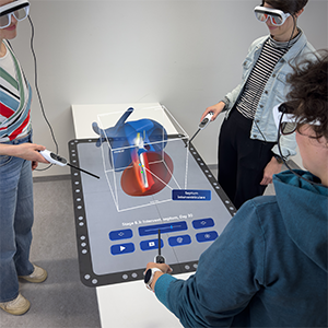
Schott, D; Heinrich, F; Kunz, M; Mandel, J; Albrecht, A; Braun-Dullaeus, R; Hansen, C
CardioCoLab: Collaborative Learning of Embryonic Heart Anatomy in Mixed Reality Proceedings Article
In: Eurographics Workshop on Visual Computing for Biology and Medicine, 2024, (Artwork Size: 5 pages Edition: 1191 ISBN: 9783038682448 Publisher: The Eurographics Association).
@inproceedings{schott_cardiocolab_2024,

title = {CardioCoLab: Collaborative Learning of Embryonic Heart Anatomy in Mixed Reality},

author = {D Schott and F Heinrich and M Kunz and J Mandel and A Albrecht and R Braun-Dullaeus and C Hansen},

url = {https://diglib.eg.org/handle/10.2312/vcbm20241191},

doi = {10.2312/VCBM.20241191},

year  = {2024},

date = {2024-01-01},

urldate = {2024-01-01},

booktitle = {Eurographics Workshop on Visual Computing for Biology and Medicine},

abstract = {The complexity of embryonic heart development presents significant challenges for medical education, particularly in illustrating dynamic morphological changes over short time periods. Traditional teaching methods, such as 2D textbook illustrations and static models, are often insufficient for conveying these intricate processes. To address this gap, we developed a multi-user Mixed Reality (MR) system designed to enhance collaborative learning and interaction with virtual heart models. Building on previous research, we identified the needs of both students and teachers, implementing various interaction and visualization features iteratively. An evaluation with teachers and students (N = 12) demonstrated the system's effectiveness in improving engagement and understanding of embryonic heart development. The study highlights the potential of MR in medical seminar settings as a valuable addition to medical education by enhancing traditional learning methods.},

note = {Artwork Size: 5 pages

Edition: 1191

ISBN: 9783038682448

Publisher: The Eurographics Association},

keywords = {},

pubstate = {published},

tppubtype = {inproceedings}

}
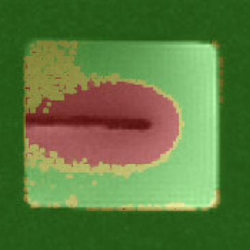
Schröer, S; Alpers, J; Gutberlet, M; Brüsch, I; Rumpel, R; Wacker, F; Hensen, B; Hansen, C
A probabilistic thermal dose model for the estimation of necrosis in MR-guided tumor ablations Journal Article
In: Medical Physics, vol. 51, no. 1, pp. 239–250, 2024, ISSN: 2473-4209, (_eprint: https://aapm.onlinelibrary.wiley.com/doi/pdf/10.1002/mp.16605).
@article{schroer_probabilistic_2024,

title = {A probabilistic thermal dose model for the estimation of necrosis in MR-guided tumor ablations},

author = {S Schröer and J Alpers and M Gutberlet and I Brüsch and R Rumpel and F Wacker and B Hensen and C Hansen},

url = {https://onlinelibrary.wiley.com/doi/abs/10.1002/mp.16605

https://pubmed.ncbi.nlm.nih.gov/37449443/, PubMed},

doi = {10.1002/mp.16605},

issn = {2473-4209},

year  = {2024},

date = {2024-01-01},

urldate = {2024-01-01},

journal = {Medical Physics},

volume = {51},

number = {1},

pages = {239–250},

abstract = {Background Monitoring minimally invasive thermo ablation procedures using magnetic resonance (MR) thermometry allows therapy of tumors even close to critical anatomical structures. Unfortunately, intraoperative monitoring remains challenging due to the necessary accuracy and real-time capability. One reason for this is the statistical error introduced by MR measurement, which causes the prediction of ablation zones to become inaccurate. Purpose In this work, we derive a probabilistic model for the prediction of ablation zones during thermal ablation procedures based on the thermal damage model CEM43. By integrating the statistical error caused by MR measurement into the conventional prediction, we hope to reduce the amount of falsely classified voxels. Methods The probabilistic CEM43 model is empirically evaluated using a polyacrilamide gel phantom and three in-vivo pig livers. Results The results show a higher accuracy in three out of four data sets, with a relative difference in Sørensen–Dice coefficient from textbackslash-3.04%textbackslash to 3.97% compared to the conventional model. Furthermore, the ablation zones predicted by the probabilistic model show a false positive rate with a relative decrease of 11.89%–30.04% compared to the conventional model. Conclusion The presented probabilistic thermal dose model might help to prevent false classification of voxels within ablation zones. This could potentially result in an increased success rate for MR-guided thermal ablation procedures. Future work may address additional error sources and a follow-up study in a more realistic clinical context.},

note = {_eprint: https://aapm.onlinelibrary.wiley.com/doi/pdf/10.1002/mp.16605},

keywords = {},

pubstate = {published},

tppubtype = {article}

}
2023
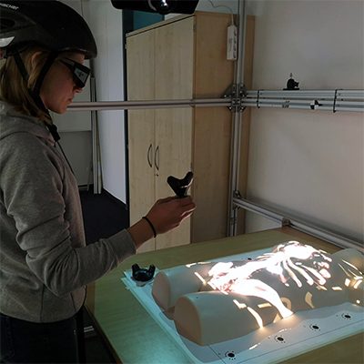
Heinrich, F; Bornemann, K; Polenz, L; Lawonn, K; Hansen, C
Clutch & Grasp: Activation gestures and grip styles for device-based interaction in medical spatial augmented reality Journal Article
In: International Journal of Human-Computer Studies, vol. 180, pp. 103117, 2023, ISSN: 1071-5819.
@article{heinrich_clutch_2023,

title = {Clutch & Grasp: Activation gestures and grip styles for device-based interaction in medical spatial augmented reality},

author = {F Heinrich and K Bornemann and L Polenz and K Lawonn and C Hansen},

url = {https://www.sciencedirect.com/science/article/pii/S107158192300126X},

doi = {10.1016/j.ijhcs.2023.103117},

issn = {1071-5819},

year  = {2023},

date = {2023-12-01},

urldate = {2023-12-01},

journal = {International Journal of Human-Computer Studies},

volume = {180},

pages = {103117},

abstract = {Presenting medical volume data using augmented reality (AR) can facilitate the identification of anatomical structures, the perception of their spatial relations and the development of mental maps compared to more commonly used monitors. However, interaction methods explored in these conventional settings may not be applicable in AR environments, or perform differently. In terms of mode activation, gestural interaction was shown to be a viable, touchless alternative to traditional input devices, which is desirable in sterile medical use cases. Therefore, we present a user study (n = 21) comparing hand and foot gestures with voice commands for the activation of interaction modes within a projector-based, spatial AR prototype to visualize medical volume data. Interaction itself was performed via hand movements captured by a data glove. Consistent, statistically significant results across measured variables suggest advantages of voice commands. In addition, a second experiment (n = 17) compared the hand-based interaction with two motion-sensitive devices held in power and in precision grip respectively. All modes were activated using voice commands. No considerable differences between tested grip styles could be determined. The findings suggest that the choice of preferable interaction devices is user and use case dependent.},

keywords = {},

pubstate = {published},

tppubtype = {article}

}
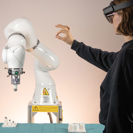
Mielke, T; Joeres, F; Schott, D; Hansen, C
Interactive Registration Methods for Augmented Reality in Robotics: A Comparative Evaluation Proceedings Article
In: 2023 IEEE International Symposium on Mixed and Augmented Reality Adjunct (ISMAR-Adjunct), pp. 501–506, IEEE, Sydney, Australia, 2023, ISBN: 979-8-3503-2891-2.
@inproceedings{mielke_interactive_2023,

title = {Interactive Registration Methods for Augmented Reality in Robotics: A Comparative Evaluation},

author = {T Mielke and F Joeres and D Schott and C Hansen},

url = {https://ieeexplore.ieee.org/document/10322246/},

doi = {10.1109/ISMAR-Adjunct60411.2023.00109},

isbn = {979-8-3503-2891-2},

year  = {2023},

date = {2023-10-01},

urldate = {2023-10-01},

booktitle = {2023 IEEE International Symposium on Mixed and Augmented Reality Adjunct (ISMAR-Adjunct)},

pages = {501–506},

publisher = {IEEE},

address = {Sydney, Australia},

abstract = {Augmented Reality (AR) visualization has shown potential for supporting intuitive and efficient human-robot interaction in a range of tasks. Since all these tasks are spatially related to the robot, the precise positioning of the AR content is critical to the applicability. However, most research has primarily focused on developing visualizations rather than exploring methods for aligning AR content in the robotic workspace. This paper aims to bridge this gap by implementing and comparing different interactive registration methods, including two point-based and one manual approach. We comparatively evaluated these registration methods in a user study (n=21), measuring registration accuracy, duration, and subjective user feedback. Our results indicate that the point-based methods outperform the manual approach in terms of both accuracy and perceived workload. Furthermore, participants achieved significantly faster performance with a point-based approach using physically defined registration points compared to a point-based approach using markers attached to the robot.},

keywords = {},

pubstate = {published},

tppubtype = {inproceedings}

}
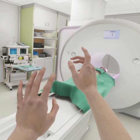
Schott, D; Heinrich, F; Stallmeister, L; Moritz, J; Hensen, B; Hansen, C
Is this the vReal Life? Manipulating Visual Fidelity of Immersive Environments for Medical Task Simulation Proceedings Article
In: 2023 IEEE International Symposium on Mixed and Augmented Reality (ISMAR), pp. 1171–1180, IEEE, Sydney, Australia, 2023, ISBN: 979-8-3503-2838-7.
@inproceedings{schott_is_2023,

title = {Is this the vReal Life? Manipulating Visual Fidelity of Immersive Environments for Medical Task Simulation},

author = {D Schott and F Heinrich and L Stallmeister and J Moritz and B Hensen and C Hansen},

url = {https://ieeexplore.ieee.org/document/10316533/},

doi = {10.1109/ISMAR59233.2023.00134},

isbn = {979-8-3503-2838-7},

year  = {2023},

date = {2023-10-01},

urldate = {2023-10-01},

booktitle = {2023 IEEE International Symposium on Mixed and Augmented Reality (ISMAR)},

pages = {1171–1180},

publisher = {IEEE},

address = {Sydney, Australia},

abstract = {Recent developments and research advances contribute to an ever-increasing trend towards quality levels close to what we experience in reality. In this work, we investigate how different degrees of these quality characteristics affect user performance, qualia of user experience (UX), and sense of presence in an example medical task. To this end, a two-way within-subjects design user study was conducted, in which three different levels of visual fidelity were compared. In addition, two different interaction modalities were considered: (1) the use of conventional VR controllers and (2) natural hand interaction using 3D-printed, spatially-registered replicas of medical devices, to interact with their virtual representations. Consistent results indicate that higher degrees of visual fidelity evoke a higher sense of presence and UX. However, user performance was less affected. Moreover, no differences were detected between both interaction modalities for the examined task. Future work should investigate the discovered interaction effects between quality levels and interaction modalities in more detail and examine whether these results can be reproduced in tasks that require more precision. This work provides insights into the implications to consider when studying interactions in VR and paves the way for investigations into early phases of medical product development and workflow analysis.},

keywords = {},

pubstate = {published},

tppubtype = {inproceedings}

}
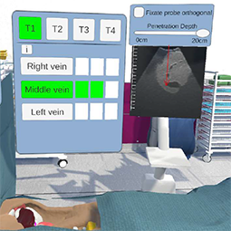
Allgaier, M; Huettl, F; Hanke, L; Lang, H; Huber, T; Preim, B; Saalfeld, S; Hansen, C
LiVRSono – Virtual Reality Training with Haptics for Intraoperative Ultrasound Proceedings Article
In: 2023 IEEE International Symposium on Mixed and Augmented Reality (ISMAR), pp. 980–989, IEEE, Sydney, Australia, 2023, ISBN: 979-8-3503-2838-7.
@inproceedings{allgaier_livrsono_2023,

title = {LiVRSono - Virtual Reality Training with Haptics for Intraoperative Ultrasound},

author = {M Allgaier and F Huettl and L Hanke and H Lang and T Huber and B Preim and S Saalfeld and C Hansen},

url = {https://ieeexplore.ieee.org/document/10316488/},

doi = {10.1109/ISMAR59233.2023.00114},

isbn = {979-8-3503-2838-7},

year  = {2023},

date = {2023-10-01},

urldate = {2023-10-01},

booktitle = {2023 IEEE International Symposium on Mixed and Augmented Reality (ISMAR)},

pages = {980–989},

publisher = {IEEE},

address = {Sydney, Australia},

abstract = {One of the biggest challenges in using ultrasound (US) is learning to create a spatial mental model of the interior of the scanned object based on the US image and the probe position. As intraoperative ultrasound (IOUS) cannot be easily trained on patients, we present LiVRSono, an immersive VR application to train this skill. The immersive environment, including an US simulation with patientspecific data as well as haptics to support hand-eye coordination, provides a realistic setting. Four clinically relevant training scenarios were identified based on the described learning goal and the workflow of IOUS for liver. The realism of the setting and the training scenarios were evaluated with eleven physicians, of which six participants are experts in IOUS for liver and five participants are potential users of the training system. The setting, handling of the US probe, and US image were considered realistic enough for the learning goal. Regarding the haptic feedback, a limitation is the restricted workspace of the input device. Three of the four training scenarios were rated as meaningful and effective. A pilot study regarding learning outcome shows positive results, especially with respect to confidence and perceived competence. Besides the drawbacks of the input device, our training system provides a realistic learning environment with meaningful scenarios to train the creation of a mental 3D model when performing IOUS. We also identified important improvements to the training scenarios to further enhance the training experience.},

keywords = {},

pubstate = {published},

tppubtype = {inproceedings}

}
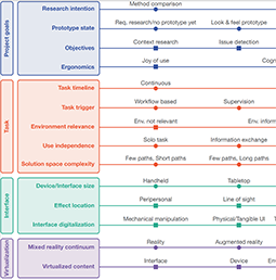
Schott, D; Moritz, J; Hansen, C; Joeres, F
The UUXR-Framework: A Draft Classification for Using Extended Reality in Usability and User Experience Research Proceedings Article
In: 2023 IEEE International Symposium on Mixed and Augmented Reality Adjunct (ISMAR-Adjunct), pp. 460–465, IEEE, Sydney, Australia, 2023, ISBN: 979-8-3503-2891-2.
@inproceedings{schott_uuxr-framework_2023,

title = {The UUXR-Framework: A Draft Classification for Using Extended Reality in Usability and User Experience Research},

author = {D Schott and J Moritz and C Hansen and F Joeres},

url = {https://ieeexplore.ieee.org/document/10322234/},

doi = {10.1109/ISMAR-Adjunct60411.2023.00100},

isbn = {979-8-3503-2891-2},

year  = {2023},

date = {2023-10-01},

urldate = {2023-10-01},

booktitle = {2023 IEEE International Symposium on Mixed and Augmented Reality Adjunct (ISMAR-Adjunct)},

pages = {460–465},

publisher = {IEEE},

address = {Sydney, Australia},

abstract = {Conducting human-centered evaluations in extended reality (XR) environments is a growing trend in user research and usability engineering. However, there has been little to no systematic investigation of the emerging methods in this field published to date. The motivation behind our work is to explore and classify strategies and methods for utilizing XR technologies in the context of usability and user experience (UUX) activities. This paper proposes a draft classification framework for the use of XR technologies in UUX activities, combining an informal exploration of relevant literature with established UUX methods. Within this framework, we propose 12 dimensions that we consider potentially relevant for determining whether and how the use of XR technologies can benefit product development and user research. To evaluate the structure and phrasing of our proposed dimensions, we conducted an initial evaluation with UUX professionals (N = 11). We believe that our dimensions form an early-stage foundation for future guidelines aimed at UUX researchers. The framework serves as a tool for assessing different levels of virtualization in UUX work and facilitating knowledge transfer between academia and industry.},

keywords = {},

pubstate = {published},

tppubtype = {inproceedings}

}
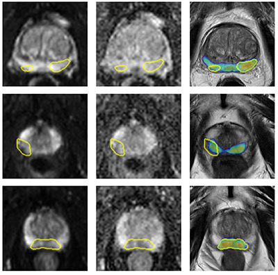
Bashkanov, O; Rak, M; Meyer, A; Engelage, L; Lumiani, A; Muschter, R; Hansen, C
Automatic detection of prostate cancer grades and chronic prostatitis in biparametric MRI Journal Article
In: Computer Methods and Programs in Biomedicine, vol. 239, pp. 107624, 2023, ISSN: 0169-2607.
@article{bashkanov_automatic_2023,

title = {Automatic detection of prostate cancer grades and chronic prostatitis in biparametric MRI},

author = {O Bashkanov and M Rak and A Meyer and L Engelage and A Lumiani and R Muschter and C Hansen},

url = {https://www.sciencedirect.com/science/article/pii/S0169260723002894

https://pubmed.ncbi.nlm.nih.gov/37271051/, PubMed},

doi = {10.1016/j.cmpb.2023.107624},

issn = {0169-2607},

year  = {2023},

date = {2023-09-01},

urldate = {2023-09-01},

journal = {Computer Methods and Programs in Biomedicine},

volume = {239},

pages = {107624},

abstract = {Background and objective:With emerging evidence to improve prostate cancer (PCa) screening, multiparametric magnetic prostate imaging is becoming an essential noninvasive component of the diagnostic routine. Computer-aided diagnostic (CAD) tools powered by deep learning can help radiologists interpret multiple volumetric images. In this work, our objective was to examine promising methods recently proposed in the multigrade prostate cancer detection task and to suggest practical considerations regarding model training in this context. Methods:We collected 1647 fine-grained biopsy-confirmed findings, including Gleason scores and prostatitis, to form a training dataset. In our experimental framework for lesion detection, all models utilized 3D nnU-Net architecture that accounts for anisotropy in the MRI data. First, we explore an optimal range of b-values for diffusion-weighted imaging (DWI) modality and its effect on the detection of clinically significant prostate cancer (csPCa) and prostatitis using deep learning, as the optimal range is not yet clearly defined in this domain. Next, we propose a simulated multimodal shift as a data augmentation technique to compensate for the multimodal shift present in the data. Third, we study the effect of incorporating the prostatitis class alongside cancer-related findings at three different granularities of the prostate cancer class (coarse, medium, and fine) and its impact on the detection rate of the target csPCa. Furthermore, ordinal and one-hot encoded (OHE) output formulations were tested. Results: An optimal model configuration with fine class granularity (prostatitis included) and OHE has scored the lesion-wise partial Free-Response Receiver Operating Characteristic (FROC) area under the curve (AUC) of 1.94 (CI 95%: 1.76–2.11) and patient-wise ROC AUC of 0.874 (CI 95%: 0.793–0.938) in the detection of csPCa. Inclusion of the auxiliary prostatitis class has demonstrated a stable relative improvement in specificity at a false positive rate (FPR) of 1.0 per patient, with an increase of 3%, 7%, and 4% for coarse, medium, and fine class granularities. Conclusions: This paper examines several configurations for model training in the biparametric MRI setup and proposes optimal value ranges. It also shows that the fine-grained class configuration, including prostatitis, is beneficial for detecting csPCa. The ability to detect prostatitis in all low-risk cancer lesions suggests the potential to improve the quality of the early diagnosis of prostate diseases. It also implies an improved interpretability of the results by the radiologist.},

keywords = {},

pubstate = {published},

tppubtype = {article}

}
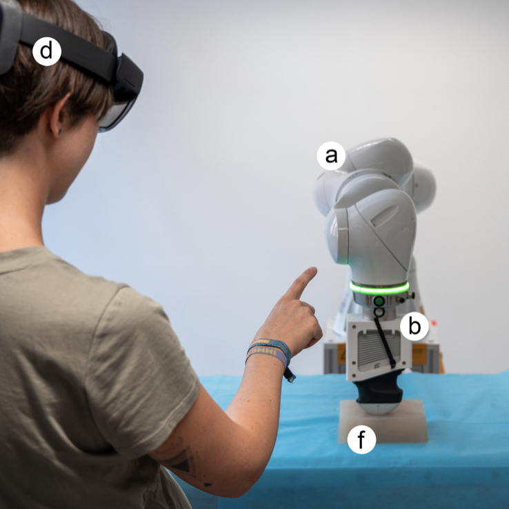
Schreiter, J; Mielke, T; Schott, D; Thormann, M; Omari, J; Pech, M; Hansen, C
A multimodal user interface for touchless control of robotic ultrasound Journal Article
In: International Journal of Computer Assisted Radiology and Surgery, vol. 18, no. 8, pp. 1429–1436, 2023, ISSN: 1861-6429.
@article{schreiter_multimodal_2022,

title = {A multimodal user interface for touchless control of robotic ultrasound},

author = {J Schreiter and T Mielke and D Schott and M Thormann and J Omari and M Pech and C Hansen},

url = {https://link.springer.com/10.1007/s11548-022-02810-0

https://pubmed.ncbi.nlm.nih.gov/36565368/, PubMed},

doi = {10.1007/s11548-022-02810-0},

issn = {1861-6429},

year  = {2023},

date = {2023-08-01},

urldate = {2023-08-01},

journal = {International Journal of Computer Assisted Radiology and Surgery},

volume = {18},

number = {8},

pages = {1429–1436},

abstract = {Purpose Past research contained the investigation and development of robotic ultrasound. In this context, interfaces which allow for interaction with the robotic system are of paramount importance. Few researchers have addressed the issue of developing non-tactile interaction approaches, although they could be beneficial for maintaining sterility during medical procedures. Interaction could be supported by multimodality, which has the potential to enable intuitive and natural interaction. To assess the feasibility of multimodal interaction for non-tactile control of a co-located robotic ultrasound system, a novel human–robot interaction concept was developed.

Methods The medical use case of needle-based interventions under hybrid computed tomography and ultrasound imaging was analyzed by interviewing four radiologists. From the resulting workflow, interaction tasks were derived which include human–robot interaction. Based on this, characteristics of a multimodal, touchless human–robot interface were elaborated, suitable interaction modalities were identified, and a corresponding interface was developed, which was thereafter evaluated in a user study with eight participants.

Results The implemented interface includes voice commands, combined with hand gesture control for discrete control and navigation interaction of the robotic US probe, respectively. The interaction concept was evaluated by the users in the form of a quantitative questionnaire with a average usability. Qualitative analysis of interview results revealed user satisfaction with the implemented interaction methods and potential improvements to the system.

Conclusion A multimodal, touchless interaction concept for a robotic US for the use case of needle-based procedures in interventional radiology was developed, incorporating combined voice and hand gesture control. Future steps will include the integration of a solution for the missing haptic feedback and the evaluation of its clinical suitability.},

keywords = {},

pubstate = {published},

tppubtype = {article}

}
Methods The medical use case of needle-based interventions under hybrid computed tomography and ultrasound imaging was analyzed by interviewing four radiologists. From the resulting workflow, interaction tasks were derived which include human–robot interaction. Based on this, characteristics of a multimodal, touchless human–robot interface were elaborated, suitable interaction modalities were identified, and a corresponding interface was developed, which was thereafter evaluated in a user study with eight participants.
Results The implemented interface includes voice commands, combined with hand gesture control for discrete control and navigation interaction of the robotic US probe, respectively. The interaction concept was evaluated by the users in the form of a quantitative questionnaire with a average usability. Qualitative analysis of interview results revealed user satisfaction with the implemented interaction methods and potential improvements to the system.
Conclusion A multimodal, touchless interaction concept for a robotic US for the use case of needle-based procedures in interventional radiology was developed, incorporating combined voice and hand gesture control. Future steps will include the integration of a solution for the missing haptic feedback and the evaluation of its clinical suitability.
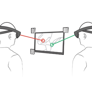
Huettl, F; Heinrich, F; Boedecker, C; Vradelis, L; Ludt, A; Kneist, W; Lang, H; Hansen, C; Huber, T
In: Journal of the American College of Surgeons, vol. 237, no. 2, pp. 292, 2023, ISSN: 1879-1190.
@article{huettl_real-time_2023,

title = {Real-Time Augmented Reality Annotation for Surgical Education during Laparoscopic Surgery: Results from a Single-Center Randomized Controlled Trial and Future Aspects},

author = {F Huettl and F Heinrich and C Boedecker and L Vradelis and A Ludt and W Kneist and H Lang and C Hansen and T Huber},

url = {https://journals.lww.com/journalacs/abstract/2023/08000/real_time_augmented_reality_annotation_for.20.aspx

https://pubmed.ncbi.nlm.nih.gov/37042553/, PubMed},

doi = {10.1097/XCS.0000000000000712},

issn = {1879-1190},

year  = {2023},

date = {2023-08-01},

urldate = {2023-08-01},

journal = {Journal of the American College of Surgeons},

volume = {237},

number = {2},

pages = {292},

abstract = {Background: We developed an interactive augmented reality tool (HoloPointer) that enables real-time annotation on a laparoscopy monitor for intraoperative guidance. This application operates exclusively via verbal commands and head movements to ensure a sterile workflow. Study design: Purpose of this randomized controlled clinical trial was to evaluate the integration of this new technology into the operating room. This prospective single-center study included 32 elective laparoscopic cholecystectomies (29 surgical teams, 15 trainees, 13 trainers). Primary objectives and assessment measures was the HoloPointer's influence on surgical performance (subjective assessment, global operative assessment of laparoscopic skills - GOALS, and Critical View of Safety -CVS). Secondary objectives and outcome variables were its influence on operation time, quality of assistance (5 point likert-scale), and user-friendliness (System Usability Scale - SUS, 0-100 points). Results: Gestural corrections were reduced by 59.4% (4.6 SD 8.1 vs. 1.9 SD 4.7; p > 0.05) and verbal corrections by 36.1% (17.8 SD 12.9 vs. 11.4 SD 8.1; p > 0.05). Subjective surgical performance could be improved by 84,6% of participants. No statistically significant differences were observed for objective parameters GOALS, CVS and operation time. In the SUS, the application achieved an average score of 72.5 SD 16.3 (good user-friendliness). Of the participants, 69.2% wanted to use the HoloPointer more frequently. Conclusion: The majority of trainees had improved their surgical performance using the HoloPointer in elective laparoscopic cholecystectomies, and the rate of classic but potentially misleading corrections was noticeably reduced. The HoloPointer has the potential to improve education in minimally invasive surgery.},

keywords = {},

pubstate = {published},

tppubtype = {article}

}

Schwenderling, L; Kleinau, A; Herbrich, W; Kasireddy, H; Heinrich, F; Hansen, C
Activation modes for gesture-based interaction with a magic lens in AR anatomy visualisation Journal Article
In: Computer Methods in Biomechanics and Biomedical Engineering: Imaging & Visualization, vol. 11, no. 4, pp. 1243–1250, 2023, ISSN: 2168-1163, (Publisher: Taylor & Francis).
@article{schwenderling_activation_2023,

title = {Activation modes for gesture-based interaction with a magic lens in AR anatomy visualisation},

author = {L Schwenderling and A Kleinau and W Herbrich and H Kasireddy and F Heinrich and C Hansen},

url = {https://doi.org/10.1080/21681163.2022.2157749},

doi = {10.1080/21681163.2022.2157749},

issn = {2168-1163},

year  = {2023},

date = {2023-07-01},

urldate = {2023-07-01},

journal = {Computer Methods in Biomechanics and Biomedical Engineering: Imaging & Visualization},

volume = {11},

number = {4},

pages = {1243–1250},

abstract = {Learning human anatomy is key for health-related education and often requires expensive and time-consuming cadaver dissection courses. Augmented reality (AR) for the representation of spatially registered 3D models can be used as a low-cost and flexible alternative. However, suitable visualisation and interaction approaches are needed to display multilayered anatomy data. This paper features a spherical volumetric AR Magic Lens controlled by mid-air hand gestures to explore the human anatomy on a phantom. Defining how gestures control associated actions is important for intuitive interaction. Therefore, two gesture activation modes were investigated in a user study (n = 24). Performing the gestures once to toggle actions showed a higher interaction count since an additional stop gesture was used. Holding the gestures was favoured in the qualitative feedback. Both modes showed similar performance in terms of accuracy and task completion time. Overall, direct gesture manipulation of a magic lens for anatomy visualisation is, thus, recommended.},

note = {Publisher: Taylor & Francis},

keywords = {},

pubstate = {published},

tppubtype = {article}

}
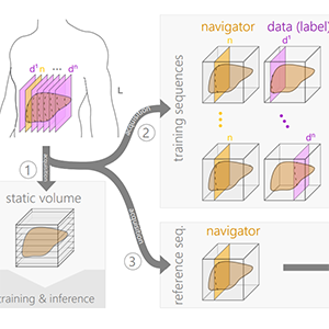
Gulamhussene, G; Rak, M; Bashkanov, O; Joeres, F; Omari, J; Pech, M; Hansen, C
Transfer-learning is a key ingredient to fast deep learning-based 4D liver MRI reconstruction Journal Article
In: Scientific Reports, vol. 13, no. 1, pp. 11227, 2023, ISSN: 2045-2322, (Publisher: Nature Publishing Group).
@article{gulamhussene_transfer-learning_2023,

title = {Transfer-learning is a key ingredient to fast deep learning-based 4D liver MRI reconstruction},

author = {G Gulamhussene and M Rak and O Bashkanov and F Joeres and J Omari and M Pech and C Hansen},

url = {https://www.nature.com/articles/s41598-023-38073-1

https://pubmed.ncbi.nlm.nih.gov/37433827/, PubMed},

doi = {10.1038/s41598-023-38073-1},

issn = {2045-2322},

year  = {2023},

date = {2023-07-01},

urldate = {2023-07-01},

journal = {Scientific Reports},

volume = {13},

number = {1},

pages = {11227},

abstract = {Time-resolved volumetric magnetic resonance imaging (4D MRI) could be used to address organ motion in image-guided interventions like tumor ablation. Current 4D reconstruction techniques are unsuitable for most interventional settings because they are limited to specific breathing phases, lack temporal/spatial resolution, and have long prior acquisitions or reconstruction times. Deep learning-based (DL) 4D MRI approaches promise to overcome these shortcomings but are sensitive to domain shift. This work shows that transfer learning (TL) combined with an ensembling strategy can help alleviate this key challenge. We evaluate four approaches: pre-trained models from the source domain, models directly trained from scratch on target domain data, models fine-tuned from a pre-trained model and an ensemble of fine-tuned models. For that the data base was split into 16 source and 4 target domain subjects. Comparing ensemble of fine-tuned models (N = 10) with directly learned models, we report significant improvements (P < 0.001) of the root mean squared error (RMSE) of up to 12% and the mean displacement (MDISP) of up to 17.5%. The smaller the target domain data amount, the larger the effect. This shows that TL + Ens significantly reduces beforehand acquisition time and improves reconstruction quality, rendering it a key component in making 4D MRI clinically feasible for the first time in the context of 4D organ motion models of the liver and beyond.},

note = {Publisher: Nature Publishing Group},

keywords = {},

pubstate = {published},

tppubtype = {article}

}
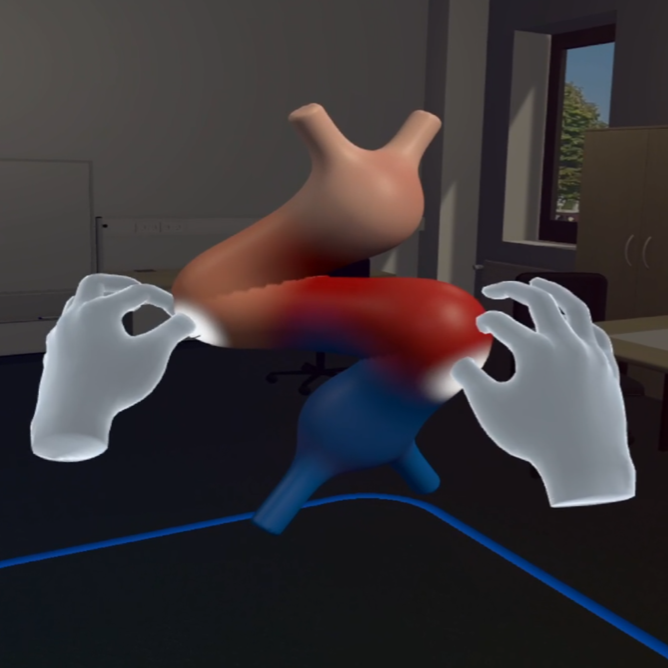
Schott, D; Kunz, M; Wunderling, T; Heinrich, F; Braun-Dullaeus, R; Hansen, C
CardioGenesis4D: Interactive Morphological Transitions of Embryonic Heart Development in a Virtual Learning Environment Journal Article
In: IEEE Transactions on Visualization and Computer Graphics, vol. 29, no. 5, pp. 2615–2625, 2023, ISSN: 1941-0506.
@article{schott_cardiogenesis4d_2023,

title = {CardioGenesis4D: Interactive Morphological Transitions of Embryonic Heart Development in a Virtual Learning Environment},

author = {D Schott and M Kunz and T Wunderling and F Heinrich and R Braun-Dullaeus and C Hansen},

url = {https://ieeexplore.ieee.org/document/10049681},

doi = {10.1109/TVCG.2023.3247110},

issn = {1941-0506},

year  = {2023},

date = {2023-05-01},

urldate = {2023-05-01},

journal = {IEEE Transactions on Visualization and Computer Graphics},

volume = {29},

number = {5},

pages = {2615–2625},

abstract = {In the embryonic human heart, complex dynamic shape changes take place in a short period of time on a microscopic scale, making this development difficult to visualize. However, spatial understanding of these processes is essential for students and future cardiologists to properly diagnose and treat congenital heart defects. Following a user centered approach, the most crucial embryological stages were identified and translated into a virtual reality learning environment (VRLE) to enable the understanding of the morphological transitions of these stages through advanced interactions. To address individual learning types, we implemented different features and evaluated the application regarding usability, perceived task load, and sense of presence in a user study. We also assessed spatial awareness and knowledge gain, and finally obtained feedback from domain experts. Overall, students and professionals rated the application positively. To minimize distraction from interactive learning content, such VRLEs should consider features for different learning types, allow for gradual habituation, and at the same time provide enough playful stimuli. Our work previews how VR can be integrated into a cardiac embryology education curriculum.},

keywords = {},

pubstate = {published},

tppubtype = {article}

}
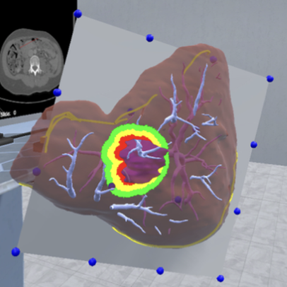
Chheang, V; Bruggernann, R; Preim, B; Hansen, C
Virtual Resection Planning using Bezier Surface Interactions in Collaborative VR Environments Proceedings Article
In: 2023 IEEE Conference on Virtual Reality and 3D User Interfaces Abstracts and Workshops (VRW), pp. 166–169, 2023.
@inproceedings{chheang_virtual_2023,

title = {Virtual Resection Planning using Bezier Surface Interactions in Collaborative VR Environments},

author = {V Chheang and R Bruggernann and B Preim and C Hansen},

url = {https://ieeexplore.ieee.org/document/10108900},

doi = {10.1109/VRW58643.2023.00041},

year  = {2023},

date = {2023-03-01},

urldate = {2023-03-01},

booktitle = {2023 IEEE Conference on Virtual Reality and 3D User Interfaces Abstracts and Workshops (VRW)},

pages = {166–169},

abstract = {The use of virtual reality (VR) has been proposed for collaborative planning or training scenarios to aid surgeons in preparing surgical interventions. In this paper, we explore the usage of cubic Bezier surfaces and their respective interactions in collaborative VR environments to define atypical resection surfaces for planning abdominal organ tumor resections. We conducted a pilot study (n = 10) to evaluate the usability of the Bezier surface interaction compared to a free deformation approach using the example of liver surgery planning. Moreover, we showed the prototype to an expert and conducted an interview to collect clinical feedback. The results show potential benefits of both interaction techniques that could be essential for virtual resection planning and training. The expert highlighted that combining both techniques could speed up the workflow and provide various options for planning.},

keywords = {},

pubstate = {published},

tppubtype = {inproceedings}

}
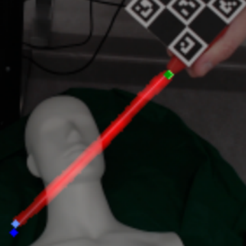
Gulamhussene, G; Spiegel, J; Das, A; Rak, M; Hansen, C
Deep Learning-based Marker-less Pose Estimation of Interventional Tools using Surrogate Keypoints Proceedings Article
In: Deserno, T; Handels, H; Maier, A; Maier-Hein, K; Palm, C; Tolxdorff, T (Ed.): Bildverarbeitung für die Medizin 2023, pp. 292–298, Springer Fachmedien, Wiesbaden, 2023, ISBN: 978-3-658-41657-7.
@inproceedings{gulamhussene_deep_2023,

title = {Deep Learning-based Marker-less Pose Estimation of Interventional Tools using Surrogate Keypoints},

author = {G Gulamhussene and J Spiegel and A Das and M Rak and C Hansen},

editor = {T Deserno and H Handels and A Maier and K Maier-Hein and C Palm and T Tolxdorff},

doi = {10.1007/978-3-658-41657-7_63},

isbn = {978-3-658-41657-7},

year  = {2023},

date = {2023-01-01},

urldate = {2023-01-01},

booktitle = {Bildverarbeitung für die Medizin 2023},

pages = {292–298},

publisher = {Springer Fachmedien},

address = {Wiesbaden},

abstract = {Estimating the position of an intervention needle is an important ability in computer-assisted interventions. Currently, such pose estimations rely either on radiation-intensive CT imaging or need additional optical markers which add overhead to the clinical workflow. We propose a novel deep-learning-based technique for pose estimation of interventional tools which relies on detecting visible features on the tool itself without additional markers.We also propose a novel and fast pipeline for creating vast amounts of robustly labeled and markerless ground truth data for training such neural networks. Initial evaluations suggest that with needle base and needle tip localization errors of about 1 and 4 cm, Our approach can yield a search corridor that can be used to find the needle in a low-dose CT image, reducing radiation exposure.},

keywords = {},

pubstate = {published},

tppubtype = {inproceedings}

}
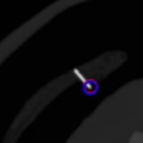
Gulamhussene, G; Das, A; Spiegel, J; Punzet, D; Rak, M; Hansen, C
Needle Tip Tracking During CT-guided Interventions using Fuzzy Segmentation Proceedings Article
In: Deserno, T; Handels, H; Maier, A; Maier-Hein, K; Palm, C; Tolxdorff, T (Ed.): Bildverarbeitung für die Medizin 2023, pp. 285–291, Springer Fachmedien, Wiesbaden, 2023, ISBN: 978-3-658-41657-7.
@inproceedings{gulamhussene_needle_2023,

title = {Needle Tip Tracking During CT-guided Interventions using Fuzzy Segmentation},

author = {G Gulamhussene and A Das and J Spiegel and D Punzet and M Rak and C Hansen},

editor = {T Deserno and H Handels and A Maier and K Maier-Hein and C Palm and T Tolxdorff},

doi = {10.1007/978-3-658-41657-7_62},

isbn = {978-3-658-41657-7},

year  = {2023},

date = {2023-01-01},

urldate = {2023-01-01},

booktitle = {Bildverarbeitung für die Medizin 2023},

pages = {285–291},

publisher = {Springer Fachmedien},

address = {Wiesbaden},

abstract = {CT-guided interventions are standard practice for radiologists to treat lesions in various parts of the human body. In this context, accurate tracking of instruments is of paramount importance for the safety of the procedure and helps radiologists avoid unintended damage to adjacent organs. In this work, a novel method for the estimation of 3D needle tip coordinates in a CT volume using only two 2D projections in an interventional setting is proposed. The method applies a deep learning model for the fuzzy segmentation of the region containing the tip on 2D projections and automatically extracts the position of the tip. A simple UNet achieves a Dice score of 0.9906 for the fuzzy segmentation and an average euclidean distance of 2.96 mm for the needle tip regression task.},

keywords = {},

pubstate = {published},

tppubtype = {inproceedings}

}
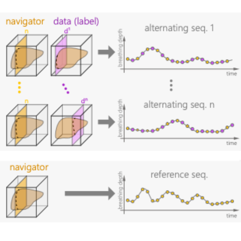
Gulamhussene, G; Bashkanov, O; Omari, J; Pech, M; Hansen, C; Rak, M
Using Training Samples as Transitive Information Bridges in Predicted 4D MRI Proceedings Article
In: Xue, Z; Antani, S; Zamzmi, G; Yang, F; Rajaraman, S; Huang, S; Linguraru, M; Liang, Z (Ed.): Medical Image Learning with Limited and Noisy Data, pp. 237–245, Springer Nature Switzerland, Cham, 2023, ISBN: 978-3-031-44917-8.
@inproceedings{gulamhussene_using_2023,

title = {Using Training Samples as Transitive Information Bridges in Predicted 4D MRI},

author = {G Gulamhussene and O Bashkanov and J Omari and M Pech and C Hansen and M Rak},

editor = {Z Xue and S Antani and G Zamzmi and F Yang and S Rajaraman and S Huang and M Linguraru and Z Liang},

doi = {10.1007/978-3-031-44917-8_23},

isbn = {978-3-031-44917-8},

year  = {2023},

date = {2023-01-01},

urldate = {2023-01-01},

booktitle = {Medical Image Learning with Limited and Noisy Data},

pages = {237–245},

publisher = {Springer Nature Switzerland},

address = {Cham},

abstract = {The lack of real-time techniques for monitoring respiratory motion impairs the development of guidance systems for image-guided interventions. Recent works show that U-Net based real-time 4D MRI prediction methods are promising, but prone to bad image quality when small training data sets and inputs with multiple MR contrast are used. To overcome this problem, we propose a more efficient use of the spare training data and re-utilize 2D training samples as a secondary input for construction of transitive information bridges between the navigator slice primary input and the data slice prediction. We thus remove the need for a separate 3D breath-hold MRI with different MR contrast as the secondary input. Results show that our novel construction leads to improved prediction quality with very sparse training data, with a significant decrease in root mean squared error (RMSE) from 0.3 to 0.27 (p$$<2.2eˆ-16$$<2.2e-16},

keywords = {},

pubstate = {published},

tppubtype = {inproceedings}

}
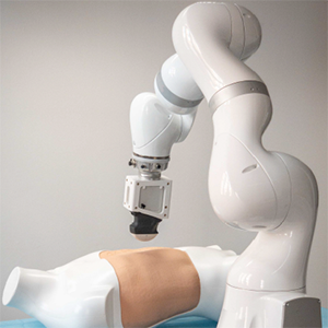
Urrutia, R; Espejo, D; Evens, N; Guerra, M; Sühn, T; Boese, A; Hansen, C; Fuentealba, P; Illanes, A; Poblete, V
Clustering Methods for Vibro-Acoustic Sensing Features as a Potential Approach to Tissue Characterisation in Robot-Assisted Interventions Journal Article
In: Sensors, vol. 23, no. 23, pp. 9297, 2023, ISSN: 1424-8220, (Publisher: Multidisciplinary Digital Publishing Institute).
@article{urrutia_clustering_2023,

title = {Clustering Methods for Vibro-Acoustic Sensing Features as a Potential Approach to Tissue Characterisation in Robot-Assisted Interventions},

author = {R Urrutia and D Espejo and N Evens and M Guerra and T Sühn and A Boese and C Hansen and P Fuentealba and A Illanes and V Poblete},

url = {https://www.mdpi.com/1424-8220/23/23/9297

https://pubmed.ncbi.nlm.nih.gov/38067671/, PubMed},

doi = {10.3390/s23239297},

issn = {1424-8220},

year  = {2023},

date = {2023-01-01},

urldate = {2023-01-01},

journal = {Sensors},

volume = {23},

number = {23},

pages = {9297},

abstract = {This article provides a comprehensive analysis of the feature extraction methods applied to vibro-acoustic signals (VA signals) in the context of robot-assisted interventions. The primary objective is to extract valuable information from these signals to understand tissue behaviour better and build upon prior research. This study is divided into three key stages: feature extraction using the Cepstrum Transform (CT), Mel-Frequency Cepstral Coefficients (MFCCs), and Fast Chirplet Transform (FCT); dimensionality reduction employing techniques such as Principal Component Analysis (PCA), t-Distributed Stochastic Neighbour Embedding (t-SNE), and Uniform Manifold Approximation and Projection (UMAP); and, finally, classification using a nearest neighbours classifier. The results demonstrate that using feature extraction techniques, especially the combination of CT and MFCC with dimensionality reduction algorithms, yields highly efficient outcomes. The classification metrics (Accuracy, Recall, and F1-score) approach 99%, and the clustering metric is 0.61. The performance of the CT–UMAP combination stands out in the evaluation metrics.},

note = {Publisher: Multidisciplinary Digital Publishing Institute},

keywords = {},

pubstate = {published},

tppubtype = {article}

}
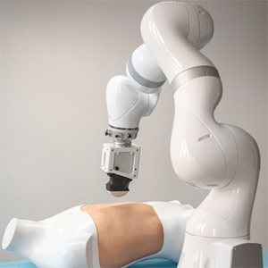
Sühn, T; Esmaeili, N; Mattepu, S; Spiller, M; Boese, A; Urrutia, R; Poblete, V; Hansen, C; Lohmann, C; Illanes, A; Friebe, M
Vibro-Acoustic Sensing of Instrument Interactions as a Potential Source of Texture-Related Information in Robotic Palpation Journal Article
In: Sensors, vol. 23, no. 6, pp. 3141, 2023, ISSN: 1424-8220, (Publisher: Multidisciplinary Digital Publishing Institute).
@article{suhn_vibro-acoustic_2023,

title = {Vibro-Acoustic Sensing of Instrument Interactions as a Potential Source of Texture-Related Information in Robotic Palpation},

author = {T Sühn and N Esmaeili and S Mattepu and M Spiller and A Boese and R Urrutia and V Poblete and C Hansen and C Lohmann and A Illanes and M Friebe},

url = {https://www.mdpi.com/1424-8220/23/6/3141

https://pubmed.ncbi.nlm.nih.gov/36991854/, PubMed},

doi = {10.3390/s23063141},

issn = {1424-8220},

year  = {2023},

date = {2023-01-01},

urldate = {2023-01-01},

journal = {Sensors},

volume = {23},

number = {6},

pages = {3141},

abstract = {The direct tactile assessment of surface textures during palpation is an essential component of open surgery that is impeded in minimally invasive and robot-assisted surgery. When indirectly palpating with a surgical instrument, the structural vibrations from this interaction contain tactile information that can be extracted and analysed. This study investigates the influence of the parameters contact angle α and velocity v→ on the vibro-acoustic signals from this indirect palpation. A 7-DOF robotic arm, a standard surgical instrument, and a vibration measurement system were used to palpate three different materials with varying α and v→. The signals were processed based on continuous wavelet transformation. They showed material-specific signatures in the time–frequency domain that retained their general characteristic for varying α and v→. Energy-related and statistical features were extracted, and supervised classification was performed, where the testing data comprised only signals acquired with different palpation parameters than for training data. The classifiers support vector machine and k-nearest neighbours provided 99.67% and 96.00% accuracy for the differentiation of the materials. The results indicate the robustness of the features against variations in the palpation parameters. This is a prerequisite for an application in minimally invasive surgery but needs to be confirmed in realistic experiments with biological tissues.},

note = {Publisher: Multidisciplinary Digital Publishing Institute},

keywords = {},

pubstate = {published},

tppubtype = {article}

}
2022
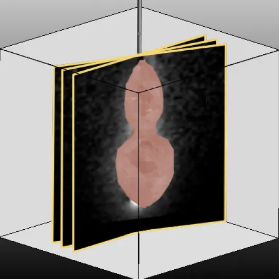
Alpers, J; Rötzer, M; Gutberlet, M; Wacker, F; Hensen, B; Hansen, C
Adaptive simulation of 3D thermometry maps for interventional MR-guided tumor ablation using Pennes’ bioheat equation and isotherms Journal Article
In: Scientific Reports, vol. 12, no. 1, pp. 20356, 2022, ISSN: 2045-2322.
@article{alpers_adaptive_2022,

title = {Adaptive simulation of 3D thermometry maps for interventional MR-guided tumor ablation using Pennes’ bioheat equation and isotherms},

author = {J Alpers and M Rötzer and M Gutberlet and F Wacker and B Hensen and C Hansen},

url = {https://www.nature.com/articles/s41598-022-24911-1

https://pubmed.ncbi.nlm.nih.gov/36437405/, PubMed},

doi = {10.1038/s41598-022-24911-1},

issn = {2045-2322},

year  = {2022},

date = {2022-11-01},

urldate = {2022-11-01},

journal = {Scientific Reports},

volume = {12},

number = {1},

pages = {20356},

abstract = {Abstract

 Minimally-invasive thermal ablation procedures have become clinically accepted treatment options for tumors and metastases. Continuous and reliable monitoring of volumetric heat distribution promises to be an important condition for successful outcomes. In this work, an adaptive bioheat transfer simulation of 3D thermometry maps is presented. Pennes’ equation model is updated according to temperature maps generated by uniformly distributed 2D MR phase images rotated around the main axis of the applicator. The volumetric heat diffusion and the resulting shape of the ablation zone can be modelled accurately without introducing a specific heat source term. Filtering the temperature maps by extracting isotherms reduces artefacts and noise, compresses information of the measured data and adds physical a priori knowledge. The inverse heat transfer for estimating values of the simulated tissue and heating parameters is done by reducing the sum squared error between these isotherms and the 3D simulation. The approach is evaluated on data sets consisting of 13 ex vivo bio protein phantoms, including six perfusion phantoms with simulated heat sink effects. Results show an overall average Dice score of 0.89 ± 0.04 (SEM < 0.01). The optimization of the parameters takes 1.05 ± 0.26 s for each acquired image. Future steps should consider the local optimization of the simulation parameters instead of a global one to better detect heat sinks without a priori knowledge. In addition, the use of a proper Kalman filter might increase robustness and accuracy if combined with our method.},

keywords = {},

pubstate = {published},

tppubtype = {article}

}
Minimally-invasive thermal ablation procedures have become clinically accepted treatment options for tumors and metastases. Continuous and reliable monitoring of volumetric heat distribution promises to be an important condition for successful outcomes. In this work, an adaptive bioheat transfer simulation of 3D thermometry maps is presented. Pennes’ equation model is updated according to temperature maps generated by uniformly distributed 2D MR phase images rotated around the main axis of the applicator. The volumetric heat diffusion and the resulting shape of the ablation zone can be modelled accurately without introducing a specific heat source term. Filtering the temperature maps by extracting isotherms reduces artefacts and noise, compresses information of the measured data and adds physical a priori knowledge. The inverse heat transfer for estimating values of the simulated tissue and heating parameters is done by reducing the sum squared error between these isotherms and the 3D simulation. The approach is evaluated on data sets consisting of 13 ex vivo bio protein phantoms, including six perfusion phantoms with simulated heat sink effects. Results show an overall average Dice score of 0.89 ± 0.04 (SEM < 0.01). The optimization of the parameters takes 1.05 ± 0.26 s for each acquired image. Future steps should consider the local optimization of the simulation parameters instead of a global one to better detect heat sinks without a priori knowledge. In addition, the use of a proper Kalman filter might increase robustness and accuracy if combined with our method.

Schwenderling, L; Heinrich, F; Hansen, C
Augmented reality visualization of automated path planning for percutaneous interventions: a phantom study Journal Article
In: International Journal of Computer Assisted Radiology and Surgery, vol. 17, no. 11, pp. 2071–2079, 2022, ISSN: 1861-6429.
@article{schwenderling_augmented_2022,

title = {Augmented reality visualization of automated path planning for percutaneous interventions: a phantom study},

author = {L Schwenderling and F Heinrich and C Hansen},

url = {https://doi.org/10.1007/s11548-022-02690-4

https://pubmed.ncbi.nlm.nih.gov/35737284/, PubMed},

doi = {10.1007/s11548-022-02690-4},

issn = {1861-6429},

year  = {2022},

date = {2022-11-01},

urldate = {2022-11-01},

journal = {International Journal of Computer Assisted Radiology and Surgery},

volume = {17},

number = {11},

pages = {2071–2079},

abstract = {Insertion point identification is a major challenge for percutaneous interventions. Planning in 2D slice image data is time-consuming and inefficient. Automated path planning can help to overcome these challenges. However, the setup of the intervention room is difficult to consider. In addition, transferring the insertion point to the skin is often prone to error. Therefore, a visualization for an automated path planning was implemented.},

keywords = {},

pubstate = {published},

tppubtype = {article}

}
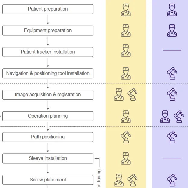
Schreiter, J; Schott, D; Schwenderling, L; Hansen, C; Heinrich, F; Joeres, F
AR-Supported Supervision of Conditional Autonomous Robots: Considerations for Pedicle Screw Placement in the Future Journal Article
In: Journal of Imaging, vol. 8, no. 10, pp. 255, 2022, ISSN: 2313-433X, (Publisher: Multidisciplinary Digital Publishing Institute).
@article{schreiter_ar-supported_2022,

title = {AR-Supported Supervision of Conditional Autonomous Robots: Considerations for Pedicle Screw Placement in the Future},

author = {J Schreiter and D Schott and L Schwenderling and C Hansen and F Heinrich and F Joeres},

url = {https://www.mdpi.com/2313-433X/8/10/255

https://pubmed.ncbi.nlm.nih.gov/36286350/, PubMed},

doi = {10.3390/jimaging8100255},

issn = {2313-433X},

year  = {2022},

date = {2022-10-01},

urldate = {2022-10-01},

journal = {Journal of Imaging},

volume = {8},

number = {10},

pages = {255},

abstract = {Robotic assistance is applied in orthopedic interventions for pedicle screw placement (PSP). While current robots do not act autonomously, they are expected to have higher autonomy under surgeon supervision in the mid-term. Augmented reality (AR) is promising to support this supervision and to enable human–robot interaction (HRI). To outline a futuristic scenario for robotic PSP, the current workflow was analyzed through literature review and expert discussion. Based on this, a hypothetical workflow of the intervention was developed, which additionally contains the analysis of the necessary information exchange between human and robot. A video see-through AR prototype was designed and implemented. A robotic arm with an orthopedic drill mock-up simulated the robotic assistance. The AR prototype included a user interface to enable HRI. The interface provides data to facilitate understanding of the robot’s ”intentions”, e.g., patient-specific CT images, the current workflow phase, or the next planned robot motion. Two-dimensional and three-dimensional visualization illustrated patient-specific medical data and the drilling process. The findings of this work contribute a valuable approach in terms of addressing future clinical needs and highlighting the importance of AR support for HRI.},

note = {Publisher: Multidisciplinary Digital Publishing Institute},

keywords = {},

pubstate = {published},

tppubtype = {article}

}
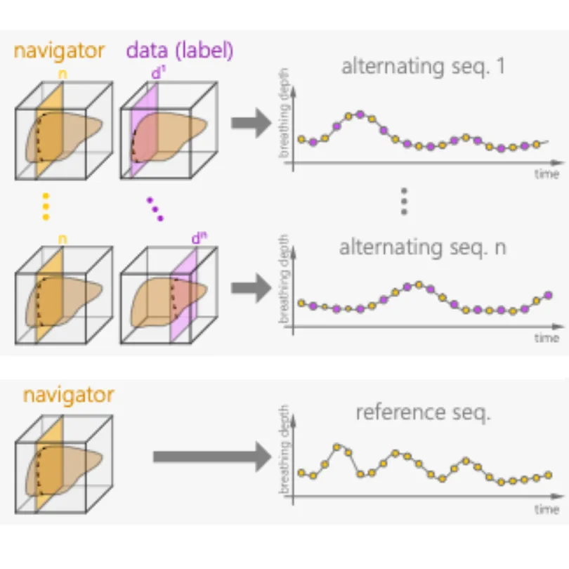
Gulamhussene, G; Meyer, A; Rak, M; Bashkanov, O; Omari, J; Pech, M; Hansen, C
Predicting 4D liver MRI for MR-guided interventions Journal Article
In: Computerized Medical Imaging and Graphics, vol. 101, pp. 102122, 2022, ISSN: 0895-6111.
@article{gulamhussene_predicting_2022,

title = {Predicting 4D liver MRI for MR-guided interventions},

author = {G Gulamhussene and A Meyer and M Rak and O Bashkanov and J Omari and M Pech and C Hansen},

url = {https://www.sciencedirect.com/science/article/pii/S0895611122000921

https://pubmed.ncbi.nlm.nih.gov/36122484/, PubMed},

doi = {10.1016/j.compmedimag.2022.102122},

issn = {0895-6111},

year  = {2022},

date = {2022-10-01},

urldate = {2022-10-01},

journal = {Computerized Medical Imaging and Graphics},

volume = {101},

pages = {102122},

abstract = {Organ motion poses an unresolved challenge in image-guided interventions like radiation therapy, biopsies or tumor ablation. In the pursuit of solving this problem, the research field of time-resolved volumetric magnetic resonance imaging (4D MRI) has evolved. However, current techniques are unsuitable for most interventional settings because they lack sufficient temporal and/or spatial resolution or have long acquisition times. In this work, we propose a novel approach for real-time, high-resolution 4D MRI with large fields of view for MR-guided interventions. To this end, we propose a network-agnostic, end-to-end trainable, deep learning formulation that enables the prediction of a 4D liver MRI with respiratory states from a live 2D navigator MRI. Our method can be used in two ways: First, it can reconstruct high quality fast (near real-time) 4D MRI with high resolution (209×128×128 matrix size with isotropic 1.8mm voxel size and 0.6s/volume) given a dynamic interventional 2D navigator slice for guidance during an intervention. Second, it can be used for retrospective 4D reconstruction with a temporal resolution of below 0.2s/volume for motion analysis and use in radiation therapy. We report a mean target registration error (TRE) of 1.19±0.74mm, which is below voxel size. We compare our results with a state-of-the-art retrospective 4D MRI reconstruction. Visual evaluation shows comparable quality. We compare different network architectures within our formulation. We show that small training sizes with short acquisition times down to 2 min can already achieve promising results and 24 min are sufficient for high quality results. Because our method can be readily combined with earlier time reducing methods, acquisition time can be further decreased while also limiting quality loss. We show that an end-to-end, deep learning formulation is highly promising for 4D MRI reconstruction.},

keywords = {},

pubstate = {published},

tppubtype = {article}

}
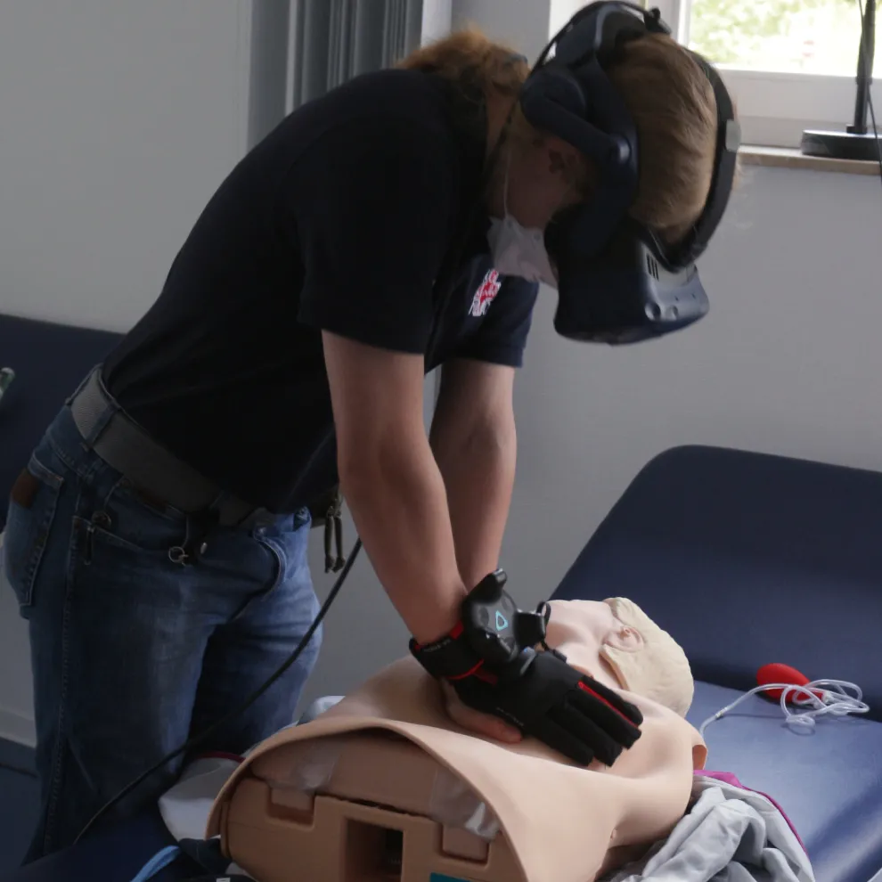
Bublak, T; Bofferding, M; Olson, C; Jonas, H; Rademeier, J; Hansen, C
A Virtual Environment for Emergency Ultrasound Training during Cardiopulmonary Resuscitation Proceedings Article
In: Proceedings of Mensch und Computer 2022, pp. 608–610, Association for Computing Machinery, New York, NY, USA, 2022, ISBN: 978-1-4503-9690-5.
@inproceedings{bublak_virtual_2022,

title = {A Virtual Environment for Emergency Ultrasound Training during Cardiopulmonary Resuscitation},

author = {T Bublak and M Bofferding and C Olson and H Jonas and J Rademeier and C Hansen},

url = {https://doi.org/10.1145/3543758.3547516},

doi = {10.1145/3543758.3547516},

isbn = {978-1-4503-9690-5},

year  = {2022},

date = {2022-09-01},

urldate = {2022-09-01},

booktitle = {Proceedings of Mensch und Computer 2022},

pages = {608–610},

publisher = {Association for Computing Machinery},

address = {New York, NY, USA},

series = {MuC '22},

abstract = {We propose a virtual reality (VR) communication training environment to train cardiopulmonary resuscitation (CPR) and ultrasound (US) imaging simultaneously. Existing simulators only allow separate training of the two procedures. Our method separates the users spatially, as they each work on their own manikin. One manikin is part of a US simulator commonly used in medical training, and the other manikin is a CPR manikin. Our VR application simulates the users working on the same virtual patient. This allows for an immersive training experience in which communication skills, which are especially important in such critical situations, can be practiced by medical professionals. The prototype has the potential to provide an important foundation for advanced VR-based emergency medical training.},

keywords = {},

pubstate = {published},

tppubtype = {inproceedings}

}
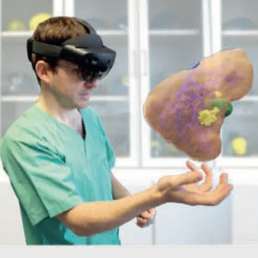
Huber, T; Huettl, F; Hanke, L; Vradelis, L; Heinrich, F; Hansen, C; Boedecker, C; Lang, H
Liver Surgery 4.0 – Planning, Volumetry, Navigation and Virtual Reality Journal Article
In: Zentralblatt für Chirurgie – Zeitschrift für Allgemeine, Viszeral-, Thorax- und Gefäßchirurgie, vol. 147, no. 04, pp. 361–368, 2022, ISSN: 0044-409X, 1438-9592.
@article{huber_liver_2022,

title = {Liver Surgery 4.0 - Planning, Volumetry, Navigation and Virtual Reality},

author = {T Huber and F Huettl and L Hanke and L Vradelis and F Heinrich and C Hansen and C Boedecker and H Lang},

url = {http://www.thieme-connect.de/DOI/DOI?10.1055/a-1844-0549

https://pubmed.ncbi.nlm.nih.gov/35793686/, PubMed},

doi = {10.1055/a-1844-0549},

issn = {0044-409X, 1438-9592},

year  = {2022},

date = {2022-08-01},

urldate = {2022-08-01},

journal = {Zentralblatt für Chirurgie - Zeitschrift für Allgemeine, Viszeral-, Thorax- und Gefäßchirurgie},

volume = {147},

number = {04},

pages = {361–368},

abstract = {Due to the optimisation of conservative treatment, the improvement of imaging methods and the continuous development of surgical techniques, the borders of resectability in liver surgery have changed significantly in recent decades.

Thanks to numerous technical developments, in particular three-dimensional segmentation, preoperative planning and orientation during the operation itself, can now be facilitated, especially in complex procedures.

New technologies such as 3D printing as well as virtual and augmented reality offer additional display options for the patients’ individual anatomy. Various intraoperative navigation options are intended to make preoperative planning available in the operating room in order to increase patient safety.

This review article is intended to provide an overview of the current state of available technologies and an outlook into the operating theatre of the future.},

keywords = {},

pubstate = {published},

tppubtype = {article}

}
Thanks to numerous technical developments, in particular three-dimensional segmentation, preoperative planning and orientation during the operation itself, can now be facilitated, especially in complex procedures.
New technologies such as 3D printing as well as virtual and augmented reality offer additional display options for the patients’ individual anatomy. Various intraoperative navigation options are intended to make preoperative planning available in the operating room in order to increase patient safety.
This review article is intended to provide an overview of the current state of available technologies and an outlook into the operating theatre of the future.
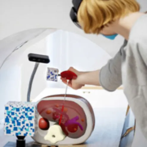
Schott, D; Heinrich, F; Stallmeister, L; Hansen, C
Exploring object and multi-target instrument tracking for AR-guided interventions Journal Article
In: Current Directions in Biomedical Engineering, vol. 8, no. 1, pp. 74–77, 2022, ISSN: 2364-5504, (Publisher: De Gruyter).
@article{schott_exploring_2022,

title = {Exploring object and multi-target instrument tracking for AR-guided interventions},

author = {D Schott and F Heinrich and L Stallmeister and C Hansen},

url = {https://www.degruyterbrill.com/document/doi/10.1515/cdbme-2022-0019/html},

doi = {10.1515/cdbme-2022-0019},

issn = {2364-5504},

year  = {2022},

date = {2022-07-01},

urldate = {2022-07-01},

journal = {Current Directions in Biomedical Engineering},

volume = {8},

number = {1},

pages = {74–77},

abstract = {The rapid development of available hard- and software for computer-assisted or augmented reality (AR) guided interventions creates a need for fast and inexpensive prototyping environments. However, intraoperative tracking systems in particular represent a high cost threshold. Therefore, this work presents a low-cost tracking method based on a conventional RGB camera. Here, a combined approach of multiple image targets and 3D object target recognition is implemented. The system is evaluated with a systematic accuracy assessment analyzing a total of 385 3D positions. On average, a deviation of 15,69+-9,95 mm was measured. In addition, a prototypical AR-based needle navigation visualization was developed using Microsoft HoloLens 2. This system’s feasibility and usability was evaluated positively in a pilot study (n=3).},

note = {Publisher: De Gruyter},

keywords = {},

pubstate = {published},

tppubtype = {article}

}
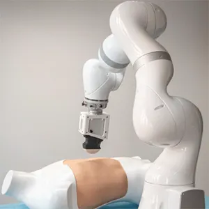
Schreiter, J; Semshchikov, V; Hanses, M; Elkmann, N; Hansen, C
Towards a real-time control of robotic ultrasound using haptic force feedback Journal Article
In: Current Directions in Biomedical Engineering, vol. 8, no. 1, pp. 81–84, 2022, ISSN: 2364-5504, (Publisher: De Gruyter).
@article{schreiter_towards_2022,

title = {Towards a real-time control of robotic ultrasound using haptic force feedback},

author = {J Schreiter and V Semshchikov and M Hanses and N Elkmann and C Hansen},

url = {https://www.degruyterbrill.com/document/doi/10.1515/cdbme-2022-0021/html},

doi = {10.1515/cdbme-2022-0021},

issn = {2364-5504},

year  = {2022},

date = {2022-07-01},

urldate = {2022-07-01},

journal = {Current Directions in Biomedical Engineering},

volume = {8},

number = {1},

pages = {81–84},

abstract = {Ultrasound is a widely used imaging technique and is appreciated for its non-invasiveness, absence of radiation, widespread availability, and compact equipment. Ergonomic difficulties in manual handling of the probe could be enhanced by a robotic controlled ultrasound. The paper addresses the development of such a system which enables remote operation of a ultrasound probe and includes haptic force feedback as well as video conferencing components for visual feedback. The development process followed a user-centered approach by investigating needs of potential end-users. Preliminary results demonstrated the functionality of the developed system for generating medical image data under laboratory conditions.},

note = {Publisher: De Gruyter},

keywords = {},

pubstate = {published},

tppubtype = {article}

}
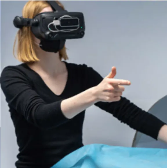
Schott, D; Heinrich, F; Labsch, D; Hensen, B; Hansen, C
Towards multimodal interaction for needlebased procedures in a virtual radiology suite Journal Article
In: Current Directions in Biomedical Engineering, vol. 8, no. 1, pp. 70–73, 2022, ISSN: 2364-5504, (Publisher: De Gruyter).
@article{schott_towards_2022,

title = {Towards multimodal interaction for needlebased procedures in a virtual radiology suite},

author = {D Schott and F Heinrich and D Labsch and B Hensen and C Hansen},

url = {https://www.degruyterbrill.com/document/doi/10.1515/cdbme-2022-0018/html},

doi = {10.1515/cdbme-2022-0018},

issn = {2364-5504},

year  = {2022},

date = {2022-07-01},

urldate = {2022-07-01},

journal = {Current Directions in Biomedical Engineering},

volume = {8},

number = {1},

pages = {70–73},

abstract = {Touchless interaction is popular in the medical domain because it maintains sterility and ensures physicians’ autonomy. Evaluating these technologies, however, proves difficult due to technical and human hurdles. Virtual reality leaves these limitations behind and allows for the exploration of promising concepts by simulating an environment and the interactions that takes place within it.We present a virtual radiology suite in the context of needle-based MR-interventions to evaluate touchless interactions. Hand and foot inputs were implemented on a custom interface and evaluated in a user study (n= 16). Results show that activating the system and manipulating values was faster with foot input. However, multimodal interaction is preferable because it is less demanding.},

note = {Publisher: De Gruyter},

keywords = {},

pubstate = {published},

tppubtype = {article}

}
Alpers, J; Hensen, B; Rötzer, M; Reimert, D; Gerlach, T; Vick, R; Gutberlet, M; Wacker, F; Hansen, C
Comparison study of reconstruction algorithms for volumetric necrosis maps from 2D multi-slice GRE thermometry images Journal Article
In: Scientific Reports, vol. 12, no. 1, pp. 11509, 2022, ISSN: 2045-2322.
@article{alpers_comparison_2022,

title = {Comparison study of reconstruction algorithms for volumetric necrosis maps from 2D multi-slice GRE thermometry images},

author = {J Alpers and B Hensen and M Rötzer and D Reimert and T Gerlach and R Vick and M Gutberlet and F Wacker and C Hansen},

url = {https://www.nature.com/articles/s41598-022-15712-7

https://pubmed.ncbi.nlm.nih.gov/35799055/, PubMed},

doi = {10.1038/s41598-022-15712-7},

issn = {2045-2322},

year  = {2022},

date = {2022-07-01},

urldate = {2022-07-01},

journal = {Scientific Reports},

volume = {12},

number = {1},

pages = {11509},

abstract = {Cancer is a disease which requires a significant amount of careful medical attention. For minimally‑

invasive thermal ablation procedures, the monitoring of heat distribution is one of the biggest

challenges. In this work, three approaches for volumetric heat map reconstruction (Delauney

triangulation, minimum volume enclosing ellipsoids (MVEE) and splines) are presented based on

uniformly distributed 2D MRI phase images rotated around the applicator’s main axis. We compare

them with our previous temperature interpolation method with respect to accuracy, robustness and

adaptability. All approaches are evaluated during MWA treatment on the same data sets consisting of

13 ex vivo bio protein phantoms, including six phantoms with simulated heat sink effects. Regarding

accuracy, the DSC similarity results show a strong trend towards the MVEE ( 0.80 ± 0.03) and the

splines (0.77 ± 0.04) method compared to the Delauney triangulation ( 0.75 ± 0.02) or the temperature

interpolation (0.73 ± 0.07). Robustness is increased for all three approaches and the adaptability

shows a significant trend towards the initial interpolation method and the splines. To overcome local

inhomogeneities in the acquired data, the use of adaptive simulations should be considered in the

future. In addition, the transfer to in vivo animal experiments should be considered to test for clinical

applicability.},

keywords = {},

pubstate = {published},

tppubtype = {article}

}
invasive thermal ablation procedures, the monitoring of heat distribution is one of the biggest
challenges. In this work, three approaches for volumetric heat map reconstruction (Delauney
triangulation, minimum volume enclosing ellipsoids (MVEE) and splines) are presented based on
uniformly distributed 2D MRI phase images rotated around the applicator’s main axis. We compare
them with our previous temperature interpolation method with respect to accuracy, robustness and
adaptability. All approaches are evaluated during MWA treatment on the same data sets consisting of
13 ex vivo bio protein phantoms, including six phantoms with simulated heat sink effects. Regarding
accuracy, the DSC similarity results show a strong trend towards the MVEE ( 0.80 ± 0.03) and the
splines (0.77 ± 0.04) method compared to the Delauney triangulation ( 0.75 ± 0.02) or the temperature
interpolation (0.73 ± 0.07). Robustness is increased for all three approaches and the adaptability
shows a significant trend towards the initial interpolation method and the splines. To overcome local
inhomogeneities in the acquired data, the use of adaptive simulations should be considered in the
future. In addition, the transfer to in vivo animal experiments should be considered to test for clinical
applicability.
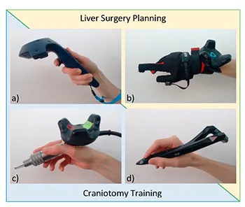
Allgaier, M; Chheang, V; Saalfeld, P; Apilla, V; Huber, T; Huettl, F; Neyazi, B; Sandalcioglu, E; Hansen, C; Preim, B; Saalfeld, P
A comparison of input devices for precise interaction tasks in VR-based surgical planning and training Journal Article
In: Computers in Biology and Medicine, vol. 145, pp. 105429, 2022, ISSN: 0010-4825.
@article{allgaier_comparison_2022,

title = {A comparison of input devices for precise interaction tasks in VR-based surgical planning and training},

author = {M Allgaier and V Chheang and P Saalfeld and V Apilla and T Huber and F Huettl and B Neyazi and E Sandalcioglu and C Hansen and B Preim and P Saalfeld},

url = {https://www.sciencedirect.com/science/article/pii/S0010482522002219

https://pubmed.ncbi.nlm.nih.gov/35349800/, PubMed},

doi = {10.1016/j.compbiomed.2022.105429},

issn = {0010-4825},

year  = {2022},

date = {2022-06-01},

urldate = {2022-06-01},

journal = {Computers in Biology and Medicine},

volume = {145},

pages = {105429},

abstract = {To exploit the potential of virtual reality (VR) in medicine, the input devices must be selected carefully due to their different benefits. In this work, input devices for common interaction tasks in medical VR planning and training are compared. Depending on the specific purpose, different requirements exist. Therefore, an appropriate trade-off between meeting task-specific requirements and having a widely applicable device has to be found. We focus on two medical use cases, liver surgery planning and craniotomy training, to cover a broad medical domain. Based on these, relevant input devices are compared with respect to their suitability for performing precise VR interaction tasks. The devices are standard VR controllers, a pen-like VR Ink, data gloves and a real craniotome, the medical instrument used for craniotomy. The input devices were quantitatively compared with respect to their performance based on different measurements. The controllers and VR Ink performed significantly better than the remaining two devices regarding precision. Qualitative data concerning task load, cybersickness, and usability and appropriateness of the devices were assessed. Although no device stands out for both applications, most participants preferred using the VR Ink, followed by the controller and finally the data gloves and craniotome. These results can guide the selection of an appropriate device for future medical VR applications.},

keywords = {},

pubstate = {published},

tppubtype = {article}

}

Heinrich, F; Schwenderling, L; Joeres, F; Hansen, C
2D versus 3D: A Comparison of Needle Navigation Concepts between Augmented Reality Display Devices Proceedings Article
In: 2022 IEEE Conference on Virtual Reality and 3D User Interfaces (VR), pp. 260–269, 2022, (ISSN: 2642-5254).
@inproceedings{heinrich_2d_2022,

title = {2D versus 3D: A Comparison of Needle Navigation Concepts between Augmented Reality Display Devices},

author = {F Heinrich and L Schwenderling and F Joeres and C Hansen},

url = {https://ieeexplore.ieee.org/document/9756753},

doi = {10.1109/VR51125.2022.00045},

year  = {2022},

date = {2022-03-01},

urldate = {2022-03-01},

booktitle = {2022 IEEE Conference on Virtual Reality and 3D User Interfaces (VR)},

pages = {260–269},

abstract = {Surgical procedures requiring needle navigation assistance suffer from complicated hand-eye coordination and are mentally demanding. Augmented reality (AR) can help overcome these issues. How-ever, only an insufficient amount of fundamental research has focused on the design and hardware selection of such AR needle navigation systems. This work contributes to this research area by presenting a user study (n=24) comparing three state-of-the-art navigation concepts displayed by an optical see-through head-mounted display and a stereoscopic projection system. A two-dimensional glyph visualization resulted in higher targeting accuracy but required more needle insertion time. In contrast, punctures guided by a three-dimensional see-through vision concept were less accurate but faster and were favored in a qualitative interview. The third concept, a static representation of the correctly positioned needle, showed too high target errors for clinical accuracy needs. This concept per-formed worse when displayed by the projection system. Besides that, no meaningful differences between the evaluated AR display devices were detected. User preferences and use case restrictions, e.g., sterility requirements, seem to be more crucial selection criteria. Future work should focus on improving the accuracy of the see-through vision concept. Until then, the glyph visualization is recommended.},

note = {ISSN: 2642-5254},

keywords = {},

pubstate = {published},

tppubtype = {inproceedings}

}
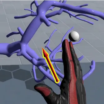
Hombeck, J; Meuschke, M; Zyla, L; Heuser, A; Toader, J; Popp, F; Bruns, C; Hansen, C; Datta, R; Lawonn, K
Evaluating Perceptional Tasks for Medicine: A Comparative User Study Between a Virtual Reality and a Desktop Application Proceedings Article
In: 2022 IEEE Conference on Virtual Reality and 3D User Interfaces (VR), pp. 514–523, 2022, (ISSN: 2642-5254).
@inproceedings{hombeck_evaluating_2022,

title = {Evaluating Perceptional Tasks for Medicine: A Comparative User Study Between a Virtual Reality and a Desktop Application},

author = {J Hombeck and M Meuschke and L Zyla and A Heuser and J Toader and F Popp and C Bruns and C Hansen and R Datta and K Lawonn},

url = {https://ieeexplore.ieee.org/abstract/document/9756759},

doi = {10.1109/VR51125.2022.00071},

year  = {2022},

date = {2022-03-01},

urldate = {2022-03-01},

booktitle = {2022 IEEE Conference on Virtual Reality and 3D User Interfaces (VR)},

pages = {514–523},

abstract = {Since for most consumers the Virtual Reality (VR) experience exceeds that of desktop applications, an increasing number of applications is being transferred from desktop to VR. Industrial and entertainment applications primarily expect for a richer consumer experience, while others, such as surgical applications, seek for improved precision over their desktop counterparts. One way to improve the performance of precision-based VR applications is to provide suitable visualizations. Today, these "suitable" visualizations are mostly transferred from desktop to VR without considering their spatial and temporal performance might change in VR. This may not lead to an optimal solution, which can be crucial for precision-based tasks. Misinterpretation of shape or distance in a surgical or pre-operative simulation can affect the chosen treatment and thus directly impact the outcome. Therefore, we evaluate the performance differences of multiple visualizations for 3D surfaces based on their shape and distance estimation for desktop and VR applications. We conducted a quantitative user study with 56 participants evaluating seven visualizations (Phong, Toon, Fresnel, Pseudo-Chromadepth, Heatmap, Isolines, and Arrow Glyphs). Our results show that the performance of each visualization varies depending on the task, system, and surface type, with VR generally providing improved results. While Isolines are able to improve distance estimation, Phong and Heatmaps are beneficial for shape estimation.},

note = {ISSN: 2642-5254},

keywords = {},

pubstate = {published},

tppubtype = {inproceedings}

}
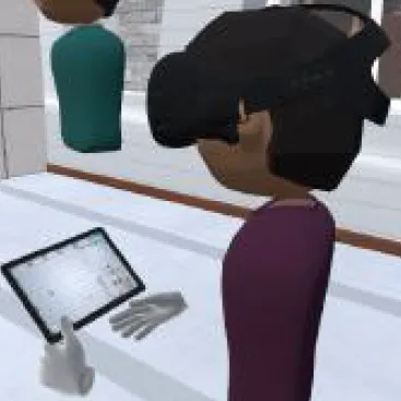
Chheang, V; Heinrich, F; Joeres, F; Saalfeld, P; Preim, B; Hansen, C
Group WiM: A Group Navigation Technique for Collaborative Virtual Reality Environments Proceedings Article
In: 2022 IEEE Conference on Virtual Reality and 3D User Interfaces Abstracts and Workshops (VRW), pp. 556–557, 2022.
@inproceedings{chheang_group_2022,

title = {Group WiM: A Group Navigation Technique for Collaborative Virtual Reality Environments},

author = {V Chheang and F Heinrich and F Joeres and P Saalfeld and B Preim and C Hansen},

url = {https://ieeexplore.ieee.org/document/9757426},

doi = {10.1109/VRW55335.2022.00129},

year  = {2022},

date = {2022-03-01},

urldate = {2022-03-01},

booktitle = {2022 IEEE Conference on Virtual Reality and 3D User Interfaces Abstracts and Workshops (VRW)},

pages = {556–557},

abstract = {In this work, we present a group World-in-Miniature (WiM) navigation technique that allows a guide to navigate a team in collaborative virtual reality (VR) environments. We evaluated the usability, discomfort, and user performance of the proposed technique compared to state-of-the-art group teleportation in a user study łeft(ntextbackslash,=textbackslash,21textbackslashright). The results show that the proposed technique induces less discomfort for the guide and has slight usability advantages. Additionally, the group WiM technique seems superior in regards to task completion time for obstructed target destination. However, it performs similarly to the group teleportation technique in direct line of sight cases. The group WiM technique provides potential benefits for effective group navigation in complex virtual environments and harder-to-reach target locations.},

keywords = {},

pubstate = {published},

tppubtype = {inproceedings}

}
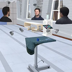
Chheang, V; Schott, D; Saalfeld, P; Vradelis, L; Huber, T; Huettl, F; Lang, H; Preim, B; Hansen, C
Towards Virtual Teaching Hospitals for Advanced Surgical Training Proceedings Article
In: 2022 IEEE Conference on Virtual Reality and 3D User Interfaces Abstracts and Workshops (VRW), pp. 410–414, 2022.
@inproceedings{chheang_towards_2022,

title = {Towards Virtual Teaching Hospitals for Advanced Surgical Training},

author = {V Chheang and D Schott and P Saalfeld and L Vradelis and T Huber and F Huettl and H Lang and B Preim and C Hansen},

url = {https://ieeexplore.ieee.org/document/9757460},

doi = {10.1109/VRW55335.2022.00089},

year  = {2022},

date = {2022-03-01},

urldate = {2022-03-01},

booktitle = {2022 IEEE Conference on Virtual Reality and 3D User Interfaces Abstracts and Workshops (VRW)},

pages = {410–414},

abstract = {Existing virtual training environments in medicine usually focus only on the training of a specific medical skill and are conducted in a single virtual room. However, medical challenges are often in the context of the planning procedure and operating room intervention. Therefore, it is crucial to provide a training environment that tackles these issues. In this paper, we present a prototype of surgical department within a virtual teaching hospital. It supports multiple users, photo-realistic avatars, and training scenarios ranging from planning procedures to laparoscopic surgery in the virtual operating room. Medical data for each virtual patient is synchronized across the different rooms, allowing users to view, analyze and train on key decision points from diagnosis to surgical incision for each patient. The environment was evaluated in a pilot study with five surgical residents and one medical student. The experts assessed the environment as an essential tool to evaluate and improve surgical planning as well as the training during critical situations of the surgical procedures. We conclude that the development of the virtual teaching hospital components described here has the potential to be a basis for future generations of medical training simulators.},

keywords = {},

pubstate = {published},

tppubtype = {inproceedings}

}

Batz, V; Lipowski, I; Morfeld, M; Hansen, C; Herzog, M
Accessible Design of Serious Games for People with Intellectual Disabilities in Inclusive Vocational Education Proceedings Article
In: Proceedings of DiGRA 2022 Conference: Bringing Worlds Together, 2022, (ISSN: 2342-9666).
@inproceedings{batz_accessible_2022,

title = {Accessible Design of Serious Games for People with Intellectual Disabilities in Inclusive Vocational Education},

author = {V Batz and I Lipowski and M Morfeld and C Hansen and M Herzog},

url = {https://dl.digra.org/index.php/dl/article/view/1372},

doi = {10.26503/dl.v2022i1.1372},

year  = {2022},

date = {2022-01-01},

urldate = {2022-01-01},

booktitle = {Proceedings of DiGRA 2022 Conference: Bringing Worlds Together},

abstract = {People with disabilities are often denied the opportunity to obtain a vocational qualification. There is a lack of an appropriate infrastructure for an inclusive education system and recognized degrees. Therefore, didactic methods for inclusive learning situations are needed. This paper describes the user-centered development and evaluation of a serious game for inclusive vocational training in kitchen professions. The theoretical teaching content in the module “hygiene” is taught to people with and without cognitive impairments using a gamified learning application. The prototype was evaluated with 22 participants when used in an inclusive teaching setting with a mixed-methods design. The learning application caused an increase in motivation and the knowledge quiz achieved good results across all target groups (15 trainee cooks, 4 trainees as kitchen assistants, 3 employees of the sheltered workshop). The results from questioning, learning quiz, observation, and video analysis form design guidelines for future digital, interactive teaching methods for inclusive vocational education.},

note = {ISSN: 2342-9666},

keywords = {},

pubstate = {published},

tppubtype = {inproceedings}

}
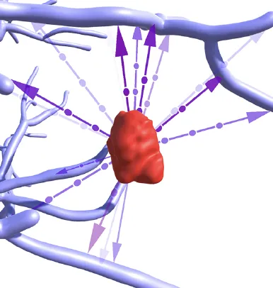
Hombeck, J; Meuschke, M; Lieb, S; Lichtenberg, N; Datta, R; Krone, M; Hansen, C; Preim, B; Lawonn, K
Distance Visualizations for Vascular Structures Proceedings Article
In: Eurographics Workshop on Visual Computing for Biology and Medicine, pp. 1-10, 2022.
@inproceedings{hombeck_distance_2022,

title = {Distance Visualizations for Vascular Structures},

author = {J Hombeck and M Meuschke and S Lieb and N Lichtenberg and R Datta and M Krone and C Hansen and B Preim and K Lawonn},

doi = {10.2312/vcbm.20221182},

year  = {2022},

date = {2022-01-01},

urldate = {2022-01-01},

booktitle = {Eurographics Workshop on Visual Computing for Biology and Medicine},

pages = {1-10},

abstract = {The role of expressive surface visualizations in rendering vascular structures has seen an increased impact over the last years. Surface visualizations provide an overview of complex anatomical structures and support treatment planning as well as medical education. To support decision-making, physicians need visualizations that depict anatomical structures and their spatial relations to each other, i.e., well perceivable visual encodings of egocentric and endocentric distances.},

keywords = {},

pubstate = {published},

tppubtype = {inproceedings}

}
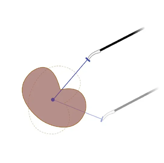
Mielke, T; Joeres, F; Hansen, C
Natural 3D Object Manipulation for Interactive Laparoscopic Augmented Reality Registration Proceedings Article
In: Chen, JYC; Fragomeni, G (Ed.): Virtual, Augmented and Mixed Reality: Design and Development, pp. 317–328, Springer International Publishing, Cham, 2022, ISBN: 978-3-031-05939-1.
@inproceedings{mielke_natural_2022,

title = {Natural 3D Object Manipulation for Interactive Laparoscopic Augmented Reality Registration},

author = {T Mielke and F Joeres and C Hansen},

editor = {JYC Chen and G Fragomeni},

doi = {10.1007/978-3-031-05939-1_21},

isbn = {978-3-031-05939-1},

year  = {2022},

date = {2022-01-01},

urldate = {2022-01-01},

booktitle = {Virtual, Augmented and Mixed Reality: Design and Development},

pages = {317–328},

publisher = {Springer International Publishing},

address = {Cham},

abstract = {Due to the growing focus on minimally invasive surgery, there is increasing interest in intraoperative software support. For example, augmented reality can be used to provide additional information. Accurate registration is required for effective support. In this work, we present a manual registration method that aims at mimicking natural manipulation of 3D objects using tracked surgical instruments. This method is compared to a point-based registration method in a simulated laparoscopic environment. Both registration methods serve as an initial alignment step prior to surface-based registration refinement. For the evaluation, we conducted a user study with 12 participants. The registration methods were compared in terms of registration accuracy, registration duration, and subjective usability feedback. No significant differences could be found with respect to the previously mentioned criteria between the manual and the point-based registration methods. Thus, the manual registration did not outperform the reference method. However, we found that our method offers qualitative advantages, which may make it more suitable for some application scenarios. Furthermore we identified possible approaches for improvement, which should be investigated in the future to strengthen possible advantages of our registration method.},

keywords = {},

pubstate = {published},

tppubtype = {inproceedings}

}
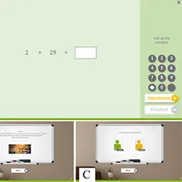
Gabele, M; Thoms, A; Schröer, S; Hußlein, S; Hansen, C
Effects and Combination of Tailored Browser-Based and Mobile Cognitive Software Training Proceedings Article
In: Jónsson, BÞ; Gurrin, C; Tran, M; Dang-Nguyen, D; Hu, A; Thanh, BHT; Huet, B (Ed.): MultiMedia Modeling, pp. 279–291, Springer International Publishing, Cham, 2022, ISBN: 978-3-030-98355-0.
@inproceedings{gabele_effects_2022,

title = {Effects and Combination of Tailored Browser-Based and Mobile Cognitive Software Training},

author = {M Gabele and A Thoms and S Schröer and S Hußlein and C Hansen},

editor = {BÞ Jónsson and C Gurrin and M Tran and D Dang-Nguyen and A Hu and BHT Thanh and B Huet},

doi = {10.1007/978-3-030-98355-0_24},

isbn = {978-3-030-98355-0},

year  = {2022},

date = {2022-01-01},

urldate = {2022-01-01},

booktitle = {MultiMedia Modeling},

pages = {279–291},

publisher = {Springer International Publishing},

address = {Cham},

abstract = {Software-based training supports maintenance or recovery of cognitive abilities. However, regular use needs a high self-motivation. For this, the individual needs of users should be taken into account during the development process. Thus, in this work, two possibilities for tailoring in combination of application, and media devices in practice are investigated in an explorative between-subject design and hypothesis-generating study (N = 68). First, the effect of a browser-based training without gamification or with assignment of gamification appropriate to mean user characteristic on training duration is investigated. Second, the effect of this training and a subsequent serious game app on perception and possible combinations of different media is investigated. The results of behavior lead to the hypothesis that with tailored gamification in browsers, users may train longer in the middle range of training duration. The results of perception show a significant difference and higher perceived value/usefulness and overall rating, as well as the assumption of a higher effect in computer and mobile training in the group with tailored gamification. This could therefore provide a basis for combinations. A combination of both usage scenarios is perceived by users as most reasonable for a positive training effect. Nevertheless, there is a high variance for self-assessed usage. Thus, the results support developing multiple combinable scenarios, tailored to the user, in media devices and game integration to address intended effects, development and user needs to support the effect of cognitive software training in practice.},

keywords = {},

pubstate = {published},

tppubtype = {inproceedings}

}
2021

Wagner, S; Belger, J; Joeres, F; Thöne-Otto, A; Hansen, C; Preim, B; Saalfeld, P
iVRoad: Immersive virtual road crossing as an assessment tool for unilateral spatial neglect Journal Article
In: Computers & Graphics, vol. 99, pp. 70–82, 2021, ISSN: 0097-8493.
@article{wagner_ivroad_2021,

title = {iVRoad: Immersive virtual road crossing as an assessment tool for unilateral spatial neglect},

author = {S Wagner and J Belger and F Joeres and A Thöne-Otto and C Hansen and B Preim and P Saalfeld},

url = {https://www.sciencedirect.com/science/article/pii/S009784932100128X},

doi = {10.1016/j.cag.2021.06.013},

issn = {0097-8493},

year  = {2021},

date = {2021-10-01},

urldate = {2021-10-01},

journal = {Computers & Graphics},

volume = {99},

pages = {70–82},

abstract = {We developed a virtual road crossing assessment tool called iVRoad - immersive Virtual Road, which allows to put the patient into realistic road crossing situations and to record various parameters that can be used to quantify unilateral spatial neglect. We present a study with 18 stroke patients in which we evaluate our system with respect to usability, satisfaction, sense of presence and possible occurring cybersickness symptoms. Unilateral spatial neglect is a cognitive disturbance, often occurring after right hemispheric stroke. Conventional neuropsychological tests, such as paper-and-pencil tests, for assessing unilateral spatial neglect, often lack sensitivity. Especially in mild forms, symptoms can be seen in everyday life, but are hard to detect in formal testing. We examined patients with and without unilateral spatial neglect in order to identify parameters that could be feasible to separate these patient groups. Using everyday life tasks as a diagnostic instrument, however, is challenging because it is time-consuming, hard to control and to quantify. Computer-aided diagnostic systems are promising for analysing the behaviour of patients in detail. Modern virtual reality technology allows to place the patient in realistic situations. Especially situations in which patients often have difficulties or that are too dangerous in reality can be assessed with VR.},

keywords = {},

pubstate = {published},

tppubtype = {article}

}
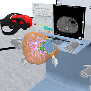
Chheang, V; Saalfeld, P; Joeres, F; Boedecker, C; Huber, T; Huettl, F; Lang, H; Preim, B; Hansen, C
A Collaborative Virtual Reality Environment for Liver Surgery Planning Journal Article
In: Computers & Graphics, vol. 99, pp. 234–246, 2021, ISSN: 0097-8493.
@article{chheang_collaborative_2021,

title = {A Collaborative Virtual Reality Environment for Liver Surgery Planning},

author = {V Chheang and P Saalfeld and F Joeres and C Boedecker and T Huber and F Huettl and H Lang and B Preim and C Hansen},

url = {https://www.sciencedirect.com/science/article/pii/S0097849321001400},

doi = {10.1016/j.cag.2021.07.009},

issn = {0097-8493},

year  = {2021},

date = {2021-10-01},

urldate = {2021-10-01},

journal = {Computers & Graphics},

volume = {99},

pages = {234–246},

abstract = {Surgical planning software is a key component in the treatment of tumor diseases. However, desktop-based systems provide only limited visualization and interaction opportunities. Moreover, collaborative planning among members of a surgical team is only possible to a limited extent. In this work, a collaborative virtual reality (VR) environment to assist liver surgeons in tumor surgery planning is presented. Our aim is to improve virtual resection planning between surgeons in a remote or co-located environment. The system allows surgeons to define and adjust virtual resections on patient-specific organ 3D surfaces and 2D image slices. Changes on both modalities are synchronized, which will enable surgeons to iterate and refine the resection surfaces quickly. In addition, a real-time risk map visualization is presented that displays safety margins around tumors. An evaluation performed by liver surgeons provides information on potential benefits, such as the possibility to visualize complex cases and assessing the safety-critical areas, applicability, and limitations for further improvement.},

keywords = {},

pubstate = {published},

tppubtype = {article}

}

Sabieleish, M; Heryan, K; Boese, A; Hansen, C; Friebe, M; Illanes, A
Study of Needle Punctures into Soft Tissue Through Audio and Force Sensing: Can Audio Be a Simple Alternative for Needle Guidance? Journal Article
In: International Journal of Computer Assisted Radiology and Surgery, vol. 16, no. 10, pp. 1683–1697, 2021, ISSN: 1861-6410, 1861-6429.
@article{sabieleish_study_2021,

title = {Study of Needle Punctures into Soft Tissue Through Audio and Force Sensing: Can Audio Be a Simple Alternative for Needle Guidance?},

author = {M Sabieleish and K Heryan and A Boese and C Hansen and M Friebe and A Illanes},

url = {https://link.springer.com/10.1007/s11548-021-02479-x

https://pubmed.ncbi.nlm.nih.gov/34652603/, PubMed},

doi = {10.1007/s11548-021-02479-x},

issn = {1861-6410, 1861-6429},

year  = {2021},

date = {2021-10-01},

urldate = {2021-10-01},

journal = {International Journal of Computer Assisted Radiology and Surgery},

volume = {16},

number = {10},

pages = {1683–1697},

abstract = {Purpose Percutaneous needle insertion is one of the most common minimally invasive procedures. The clinician’s experience and medical imaging support are essential to the procedure’s safety. However, imaging comes with inaccuracies due to artifacts, and therefore sensor-based solutions were proposed to improve accuracy. However, sensors are usually embedded in the needle tip, leading to design limitations. A novel concept was proposed for capturing tip–tissue interaction information through audio sensing, showing promising results for needle guidance. This work demonstrates that this audio approach can provide important puncture information by comparing audio and force signal dynamics during insertion.

Methods An experimental setup for inserting a needle into soft tissue was prepared. Audio and force signals were synchronously recorded at four different insertion velocities, and a dataset of 200 recordings was acquired. Indicators related to different aspects of the force and audio were compared through signal-to-signal and event-to-event correlation analysis.

Results High signal-to-signal correlations between force and audio indicators regardless of the insertion velocity were obtained. The force curvature indicator obtained the best correlation performances to audio with more than 70% of the correlations higher than 0.6. The event-to-event correlation analysis shows that a puncture event in the force is generally identifiable in audio and that their intensities firmly related.

Conclusions Audio contains valuable information for monitoring needle tip/tissue interaction. Significant dynamics obtained from a well-known sensor as force can also be extracted from audio, regardless of insertion velocities.},

keywords = {},

pubstate = {published},

tppubtype = {article}

}
Methods An experimental setup for inserting a needle into soft tissue was prepared. Audio and force signals were synchronously recorded at four different insertion velocities, and a dataset of 200 recordings was acquired. Indicators related to different aspects of the force and audio were compared through signal-to-signal and event-to-event correlation analysis.
Results High signal-to-signal correlations between force and audio indicators regardless of the insertion velocity were obtained. The force curvature indicator obtained the best correlation performances to audio with more than 70% of the correlations higher than 0.6. The event-to-event correlation analysis shows that a puncture event in the force is generally identifiable in audio and that their intensities firmly related.
Conclusions Audio contains valuable information for monitoring needle tip/tissue interaction. Significant dynamics obtained from a well-known sensor as force can also be extracted from audio, regardless of insertion velocities.

Heinrich, F; Schwenderling, L; Streuber, M; Bornemann, K; Lawonn, K; Hansen, C
Effects of Surface Visualizations on Depth Perception in Projective Augmented Reality Proceedings Article
In: 2021 IEEE 2nd International Conference on Human-Machine Systems (ICHMS), pp. 1–6, 2021.
@inproceedings{heinrich_effects_2021,

title = {Effects of Surface Visualizations on Depth Perception in Projective Augmented Reality},

author = {F Heinrich and L Schwenderling and M Streuber and K Bornemann and K Lawonn and C Hansen},

url = {https://ieeexplore.ieee.org/document/9582452},

doi = {10.1109/ICHMS53169.2021.9582452},

year  = {2021},

date = {2021-09-01},

urldate = {2021-09-01},

booktitle = {2021 IEEE 2nd International Conference on Human-Machine Systems (ICHMS)},

pages = {1–6},

abstract = {Depth perception is a common issue in augmented reality (AR). Projective AR, where the spatial relations between the projection surface and displayed virtual contents need to be represented properly, is particularly affected. This is crucial in the medical domain, e.g., for the distances between the patient’s skin and projected inner anatomical structures, but not much research was conducted in this context before. To this end, this work investigates the applicability of surface visualization techniques to support the perception of spatial relations in projective AR. Four methods previously explored in different domains were combined with the projection of inner anatomical structures on a human torso phantom. They were evaluated in a comparative user study (n=21) with respect to a distance estimation and a sorting task. Measures included Task completion time, accuracy, total Head movement and Confidence of the participants. Consistent results across variables show advantages of more occluding surface visualizations for the distance estimation task. Opposite results were obtained for the sorting task. This suggests that the amount of needed surface preservation depends on the use case and individual occlusion compromises need to be explored in future work.},

keywords = {},

pubstate = {published},

tppubtype = {inproceedings}

}
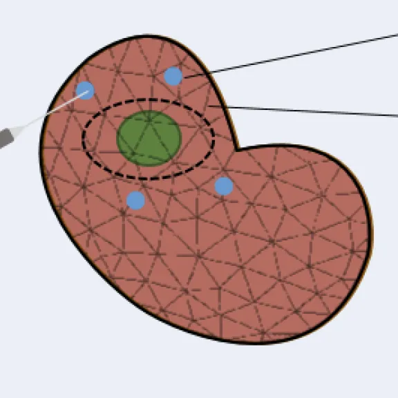
Joeres, F; Mielke, T; Hansen, C
Laparoscopic Augmented Reality Registration for Oncological Resection Site Repair Journal Article
In: International Journal of Computer Assisted Radiology and Surgery, vol. 16, no. 9, pp. 1577–1586, 2021, ISSN: 1861-6429.
@article{joeres_laparoscopic_2021,

title = {Laparoscopic Augmented Reality Registration for Oncological Resection Site Repair},

author = {F Joeres and T Mielke and C Hansen},

url = {https://doi.org/10.1007/s11548-021-02336-x

https://pubmed.ncbi.nlm.nih.gov/33797689/, PubMed},

doi = {10.1007/s11548-021-02336-x},

issn = {1861-6429},

year  = {2021},

date = {2021-09-01},

urldate = {2021-09-01},

journal = {International Journal of Computer Assisted Radiology and Surgery},

volume = {16},

number = {9},

pages = {1577–1586},

abstract = {Resection site repair during laparoscopic oncological surgery (e.g. laparoscopic partial nephrectomy) poses some unique challenges and opportunities for augmented reality (AR) navigation support. This work introduces an AR registration workflow that addresses the time pressure that is present during resection site repair.},

keywords = {},

pubstate = {published},

tppubtype = {article}

}

Schwenderling, L; Hansen, C; Heinrich, F
AR Visualization of Automated Access Path Planning for Percutaneous Interventions Journal Article
In: Current Directions in Biomedical Engineering, vol. 7, no. 1, pp. 48–52, 2021, ISSN: 2364-5504, (Publisher: De Gruyter).
@article{schwenderling_ar_2021,

title = {AR Visualization of Automated Access Path Planning for Percutaneous Interventions},

author = {L Schwenderling and C Hansen and F Heinrich},

url = {https://www.degruyterbrill.com/document/doi/10.1515/cdbme-2021-1011/html},

doi = {10.1515/cdbme-2021-1011},

issn = {2364-5504},

year  = {2021},

date = {2021-08-01},

urldate = {2021-08-01},

journal = {Current Directions in Biomedical Engineering},

volume = {7},

number = {1},

pages = {48–52},

abstract = {Minimally invasive interventions, e.g., percutaneous needle interventions, have many advantages compared to traditional surgery. However, they may require complex and time-consuming planning with experience-dependent success. Automated access path planning is faster and more consistent but individual preferences and situational circumstances are not considered. To this end, displaying the path planning results directly on the patient’s skin, using projector-based augmented reality (AR), was investigated. A constraint-based path planning was implemented to evaluate the quality of every path, taking into account risk structures and path length. A visualization was developed to display the results on the skin and to allow for path selection. The choice of the path followed by a navigated insertion was evaluated in a pilot study (n=5), considering four levels of the visualization with different amounts of displayed insertion points. Participants stated that they preferred to have multiple potential puncture points displayed. However, the results for the considered variables show only small differences. Overall, it has been shown that projectorbased AR visualization of automated access path planning is possible and enables individual, situation-adapted insertion point selection. More research is required to further explore optimal display of paths.},

note = {Publisher: De Gruyter},

keywords = {},

pubstate = {published},

tppubtype = {article}

}
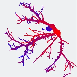
Heinrich, F; Apilla, V; Lawonn, K; Hansen, C; Preim, B; Meuschke, M
Estimating Depth Information of Vascular Models: A Comparative User Study Between a Virtual Reality and a Desktop Application Journal Article
In: Computers & Graphics, vol. 98, pp. 210–217, 2021, ISSN: 0097-8493.
@article{heinrich_estimating_2021,

title = {Estimating Depth Information of Vascular Models: A Comparative User Study Between a Virtual Reality and a Desktop Application},

author = {F Heinrich and V Apilla and K Lawonn and C Hansen and B Preim and M Meuschke},

url = {https://www.sciencedirect.com/science/article/pii/S0097849321001138},

doi = {10.1016/j.cag.2021.05.014},

issn = {0097-8493},

year  = {2021},

date = {2021-08-01},

urldate = {2021-08-01},

journal = {Computers & Graphics},

volume = {98},

pages = {210–217},

abstract = {Vascular structures are assessed, e.g., in tumor surgery to understand the influence of a planned resection on the vascular supply and venous drainage. The understanding of complex branching vascular structures may benefit from immersive virtual reality (VR) visualization. Therefore, the estimation of distance, depth and shape information is a crucial task to support diagnosis and therapy decisions. Depending on the visualization techniques used, perceptual issues can influence this process and may thus lead to false conclusions. Many studies were carried out to study depth perception for different variants of vessel visualization. However, these studies are restricted to desktop applications. Since VR exhibits specific perceptual problems, we aim at an understanding of the appropriateness of vessel visualization techniques in VR. Therefore, this paper presents a user study that investigates the effects of three commonly used visualization techniques on depth perception. The set of visualization techniques comprises Phong shading, pseudo-chromadepth and fog shading. An immersive VR setup of the study using a head-mounted display (HMD) was compared to a traditional desktop setup. Results suggest that depth judgments are less error-prone and more certain in VR than in desktop environments. Moreover, depth-enhancing visualization techniques had greater effects in the desktop study compared to the VR study.},

keywords = {},

pubstate = {published},

tppubtype = {article}

}
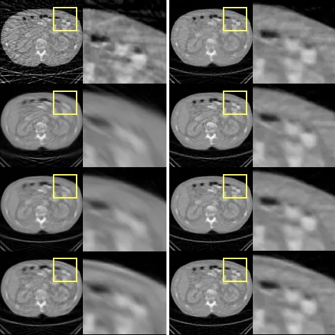
Ernst, P; Rak, M; Hansen, C; Rose, G; Nürnberger, A
Trajectory Upsampling for Sparse Conebeam Projections Using Convolutional Neural Networks Proceedings Article
In: Schramm, G; Rezaei, A; Thielemans, K; Nuyts, J (Ed.): Proceedings of the 16th Virtual International Meeting on Fully 3D Image Reconstruction in Radiology and Nuclear Medicine, pp. 285–288, 2021.
@inproceedings{ernst_trajectory_2021,

title = {Trajectory Upsampling for Sparse Conebeam Projections Using Convolutional Neural Networks},

author = {P Ernst and M Rak and C Hansen and G Rose and A Nürnberger},

editor = {G Schramm and A Rezaei and K Thielemans and J Nuyts},

url = {https://arxiv.org/abs/2110.04143},

year  = {2021},

date = {2021-07-19},

urldate = {2021-07-19},

booktitle = {Proceedings of the 16th Virtual International Meeting on Fully 3D Image Reconstruction in Radiology and Nuclear Medicine},

pages = {285–288},

abstract = {In this paper, we present an approach based on a combination of convolutional neural networks and analytical algorithms to interpolate between neighboring conebeam projections for upsampling along circular trajectories. More precisely, networks are trained to interpolate the angularly centered projection between the input projections of different angular distances. Experiments show that an analytical interpolation as additional input is more beneficial than adding more neighboring projections. Using our best model, we achieve an x8 upsampling by repeating the interpolation three times. Though not depending on a specific reconstruction algorithm, we show that FDK reconstructions substantially benefit from this upsampling for removing streak artifacts. Using this FDK reconstruction as initialization for ART is also superior to other initializations but comes with a higher computation time and therefore cannot be considered as an option in an interventional setting.},

keywords = {},

pubstate = {published},

tppubtype = {inproceedings}

}
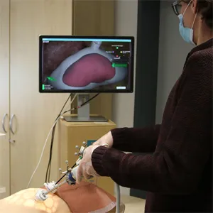
Joeres, F; Heinrich, F; Schott, D; Hansen, C
Towards Natural 3D Interaction for Laparoscopic Augmented Reality Registration Journal Article
In: Computer Methods in Biomechanics and Biomedical Engineering: Imaging & Visualization, vol. 9, no. 4, pp. 384–391, 2021, ISSN: 2168-1163, (Publisher: Taylor & Francis _eprint: https://doi.org/10.1080/21681163.2020.1834877).
@article{joeres_towards_2021,

title = {Towards Natural 3D Interaction for Laparoscopic Augmented Reality Registration},

author = {F Joeres and F Heinrich and D Schott and C Hansen},

url = {https://doi.org/10.1080/21681163.2020.1834877},

doi = {10.1080/21681163.2020.1834877},

issn = {2168-1163},

year  = {2021},

date = {2021-07-01},

urldate = {2021-07-01},

journal = {Computer Methods in Biomechanics and Biomedical Engineering: Imaging & Visualization},

volume = {9},

number = {4},

pages = {384–391},

abstract = {Augmented reality (AR) is a widely researched route for navigation support in laparoscopic surgery. Accurate registration is a crucial component for such AR systems. We introduce two methods for interactive registration that aim to be minimally invasive to the workflow and to mimic natural manipulation of 3D objects. The methods utilise spatially tracked laparoscopic tools to manipulate the virtual 3D content. We comparatively evaluated the methods against a reference, landmark-based registration method in a user study with 12 participants. We tested the methods for registration accuracy, time, and subjective usability perception. Our methods did not outperform the reference method on these parameters but showed promising results. The results indicate that our methods present no finalised solutions but that one of them is a promising approach for which we identified concrete improvement measures to be implemented in future research.},

note = {Publisher: Taylor & Francis

_eprint: https://doi.org/10.1080/21681163.2020.1834877},

keywords = {},

pubstate = {published},

tppubtype = {article}

}
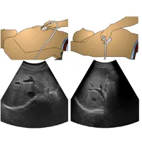
Wei, W; Haishan, X; Alpers, J; Rak, M; Hansen, C
A deep learning approach for 2D ultrasound and 3D CT/MR image registration in liver tumor ablation Journal Article
In: Computer Methods and Programs in Biomedicine, vol. 206, pp. 106117, 2021, ISSN: 0169-2607.
@article{wei_deep_2021,

title = {A deep learning approach for 2D ultrasound and 3D CT/MR image registration in liver tumor ablation},

author = {W Wei and X Haishan and J Alpers and M Rak and C Hansen},

url = {https://www.sciencedirect.com/science/article/pii/S0169260721001929

https://pubmed.ncbi.nlm.nih.gov/34022696/, PubMed},

doi = {10.1016/j.cmpb.2021.106117},

issn = {0169-2607},

year  = {2021},

date = {2021-07-01},

urldate = {2021-07-01},

journal = {Computer Methods and Programs in Biomedicine},

volume = {206},

pages = {106117},

abstract = {Background and Objective

Liver tumor ablation is often guided by ultrasound (US). Due to poor image quality, intraoperative US is fused with preoperative computed tomography or magnetic tomography (CT/MR) images to provide visual guidance. As of today, the underlying 2D US to 3D CT/MR registration problem remains a very challenging task.

Methods

We propose a novel pipeline to address this registration problem. Contrary to previous work, we do not formulate the problem as a regression task, which - for the given registration problem - achieves a low performance regarding accuracy and robustness due to the limited US soft-tissue contrast and the inter-patient variability on liver vessels. Instead, we first estimate the US probe angle roughly by using a classification network. Given this coarse initialization, we then improve the registration by formulation of the problem as a segmentation task, estimating the US plane in the 3D CT/MR through segmentation.

Results

We benchmark our approach on 1035 clinical images from 52 patients, yielding average registration errors of 11.6° and 4.7 mm, which outperforms the state of the art SVR method[1].

Conclusion

Our results show the efficiency of the proposed registration pipeline, which has potential to improve the robustness and accuracy of intraoperative patient registration.},

keywords = {},

pubstate = {published},

tppubtype = {article}

}
Liver tumor ablation is often guided by ultrasound (US). Due to poor image quality, intraoperative US is fused with preoperative computed tomography or magnetic tomography (CT/MR) images to provide visual guidance. As of today, the underlying 2D US to 3D CT/MR registration problem remains a very challenging task.
Methods
We propose a novel pipeline to address this registration problem. Contrary to previous work, we do not formulate the problem as a regression task, which – for the given registration problem – achieves a low performance regarding accuracy and robustness due to the limited US soft-tissue contrast and the inter-patient variability on liver vessels. Instead, we first estimate the US probe angle roughly by using a classification network. Given this coarse initialization, we then improve the registration by formulation of the problem as a segmentation task, estimating the US plane in the 3D CT/MR through segmentation.
Results
We benchmark our approach on 1035 clinical images from 52 patients, yielding average registration errors of 11.6° and 4.7 mm, which outperforms the state of the art SVR method[1].
Conclusion
Our results show the efficiency of the proposed registration pipeline, which has potential to improve the robustness and accuracy of intraoperative patient registration.
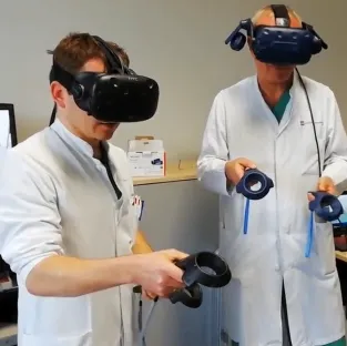
Huettl, F; Saalfeld, P; Hansen, C; Preim, B; Poplawski, A; Kneist, W; Lang, H; Huber, T
In: Annals of Translational Medicine, vol. 9, no. 13, pp. 1074, 2021, ISSN: 2305-5839.
@article{huettl_virtual_2021,

title = {Virtual Reality and 3D Printing Improve Preoperative Visualization of 3D Liver Reconstructions—Results from a Preclinical Comparison of Presentation Modalities and User’s Preference},

author = {F Huettl and P Saalfeld and C Hansen and B Preim and A Poplawski and W Kneist and H Lang and T Huber},

url = {https://www.ncbi.nlm.nih.gov/pmc/articles/PMC8339861/

https://pubmed.ncbi.nlm.nih.gov/34422986/, PubMed},

doi = {10.21037/atm-21-512},

issn = {2305-5839},

year  = {2021},

date = {2021-07-01},

urldate = {2021-07-01},

journal = {Annals of Translational Medicine},

volume = {9},

number = {13},

pages = {1074},

abstract = {Background

Preoperative three-dimensional (3D) reconstructions for liver surgery planning have been shown to be effective in reduction of blood loss and operation time. However, the role of the ‘presentation modality’ is not well investigated. We present the first study to compare 3D PDFs, 3D printed models (PR) and virtual reality (VR) 3D models with regard to anatomical orientation and personal preferences in a high volume liver surgery center.

Methods

Thirty participants, 10 medical students, 10 residents, 5 fellows and 5 hepatopancreatobiliary (HPB) experts, assigned the tumor-bearing segments of 20 different patient’s individual liver reconstructions. Liver models were presented in a random order in all modalities. Time needed to specify the tumor location was recorded. In addition, a score was calculated factoring in correct, wrong and missing segment assignments. Furthermore, standardized test/questionnaires for spatial thinking and seeing, vegetative side effects and usability were completed.

Results Participants named significantly more correct segments in VR (P=0.040) or PR (P=0.036) compared to PDF. Tumor assignment was significantly shorter with 3D PR models compared to 3D PDF (P<0.001) or VR application (P<0.001). Regardless of the modality, HPB experts were significantly faster (24±8 vs. 35±11 sec; P=0.014) and more often correct (0.87±0.12 vs. 0.83±0.15; P<0.001) than medical students. Test results for spatial thinking and seeing had no influence on time but on correctness of tumor assignment. Regarding usability and user experience the VR application achieved the highest scores without causing significant vegetative symptoms and was also the most preferred method (n=22, 73.3%) because of the multiple functions like scaling and change of transparency. Ninety percent (n=27) stated that this application can positively influence the operation planning.

Conclusions

3D PR models and 3D VR models enable a better and partially faster anatomical orientation than reconstructions presented as 3D PDFs. User’s preferred the VR application over the PR models and PDF. A prospective trial is needed to evaluate the different presentation modalities regarding intra- and postoperative outcomes.},

keywords = {},

pubstate = {published},

tppubtype = {article}

}
Preoperative three-dimensional (3D) reconstructions for liver surgery planning have been shown to be effective in reduction of blood loss and operation time. However, the role of the ‘presentation modality’ is not well investigated. We present the first study to compare 3D PDFs, 3D printed models (PR) and virtual reality (VR) 3D models with regard to anatomical orientation and personal preferences in a high volume liver surgery center.
Methods
Thirty participants, 10 medical students, 10 residents, 5 fellows and 5 hepatopancreatobiliary (HPB) experts, assigned the tumor-bearing segments of 20 different patient’s individual liver reconstructions. Liver models were presented in a random order in all modalities. Time needed to specify the tumor location was recorded. In addition, a score was calculated factoring in correct, wrong and missing segment assignments. Furthermore, standardized test/questionnaires for spatial thinking and seeing, vegetative side effects and usability were completed.
Results Participants named significantly more correct segments in VR (P=0.040) or PR (P=0.036) compared to PDF. Tumor assignment was significantly shorter with 3D PR models compared to 3D PDF (P<0.001) or VR application (P<0.001). Regardless of the modality, HPB experts were significantly faster (24±8 vs. 35±11 sec; P=0.014) and more often correct (0.87±0.12 vs. 0.83±0.15; P<0.001) than medical students. Test results for spatial thinking and seeing had no influence on time but on correctness of tumor assignment. Regarding usability and user experience the VR application achieved the highest scores without causing significant vegetative symptoms and was also the most preferred method (n=22, 73.3%) because of the multiple functions like scaling and change of transparency. Ninety percent (n=27) stated that this application can positively influence the operation planning.
Conclusions
3D PR models and 3D VR models enable a better and partially faster anatomical orientation than reconstructions presented as 3D PDFs. User’s preferred the VR application over the PR models and PDF. A prospective trial is needed to evaluate the different presentation modalities regarding intra- and postoperative outcomes.
Gabele, M; Weicker, J; Wagner, S; Thoms, A; Hußlein, S; Hansen, C
Effects and Ways of Tailored Gamification in Software-Based Training in Cognitive Rehabilitation Proceedings Article
In: Proceedings of the 29th ACM Conference on User Modeling, Adaptation and Personalization, pp. 158–168, Association for Computing Machinery, New York, NY, USA, 2021, ISBN: 978-1-4503-8366-0.
@inproceedings{gabele_effects_2021,

title = {Effects and Ways of Tailored Gamification in Software-Based Training in Cognitive Rehabilitation},

author = {M Gabele and J Weicker and S Wagner and A Thoms and S Hußlein and C Hansen},

url = {https://dl.acm.org/doi/10.1145/3450613.3456828},

doi = {10.1145/3450613.3456828},

isbn = {978-1-4503-8366-0},

year  = {2021},

date = {2021-06-01},

urldate = {2021-06-01},

booktitle = {Proceedings of the 29th ACM Conference on User Modeling, Adaptation and Personalization},

pages = {158–168},

publisher = {Association for Computing Machinery},

address = {New York, NY, USA},

series = {UMAP '21},

abstract = {A high level of motivation and frequent training are relevant in software-based rehabilitation to improve cognitive functioning after acquired brain injury. We evaluated the benefit of tailored user-centered gamification elements in a clinical study with N=83 outpatients undergoing three weeks of cognitive training in their home environment. The use of gamification in relation to the patient’s player type was explored in three steps. First, we determined the individual player types and related requests for specific game elements by means of questionnaires. Afterwards, we examined the effect of gamified training based on a non-player character and training progress within a metaphor. We considered secondly the individual perception and emotional effect and thirdly the performance based on training duration. 37 elements were requested by patients of all types, 18 elements were partially requested, and 4 elements were rejected. A comparison shows that the requested game elements partly differ between healthy persons and patients. Overall, gamification was perceived positively and gamified training leads to an increase in enjoyment compared to non-gamified training. In detail, however, there were different effects on the individual player types: socialisers experienced more enjoyment while achievers perceived higher competence throughout gamified cognitive training. Also, differences in performance in training duration were found. Within gamified training, socialisers trained significantly more than patients not primarily assigned to this type. In contrast, no significant difference was found for achievers. By showing modulating requests and effects in player types, our results support user-centered tailoring of game elements in the development of software-based cognitive training in rehabilitation.},

keywords = {},

pubstate = {published},

tppubtype = {inproceedings}

}
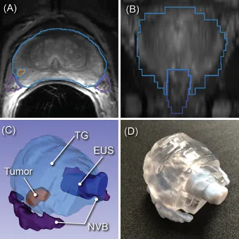
Meyer, A; Mehrtash, A; Rak, M; Bashkanov, O; Langbein, B; Ziaei, A; Kibel, A; Tempany, C; Hansen, C; Tokuda, J
Domain Adaptation for Segmentation of Critical Structures for Prostate Cancer Therapy Journal Article
In: Scientific Reports, vol. 11, no. 1, pp. 11480, 2021, ISSN: 2045-2322.
@article{meyer_domain_2021,

title = {Domain Adaptation for Segmentation of Critical Structures for Prostate Cancer Therapy},

author = {A Meyer and A Mehrtash and M Rak and O Bashkanov and B Langbein and A Ziaei and A Kibel and C Tempany and C Hansen and J Tokuda},

url = {https://www.nature.com/articles/s41598-021-90294-4

https://pubmed.ncbi.nlm.nih.gov/34075061/, PubMed},

doi = {10.1038/s41598-021-90294-4},

issn = {2045-2322},

year  = {2021},

date = {2021-06-01},

urldate = {2021-06-01},

journal = {Scientific Reports},

volume = {11},

number = {1},

pages = {11480},

abstract = {Preoperative assessment of the proximity of critical structures to the tumors is crucial in avoiding

unnecessary damage during prostate cancer treatment. A patient-specific 3D anatomical model

of those structures, namely the neurovascular bundles (NVB) and the external urethral sphincters

(EUS), can enable physicians to perform such assessments intuitively. As a crucial step to generate a

patient-specific anatomical model from preoperative MRI in a clinical routine, we propose a multi-class

automatic segmentation based on an anisotropic convolutional network. Our specific challenge is to

train the network model on a unique source dataset only available at a single clinical site and deploy it

to another target site without sharing the original images or labels. As network models trained on data

from a single source suffer from quality loss due to the domain shift, we propose a semi-supervised

domain adaptation (DA) method to refine the model’s performance in the target domain. Our DA

method combines transfer learning and uncertainty guided self-learning based on deep ensembles.

Experiments on the segmentation of the prostate, NVB, and EUS, show significant performance gain

with the combination of those techniques compared to pure TL and the combination of TL with simple

self-learning ( p < 0.005 for all structures using a Wilcoxon’s signed-rank test). Results on a different

task and data (Pancreas CT segmentation) demonstrate our method’s generic application capabilities.

Our method has the advantage that it does not require any further data from the source domain,

unlike the majority of recent domain adaptation strategies. This makes our method suitable for clinical

applications, where the sharing of patient data is restricted.},

keywords = {},

pubstate = {published},

tppubtype = {article}

}
unnecessary damage during prostate cancer treatment. A patient-specific 3D anatomical model
of those structures, namely the neurovascular bundles (NVB) and the external urethral sphincters
(EUS), can enable physicians to perform such assessments intuitively. As a crucial step to generate a
patient-specific anatomical model from preoperative MRI in a clinical routine, we propose a multi-class
automatic segmentation based on an anisotropic convolutional network. Our specific challenge is to
train the network model on a unique source dataset only available at a single clinical site and deploy it
to another target site without sharing the original images or labels. As network models trained on data
from a single source suffer from quality loss due to the domain shift, we propose a semi-supervised
domain adaptation (DA) method to refine the model’s performance in the target domain. Our DA
method combines transfer learning and uncertainty guided self-learning based on deep ensembles.
Experiments on the segmentation of the prostate, NVB, and EUS, show significant performance gain
with the combination of those techniques compared to pure TL and the combination of TL with simple
self-learning ( p < 0.005 for all structures using a Wilcoxon’s signed-rank test). Results on a different
task and data (Pancreas CT segmentation) demonstrate our method’s generic application capabilities.
Our method has the advantage that it does not require any further data from the source domain,
unlike the majority of recent domain adaptation strategies. This makes our method suitable for clinical
applications, where the sharing of patient data is restricted.
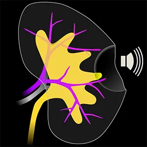
Joeres, F; Black, D; Razavizadeh, S; Hansen, C
Audiovisual AR Concepts for Laparoscopic Subsurface Structure Navigation Proceedings Article
In: Proceedings of Graphics Interface 2021, pp. 224–230, Canadian Human-Computer Communications Society, 2021.
@inproceedings{joeres_audiovisual_2021,

title = {Audiovisual AR Concepts for Laparoscopic Subsurface Structure Navigation},

author = {F Joeres and D Black and S Razavizadeh and C Hansen},

url = {https://graphicsinterface.org/proceedings/gi2021/gi2021-34/},

doi = {10.20380/GI2021.34},

year  = {2021},

date = {2021-05-28},

urldate = {2021-01-01},

booktitle = {Proceedings of Graphics Interface 2021},

pages = {224–230},

publisher = {Canadian Human-Computer Communications Society},

abstract = {The identification of subsurface structures during resection wound repair is a challenge during minimally invasive partial nephrectomy. Specifically, major blood vessels and branches of the urinary collecting system need to be localized under time pressure as target or risk structures during suture placement. This work presents concepts for AR visualization and auditory guidance based on tool position that support this task. We evaluated the concepts in a laboratory user study with a simplified, simulated task: The localization of subsurface target points in a healthy kidney phantom. We evaluated the task time, localization accuracy, and perceived workload for our concepts and a control condition without navigation support. The AR visualization improved the accuracy and perceived workload over the control condition. We observed similar, non-significant trends for the auditory display. Further, clinically realistic evaluation is pending. Our initial results indicate the potential benefits of our concepts in supporting laparoscopic resection wound repair.},

keywords = {},

pubstate = {published},

tppubtype = {inproceedings}

}
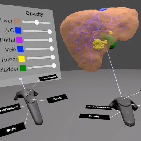
Boedecker, C; Huettl, F; Saalfeld, P; Paschold, M; Kneist, W; Baumgart, J; Preim, B; Hansen, C; Lang, H; Huber, T
Using Virtual 3D Models in Surgical Planning: Workflow of an Immersive Virtual Reality Application in Liver Surgery Journal Article
In: Langenbeck's Archives of Surgery, vol. 406, no. 3, pp. 911–915, 2021, ISSN: 1435-2451.
@article{boedecker_using_2021,

title = {Using Virtual 3D Models in Surgical Planning: Workflow of an Immersive Virtual Reality Application in Liver Surgery},

author = {C Boedecker and F Huettl and P Saalfeld and M Paschold and W Kneist and J Baumgart and B Preim and C Hansen and H Lang and T Huber},

url = {https://doi.org/10.1007/s00423-021-02127-7

https://pubmed.ncbi.nlm.nih.gov/33710462/, PubMed},

doi = {10.1007/s00423-021-02127-7},

issn = {1435-2451},

year  = {2021},

date = {2021-05-01},

urldate = {2021-05-01},

journal = {Langenbeck's Archives of Surgery},

volume = {406},

number = {3},

pages = {911–915},

abstract = {Three-dimensional (3D) surgical planning is widely accepted in liver surgery. Currently, the 3D reconstructions are usually presented as 3D PDF data on regular monitors. 3D-printed liver models are sometimes used for education and planning.},

keywords = {},

pubstate = {published},

tppubtype = {article}

}
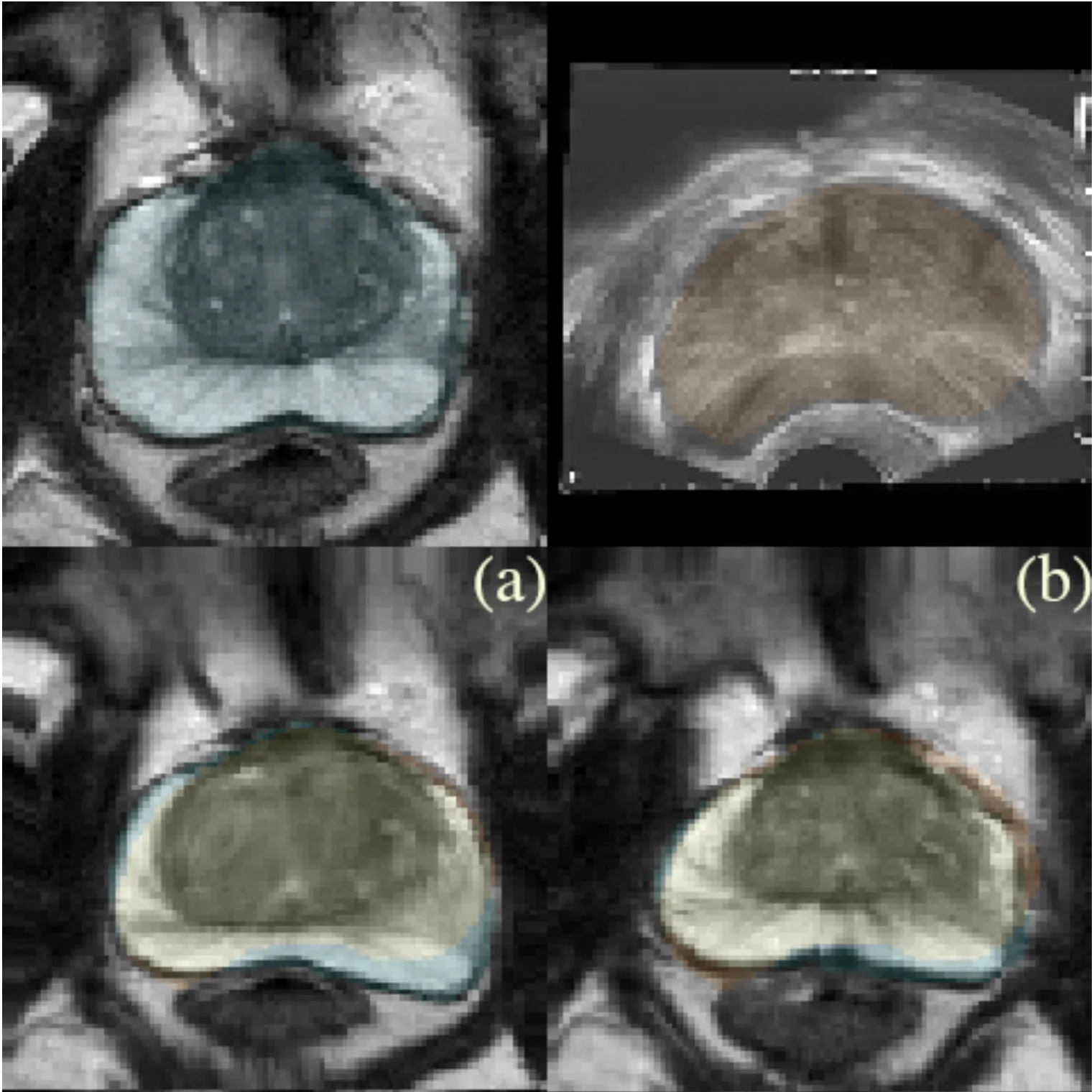
Bashkanov, O; Meyer, A; Schindele, D; Schostak, M; Tönnies, K; Hansen, C; Rak, M
Learning Multi-Modal Volumetric Prostate Registration With Weak Inter-Subject Spatial Correspondence Proceedings Article
In: 2021 IEEE 18th International Symposium on Biomedical Imaging (ISBI), pp. 1817–1821, 2021, (ISSN: 1945-8452).
@inproceedings{bashkanov_learning_2021,

title = {Learning Multi-Modal Volumetric Prostate Registration With Weak Inter-Subject Spatial Correspondence},

author = {O Bashkanov and A Meyer and D Schindele and M Schostak and K Tönnies and C Hansen and M Rak},

url = {https://ieeexplore.ieee.org/abstract/document/9433848},

doi = {10.1109/ISBI48211.2021.9433848},

year  = {2021},

date = {2021-04-01},

urldate = {2021-04-01},

booktitle = {2021 IEEE 18th International Symposium on Biomedical Imaging (ISBI)},

pages = {1817–1821},

abstract = {Recent studies demonstrated the eligibility of convolutional neural networks (CNNs) for solving the image registration problem. CNNs enable faster transformation estimation and greater generalization capability needed for better support during medical interventions. Conventional fully-supervised training requires a lot of high-quality ground truth data such as voxel-to-voxel transformations, which typically are attained in a too tedious and error-prone manner. In our work, we use weakly-supervised learning, which optimizes the model indirectly only via segmentation masks that are a more accessible ground truth than the deformation fields. Concerning the weak supervision, we investigate two segmentation similarity measures: multiscale Dice similarity coefficient (mDSC) and the similarity between segmentation-derived signed distance maps (SDMs). We show that the combination of mDSC and SDM similarity measures results in a more accurate and natural transformation pattern together with a stronger gradient coverage. Furthermore, we introduce an auxiliary input to the neural network for the prior information about the prostate location in the MR sequence, which mostly is available preoperatively. This approach significantly outperforms the standard two-input models. With weakly labelled MR-TRUS prostate data, we showed registration quality comparable to the state-of-the-art deep learning-based method.},

note = {ISSN: 1945-8452},

keywords = {},

pubstate = {published},

tppubtype = {inproceedings}

}
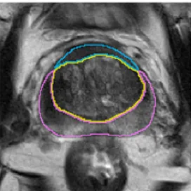
Meyer, A; Ghosh, S; Schindele, D; Schostak, M; Stober, S; Hansen, C; Rak, M
Uncertainty-Aware Temporal Self-Learning (UATS): Semi-Supervised Learning for Segmentation of Prostate Zones and Beyond Miscellaneous
2021, (arXiv:2104.03840 [eess]).
@misc{meyer_uncertainty-aware_2021,

title = {Uncertainty-Aware Temporal Self-Learning (UATS): Semi-Supervised Learning for Segmentation of Prostate Zones and Beyond},

author = {A Meyer and S Ghosh and D Schindele and M Schostak and S Stober and C Hansen and M Rak},

url = {http://arxiv.org/abs/2104.03840

https://pubmed.ncbi.nlm.nih.gov/34020751/, PubMed},

doi = {10.48550/arXiv.2104.03840},

year  = {2021},

date = {2021-04-01},

urldate = {2021-04-01},

publisher = {arXiv},

abstract = {Various convolutional neural network (CNN) based concepts have been introduced for the prostate's automatic segmentation and its coarse subdivision into transition zone (TZ) and peripheral zone (PZ). However, when targeting a fine-grained segmentation of TZ, PZ, distal prostatic urethra (DPU) and the anterior fibromuscular stroma (AFS), the task becomes more challenging and has not yet been solved at the level of human performance. One reason might be the insufficient amount of labeled data for supervised training. Therefore, we propose to apply a semi-supervised learning (SSL) technique named uncertainty-aware temporal self-learning (UATS) to overcome the expensive and time-consuming manual ground truth labeling. We combine the SSL techniques temporal ensembling and uncertainty-guided self-learning to benefit from unlabeled images, which are often readily available. Our method significantly outperforms the supervised baseline and obtained a Dice coefficient (DC) of up to 78.9% , 87.3%, 75.3%, 50.6% for TZ, PZ, DPU and AFS, respectively. The obtained results are in the range of human inter-rater performance for all structures. Moreover, we investigate the method's robustness against noise and demonstrate the generalization capability for varying ratios of labeled data and on other challenging tasks, namely the hippocampus and skin lesion segmentation. UATS achieved superiority segmentation quality compared to the supervised baseline, particularly for minimal amounts of labeled data.},

note = {arXiv:2104.03840 [eess]},

keywords = {},

pubstate = {published},

tppubtype = {misc}

}
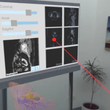
Schott, D; Saalfeld, P; Schmidt, G; Joeres, F; Boedecker, C; Huettl, F; Lang, H; Huber, T; Preim, B; Hansen, C
A VR/AR Environment for Multi-User Liver Anatomy Education Proceedings Article
In: 2021 IEEE Virtual Reality and 3D User Interfaces (VR), pp. 296–305, IEEE, Lisboa, Portugal, 2021, ISBN: 978-1-6654-1838-6.
@inproceedings{schott_vrar_2021,

title = {A VR/AR Environment for Multi-User Liver Anatomy Education},

author = {D Schott and P Saalfeld and G Schmidt and F Joeres and C Boedecker and F Huettl and H Lang and T Huber and B Preim and C Hansen},

url = {https://ieeexplore.ieee.org/document/9417662/},

doi = {10.1109/VR50410.2021.00052},

isbn = {978-1-6654-1838-6},

year  = {2021},

date = {2021-03-01},

urldate = {2021-03-01},

booktitle = {2021 IEEE Virtual Reality and 3D User Interfaces (VR)},

pages = {296–305},

publisher = {IEEE},

address = {Lisboa, Portugal},

abstract = {We present a Virtual and Augmented Reality multi-user prototype of a learning environment for liver anatomy education. Our system supports various training scenarios ranging from small learning groups to classroom-size education, where students and teachers can participate in virtual reality, augmented reality, or via desktop PCs. In an iterative development process with surgeons and teachers, a virtual organ library was created. Nineteen liver data sets were used comprising 3D surface models, 2D image data, pathology information, diagnosis and treatment decisions. These data sets can interactively be sorted and investigated individually regarding their volumetric and meta information. The three participation modes were evaluated within a user study with surgery lecturers (5) and medical students (5). We assessed the usability and presence using questionnaires. Additionally, we collected qualitative data with semistructured interviews. A total of 435 individual statements were recorded and summarized to 49 statements. The results show that our prototype is usable, induces presence, and potentially support the teaching of liver anatomy and surgery in the future.},

keywords = {},

pubstate = {published},

tppubtype = {inproceedings}

}
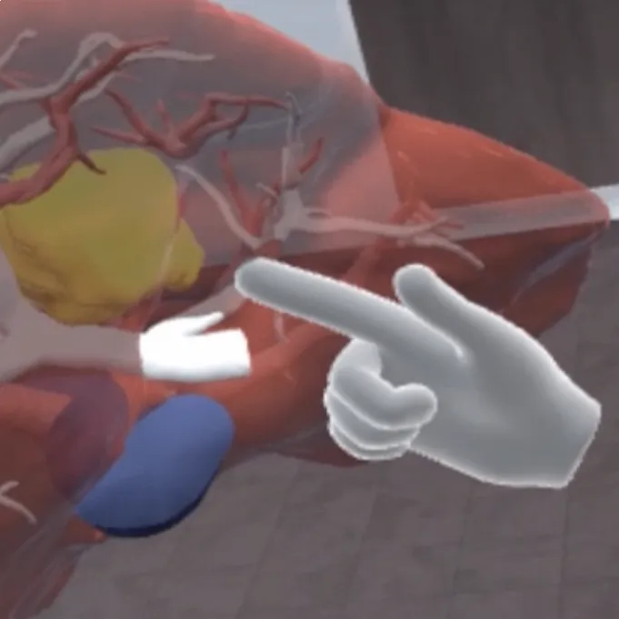
Chheang, V; Apilla, V; Saalfeld, P; Boedecker, C; Huber, T; Huettl, F; Lang, H; Preim, B; Hansen, C
Collaborative VR for Liver Surgery Planning using Wearable Data Gloves: An Interactive Demonstration Proceedings Article
In: 2021 IEEE Conference on Virtual Reality and 3D User Interfaces Abstracts and Workshops (VRW), pp. 768–768, 2021.
@inproceedings{chheang_collaborative_2021-1,

title = {Collaborative VR for Liver Surgery Planning using Wearable Data Gloves: An Interactive Demonstration},

author = {V Chheang and V Apilla and P Saalfeld and C Boedecker and T Huber and F Huettl and H Lang and B Preim and C Hansen},

url = {https://ieeexplore.ieee.org/abstract/document/9419102},

doi = {10.1109/VRW52623.2021.00268},

year  = {2021},

date = {2021-03-01},

urldate = {2021-03-01},

booktitle = {2021 IEEE Conference on Virtual Reality and 3D User Interfaces Abstracts and Workshops (VRW)},

pages = {768–768},

abstract = {Preoperative planning for liver surgery is a critical procedure to assess a potential resection and it supports surgeons to define the affected vessels and resection volume. Traditional surgical planning systems are widely used to support the planning with the usage of desktop-based 3D and 2D visualizations. However, desktop-based systems offer limited interactions and visualizations compared to virtual reality (VR) [3]. A suitable technique to support collaboration among surgeons is required. Our previous works [1], [2] illustrate that using collaborative VR is essential to enhance communication, teamwork, and over-distance collaboration. In this work, we present a collaborative VR prototype to support liver surgery planning with intuitive interactions using wearable data gloves and VR controllers. The users can explore the patient data in both 2D and 3D representations. Thereafter, a virtual resection is specified by drawing lines on the 3D model representation. The virtual re-section is further refined to keep safety margins from the tumors. Moreover, real-time risk maps are visualized to support the surgeons during the modification. Finally, the results of the virtual resection are visualized as resection volumes with their indicated amount and colors. Future work aims to conduct an extensive clinical study to compare the suitability of these input devices.},

keywords = {},

pubstate = {published},

tppubtype = {inproceedings}

}
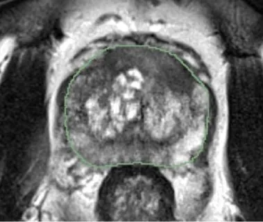
Meyer, A; Chlebus, G; Rak, M; Schindele, D; Schostak, M; Ginneken, B; Schenk, A; Meine, H; Hahn, H; Schreiber, A; Hansen, C
Anisotropic 3D Multi-Stream CNN for Accurate Prostate Segmentation from Multi-Planar MRI Journal Article
In: Computer Methods and Programs in Biomedicine, vol. 200, pp. 105821, 2021, ISSN: 01692607, (arXiv:2009.11120 [eess]).
@article{meyer_anisotropic_2021,

title = {Anisotropic 3D Multi-Stream CNN for Accurate Prostate Segmentation from Multi-Planar MRI},

author = {A Meyer and G Chlebus and M Rak and D Schindele and M Schostak and B Ginneken and A Schenk and H Meine and H Hahn and A Schreiber and C Hansen},

url = {http://arxiv.org/abs/2009.11120

https://pubmed.ncbi.nlm.nih.gov/33218704/, PubMed},

doi = {10.1016/j.cmpb.2020.105821},

issn = {01692607},

year  = {2021},

date = {2021-03-01},

urldate = {2021-03-01},

journal = {Computer Methods and Programs in Biomedicine},

volume = {200},

pages = {105821},

abstract = {Background and Objective: Accurate and reliable segmentation of the prostate gland in MR images can support the clinical assessment of prostate cancer, as well as the planning and monitoring of focal and loco-regional therapeutic interventions. Despite the availability of multi-planar MR scans due to standardized protocols, the majority of segmentation approaches presented in the literature consider the axial scans only. Methods: We propose an anisotropic 3D multi-stream CNN architecture, which processes additional scan directions to produce a higher-resolution isotropic prostate segmentation. We investigate two variants of our architecture, which work on two (dual-plane) and three (triple-plane) image orientations, respectively. We compare them with the standard baseline (single-plane) used in literature, i.e., plain axial segmentation. To realize a fair comparison, we employ a hyperparameter optimization strategy to select optimal configurations for the individual approaches. Results: Training and evaluation on two datasets spanning multiple sites obtain statistical significant improvement over the plain axial segmentation ($p<0.05$ on the Dice similarity coefficient). The improvement can be observed especially at the base ($0.898$ single-plane vs. $0.906$ triple-plane) and apex ($0.888$ single-plane vs. $0.901$ dual-plane). Conclusion: This study indicates that models employing two or three scan directions are superior to plain axial segmentation. The knowledge of precise boundaries of the prostate is crucial for the conservation of risk structures. Thus, the proposed models have the potential to improve the outcome of prostate cancer diagnosis and therapies.},

note = {arXiv:2009.11120 [eess]},

keywords = {},

pubstate = {published},

tppubtype = {article}

}
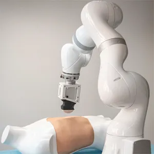
Schreiter, J; Joeres, F; March, C; Pech, M; Hansen, C
Application Potential of Robot-Guided Ultrasound During CT-Guided Interventions Proceedings Article
In: Noble, J; Aylward, S; Grimwood, A; Min, Z; Lee, S; Hu, Y (Ed.): Simplifying Medical Ultrasound, pp. 116–125, Springer International Publishing, Cham, 2021, ISBN: 978-3-030-87583-1.
@inproceedings{schreiter_application_2021,

title = {Application Potential of Robot-Guided Ultrasound During CT-Guided Interventions},

author = {J Schreiter and F Joeres and C March and M Pech and C Hansen},

editor = {J Noble and S Aylward and A Grimwood and Z Min and S Lee and Y Hu},

doi = {10.1007/978-3-030-87583-1_12},

isbn = {978-3-030-87583-1},

year  = {2021},

date = {2021-01-01},

urldate = {2021-01-01},

booktitle = {Simplifying Medical Ultrasound},

pages = {116–125},

publisher = {Springer International Publishing},

address = {Cham},

abstract = {CT-guided interventions are common practices in interventional radiology to treat oncological conditions. During these interventions, radiologists are exposed to radiation and faced with a non-ergonomic working environment. A robot-guided ultrasound (US) as a complementing imaging method for the purpose of needle guidance could help to overcome these challenges. A survey with 21 radiologists was made to analyze the application potential of US during CT-guided interventions with regard to anatomical regions to be scanned as locations of target lesions as well as specific situations during which US could complement CT imaging. The results indicate that the majority of respondents already applied US during CT-guided interventions for reasons of real-time imaging of the target lesion, organ, and needle movement as well as for lesions that are difficult to visualize in CT. Potential situations of US application were identified as out-of-plane needle insertion and puncturing lesions within the liver and subcutaneous lymph nodes. Interaction with a robot-guided US should be intuitive and include an improved sterility concept.},

keywords = {},

pubstate = {published},

tppubtype = {inproceedings}

}

Batz, V; Lipowski, I; Klaba, F; Engel, N; Weiß, V; Hansen, C; Herzog, M
The Digital Competence of Vocational Education Teachers and of Learners with and Without Cognitive Disabilities Book Section
In: Jia, W; Tang, Y; Lee, R; Herzog, M; Zhang, H; Hao, T; Wang, T (Ed.): Emerging Technologies for Education, vol. 13089, pp. 190–206, Springer International Publishing, Cham, 2021, ISBN: 978-3-030-92835-3 978-3-030-92836-0, (Series Title: Lecture Notes in Computer Science).
@incollection{jia_digital_2021,

title = {The Digital Competence of Vocational Education Teachers and of Learners with and Without Cognitive Disabilities},

author = {V Batz and I Lipowski and F Klaba and N Engel and V Weiß and C Hansen and M Herzog},

editor = {W Jia and Y Tang and R Lee and M Herzog and H Zhang and T Hao and T Wang},

url = {https://link.springer.com/10.1007/978-3-030-92836-0_17},

doi = {10.1007/978-3-030-92836-0_17},

isbn = {978-3-030-92835-3 978-3-030-92836-0},

year  = {2021},

date = {2021-01-01},

urldate = {2021-01-01},

booktitle = {Emerging Technologies for Education},

volume = {13089},

pages = {190–206},

publisher = {Springer International Publishing},

address = {Cham},

abstract = {Nowadays, digital competence is required for participation in working life, education, and social activities. Vocational education is the key to teaching and development of digital skills. Technology enhanced learning offers enormous potential for improving equal participation and reducing access barriers. In order to meet the demands for equal access to digital technologies, to a digitized labor market and to an inclusive education system, teachers and learners need to have the necessary expertise. A survey was conducted using the Digital Competency Profiler (DCP) to explore the digital competence of teachers and learners in vocational education. The items were adapted linguistically according to requirements for people with cognitive disabilities. The aim is to identify possible gaps in the development of digital competencies in three survey groups: teachers in vocational training, trainees in food occupations, and employees with disabilities of sheltered workshops. The digital technology usage habits of the test groups are analyzed and possible differences are determined. Based on an expert assessment of the DCP items, 13 relevant competencies for vocational education are defined. Overall, the participants consider their digital competence to be good. The competencies sending text messages, making phone calls and watching videos show the highest frequency and confidence in the total sample and the competencies creating documents, writing e-mails and managing online accounts the lowest. The index value social competency is particularly high in comparison to the epistemological competency. Needs for intervention are identified, such as the systematic qualification of teachers and learners as condition for digital learning in vocational education.},

note = {Series Title: Lecture Notes in Computer Science},

keywords = {},

pubstate = {published},

tppubtype = {incollection}

}

Gabele, M; Fischer, V; Steinbrügge, M; Thiemke, D; Husslein, S; Hansen, C
Potentials of a Web-Based Gamification Guidance for Knowledge Transfer Between Research and Industry Proceedings Article
In: Extended Abstracts of the 2021 Annual Symposium on Computer-Human Interaction in Play, pp. 301–307, Association for Computing Machinery, New York, NY, USA, 2021, ISBN: 978-1-4503-8356-1.
@inproceedings{gabele_potentials_2021,

title = {Potentials of a Web-Based Gamification Guidance for Knowledge Transfer Between Research and Industry},

author = {M Gabele and V Fischer and M Steinbrügge and D Thiemke and S Husslein and C Hansen},

url = {https://doi.org/10.1145/3450337.3483458},

doi = {10.1145/3450337.3483458},

isbn = {978-1-4503-8356-1},

year  = {2021},

date = {2021-01-01},

urldate = {2021-01-01},

booktitle = {Extended Abstracts of the 2021 Annual Symposium on Computer-Human Interaction in Play},

pages = {301–307},

publisher = {Association for Computing Machinery},

address = {New York, NY, USA},

series = {CHI PLAY '21},

abstract = {Research results show that gamification can influence emotions, motivation and behavior. However, in order to apply the results in the software development in industry, they need to be easily accessible. To enable a knowledge transfer, a concept and a prototype for an interactive web-based tool were designed to summarize and visualize existing research results and practical examples in a simplified way regarding the use of gamification elements. Step by step, the user defines parameters such as context of use, age, gender and user type for the target group. Based on this, effects in research results are presented simplified and sorted in four categories in pie charts. This is intended to provide users in the industry with the competence to select appropriate gamification elements for their usage. The approach was developed in a user-centered way, including pre-study interviews with seven participants from research and / or industry, and then evaluated using a clickable prototype and qualitative open interviews. Potentials for industry and research were examined. The approach was perceived as supportive in selecting appropriate gamification elements for use in industry. It may lead to faster finding of research results, promote networking and may generate a kind of meta review. The elaboration of such tools may support the interconnection between research and industry, as well as the transfer and applicability of knowledge in gamification.},

keywords = {},

pubstate = {published},

tppubtype = {inproceedings}

}
Preim, B; Saalfeld, P; Hansen, C
Virtual and Augmented Reality for Educational Anatomy Book Section
In: Uhl, J; Jorge, J; Lopes, D; Campos, P (Ed.): Digital Anatomy : Applications of Virtual, Mixed and Augmented Reality, pp. 299–324, Springer International Publishing, Cham, 2021, ISBN: 978-3-030-61905-3.
@incollection{preim_virtual_2021,

title = {Virtual and Augmented Reality for Educational Anatomy},

author = {B Preim and P Saalfeld and C Hansen},

editor = {J Uhl and J Jorge and D Lopes and P Campos},

url = {https://doi.org/10.1007/978-3-030-61905-3_16},

doi = {10.1007/978-3-030-61905-3_16},

isbn = {978-3-030-61905-3},

year  = {2021},

date = {2021-01-01},

urldate = {2021-01-01},

booktitle = {Digital Anatomy : Applications of Virtual, Mixed and Augmented Reality},

pages = {299–324},

publisher = {Springer International Publishing},

address = {Cham},

abstract = {Recent progress in VR and AR hardware enables a wide range of educational applications. Anatomy education, where the complex spatial relations of the human anatomy need to be imagined, may benefit from the immersive experience. Also the integration of virtual information and real information, e.g., muscles and bone overlaid on the user’s body, are beneficial for imaging the interplay of various anatomical structures. VR and AR systems for anatomy education compete with other media to support anatomy teaching, such as interactive 3D visualization and anatomy textbooks. We discuss the constraints that must be considered when designing VR and AR systems that enable efficient knowledge transfer.},

keywords = {},

pubstate = {published},

tppubtype = {incollection}

}
Alpers, J; Reimert, D; Rötzer, M; Gerlach, T; Gutberlet, M; Wacker, F; Hensen, B; Hansen, C
2.5D Thermometry Maps for MRI-guided Tumor Ablation Book Section
In: Medical Image Computing and Computer Assisted Intervention – MICCAI 2021, vol. 12904, pp. 311–320, Springer, Cham, 2021, (arXiv:2108.05734 [eess]).
@incollection{alpers_25d_2021,

title = {2.5D Thermometry Maps for MRI-guided Tumor Ablation},

author = {J Alpers and D Reimert and M Rötzer and T Gerlach and M Gutberlet and F Wacker and B Hensen and C Hansen},

url = {http://arxiv.org/abs/2108.05734},

doi = {10.1007/978-3-030-87202-1_30},

year  = {2021},

date = {2021-01-01},

urldate = {2021-01-01},

booktitle = {Medical Image Computing and Computer Assisted Intervention – MICCAI 2021},

volume = {12904},

pages = {311–320},

publisher = {Springer},

address = {Cham},

series = {Lecture Notes in Computer Science},

abstract = {Fast and reliable monitoring of volumetric heat distribution during MRI-guided tumor ablation is an urgent clinical need. In this work, we introduce a method for generating 2.5D thermometry maps from uniformly distributed 2D MRI phase images rotated around the applicator's main axis. The images can be fetched directly from the MR device, reducing the delay between image acquisition and visualization. For reconstruction, we use a weighted interpolation on a cylindric coordinate representation to calculate the heat value of voxels in a region of interest. A pilot study on 13 ex vivo bio protein phantoms with flexible tubes to simulate a heat sink effect was conducted to evaluate our method. After thermal ablation, we compared the measured coagulation zone extracted from the post-treatment MR data set with the output of the 2.5D thermometry map. The results show a mean Dice score of 0.75+-0.07, a sensitivity of 0.77+-0.03, and a reconstruction time within 18.02ms+-5.91ms. Future steps should address improving temporal resolution and accuracy, e.g., incorporating advanced bioheat transfer simulations.},

note = {arXiv:2108.05734 [eess]},

keywords = {},

pubstate = {published},

tppubtype = {incollection}

}
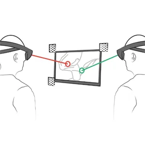
Heinrich, F; Huettl, F; Schmidt, G; Paschold, M; Kneist, W; Huber, T; Hansen, C
HoloPointer: A Virtual Augmented Reality Pointer for Laparoscopic Surgery Training Journal Article
In: International Journal of Computer Assisted Radiology and Surgery, vol. 16, no. 1, pp. 161–168, 2021, ISSN: 1861-6410, 1861-6429.
@article{heinrich_holopointer_2021,

title = {HoloPointer: A Virtual Augmented Reality Pointer for Laparoscopic Surgery Training},

author = {F Heinrich and F Huettl and G Schmidt and M Paschold and W Kneist and T Huber and C Hansen},

url = {https://link.springer.com/10.1007/s11548-020-02272-2

https://pubmed.ncbi.nlm.nih.gov/33095424/, PubMed},

doi = {10.1007/s11548-020-02272-2},

issn = {1861-6410, 1861-6429},

year  = {2021},

date = {2021-01-01},

urldate = {2021-01-01},

journal = {International Journal of Computer Assisted Radiology and Surgery},

volume = {16},

number = {1},

pages = {161–168},

abstract = {Purpose In laparoscopic surgery training, experts guide novice physicians to desired instrument positions or indicate relevant areas of interest. These instructions are usually given via verbal communication or using physical pointing devices. To facilitate a sterile work flow and to improve training, new guiding methods are needed. This work proposes to use optical see-through augmented reality to visualize an interactive virtual pointer on the laparoscopic.

Methods After an interdisciplinary development, the pointer’s applicability and feasibility for training was evaluated and it was compared to a standard condition based on verbal and gestural communication only. In this study, ten surgical trainees were guided by an experienced trainer during cholecystectomies on a laparoscopic training simulator. All trainees completed a virtual cholecystectomy with and without the interactive virtual pointer in alternating order. Measures included procedure time, economy of movement and error rates.

Results Results of standardized variables revealed significantly improved economy of movement (p 0.047) and error rates (p 0.047), as well as an overall improved user performance (Total z-score; p 0.031) in conditions using the proposed method.

Conclusion The proposed HoloPointer is a feasible and applicable tool for laparoscopic surgery training. It improved objective performance metrics without prolongation of the task completion time in this pre-clinical setup.},

keywords = {},

pubstate = {published},

tppubtype = {article}

}
Methods After an interdisciplinary development, the pointer’s applicability and feasibility for training was evaluated and it was compared to a standard condition based on verbal and gestural communication only. In this study, ten surgical trainees were guided by an experienced trainer during cholecystectomies on a laparoscopic training simulator. All trainees completed a virtual cholecystectomy with and without the interactive virtual pointer in alternating order. Measures included procedure time, economy of movement and error rates.
Results Results of standardized variables revealed significantly improved economy of movement (p 0.047) and error rates (p 0.047), as well as an overall improved user performance (Total z-score; p 0.031) in conditions using the proposed method.
Conclusion The proposed HoloPointer is a feasible and applicable tool for laparoscopic surgery training. It improved objective performance metrics without prolongation of the task completion time in this pre-clinical setup.
2020

Heinrich, F; Schwenderling, L; Joeres, F; Lawonn, K; Hansen, C
Comparison of Augmented Reality Display Techniques to Support Medical Needle Insertion Journal Article
In: IEEE Transactions on Visualization and Computer Graphics, vol. 26, no. 12, pp. 3568–3575, 2020, ISSN: 1941-0506.
@article{heinrich_comparison_2020,

title = {Comparison of Augmented Reality Display Techniques to Support Medical Needle Insertion},

author = {F Heinrich and L Schwenderling and F Joeres and K Lawonn and C Hansen},

url = {https://ieeexplore.ieee.org/abstract/document/9211732},

doi = {10.1109/TVCG.2020.3023637},

issn = {1941-0506},

year  = {2020},

date = {2020-12-01},

urldate = {2020-12-01},

journal = {IEEE Transactions on Visualization and Computer Graphics},

volume = {26},

number = {12},

pages = {3568–3575},

abstract = {Augmented reality (AR) may be a useful technique to overcome issues of conventionally used navigation systems supporting medical needle insertions, like increased mental workload and complicated hand-eye coordination. Previous research primarily focused on the development of AR navigation systems designed for specific displaying devices, but differences between employed methods have not been investigated before. To this end, a user study involving a needle insertion task was conducted comparing different AR display techniques with a monitor-based approach as baseline condition for the visualization of navigation information. A video see-through stationary display, an optical see-through head-mounted display and a spatial AR projector-camera-system were investigated in this comparison. Results suggest advantages of using projected navigation information in terms of lower task completion time, lower angular deviation and affirmative subjective participant feedback. Techniques requiring the intermediate view on screens, i.e. the stationary display and the baseline condition, showed less favorable results. Thus, benefits of providing AR navigation information compared to a conventionally used method could be identified. Significant objective measures results, as well as an identification of advantages and disadvantages of individual display techniques contribute to the development and design of improved needle navigation systems.},

keywords = {},

pubstate = {published},

tppubtype = {article}

}
Chheang, V; Fischer, V; Buggenhagen, H; Huber, T; Huettl, F; Kneist, W; Preim, B; Saalfeld, P; Hansen, C
Toward Interprofessional Team Training for Surgeons and Anesthesiologists Using Virtual Reality Journal Article
In: International Journal of Computer Assisted Radiology and Surgery, vol. 15, no. 12, pp. 2109–2118, 2020, ISSN: 1861-6429.
@article{chheang_toward_2020,

title = {Toward Interprofessional Team Training for Surgeons and Anesthesiologists Using Virtual Reality},

author = {V Chheang and V Fischer and H Buggenhagen and T Huber and F Huettl and W Kneist and B Preim and P Saalfeld and C Hansen},

url = {https://doi.org/10.1007/s11548-020-02276-y

https://pubmed.ncbi.nlm.nih.gov/33083969/, PubMed},

doi = {10.1007/s11548-020-02276-y},

issn = {1861-6429},

year  = {2020},

date = {2020-12-01},

urldate = {2020-12-01},

journal = {International Journal of Computer Assisted Radiology and Surgery},

volume = {15},

number = {12},

pages = {2109–2118},

abstract = {In this work, a virtual environment for interprofessional team training in laparoscopic surgery is proposed. Our objective is to provide a tool to train and improve intraoperative communication between anesthesiologists and surgeons during laparoscopic procedures.},

keywords = {},

pubstate = {published},

tppubtype = {article}

}
Bornemann, K; Heinrich, F; Lawonn, K; Hansen, C
Exploration of Medical Volume Data in Projective Augmented Reality: An Interactive Demonstration Proceedings Article
In: Proceedings of Mensch und Computer 2020, pp. 507–509, Association for Computing Machinery, New York, NY, USA, 2020, ISBN: 978-1-4503-7540-5.
@inproceedings{bornemann_exploration_2020,

title = {Exploration of Medical Volume Data in Projective Augmented Reality: An Interactive Demonstration},

author = {K Bornemann and F Heinrich and K Lawonn and C Hansen},

url = {https://doi.org/10.1145/3404983.3410415},

doi = {10.1145/3404983.3410415},

isbn = {978-1-4503-7540-5},

year  = {2020},

date = {2020-09-01},

urldate = {2020-09-01},

booktitle = {Proceedings of Mensch und Computer 2020},

pages = {507–509},

publisher = {Association for Computing Machinery},

address = {New York, NY, USA},

series = {MuC '20},

abstract = {This article describes the implementation and usage of a prototype to explore medical volume data in projective augmented reality (AR). To allow real-time rendering of the data, a volume ray casting algorithm is implemented on the GPU. Furthermore, the volume data is distorted to be superimposed correctly from the user's perspective. To allow exploration of the dataset, the user can use an HTC Vive Controller to show or hide structures with either clipping or windowing. In the future, the proposed prototype might be used for diagnosis, planning and computer assisted surgery. First tests indicate that this setup increases spatial understanding of the volume data compared to conventional computer monitors.},

keywords = {},

pubstate = {published},

tppubtype = {inproceedings}

}
Gulamhussene, G; Joeres, F; Rak, M; Pech, M; Hansen, C
4D MRI: Robust sorting of free breathing MRI slices for use in interventional settings Journal Article
In: PLOS ONE, vol. 15, no. 6, pp. e0235175, 2020, ISSN: 1932-6203, (Publisher: Public Library of Science).
@article{gulamhussene_4d_2020,

title = {4D MRI: Robust sorting of free breathing MRI slices for use in interventional settings},

author = {G Gulamhussene and F Joeres and M Rak and M Pech and C Hansen},

url = {https://journals.plos.org/plosone/article?id=10.1371/journal.pone.0235175

https://pubmed.ncbi.nlm.nih.gov/32569335/, PubMed},

doi = {10.1371/journal.pone.0235175},

issn = {1932-6203},

year  = {2020},

date = {2020-06-01},

urldate = {2020-06-01},

journal = {PLOS ONE},

volume = {15},

number = {6},

pages = {e0235175},

abstract = {Purpose We aim to develop a robust 4D MRI method for large FOVs enabling the extraction of irregular respiratory motion that is readily usable with all MRI machines and thus applicable to support a wide range of interventional settings. Method We propose a 4D MRI reconstruction method to capture an arbitrary number of breathing states. It uses template updates in navigator slices and search regions for fast and robust vessel cross-section tracking. It captures FOVs of 255 mm x 320 mm x 228 mm at a spatial resolution of 1.82 mm x 1.82 mm x 4mm and temporal resolution of 200ms. A total of 37 4D MRIs of 13 healthy subjects were reconstructed to validate the method. A quantitative evaluation of the reconstruction rate and speed of both the new and baseline method was performed. Additionally, a study with ten radiologists was conducted to assess the subjective reconstruction quality of both methods. Results Our results indicate improved mean reconstruction rates compared to the baseline method (79.4% vs. 45.5%) and improved mean reconstruction times (24s vs. 73s) per subject. Interventional radiologists perceive the reconstruction quality of our method as higher compared to the baseline (262.5 points vs. 217.5 points},

note = {Publisher: Public Library of Science},

keywords = {},

pubstate = {published},

tppubtype = {article}

}
Schott, D; Hatscher, B; Joeres, F; Gabele, M; Hußlein, S; Hansen, C
Lean-Interaction: Passive Image Manipulation in Concurrent Multitasking Proceedings Article
In: Proceedings of Graphics Interface 2020, pp. 404–412, Canadian Human-Computer Communications Society, 2020.
@inproceedings{schott_lean-interaction_2020,

title = {Lean-Interaction: Passive Image Manipulation in Concurrent Multitasking},

author = {D Schott and B Hatscher and F Joeres and M Gabele and S Hußlein and C Hansen},

url = {https://graphicsinterface.org/proceedings/gi2020/gi2020-40/},

doi = {10.20380/GI2020.40},

year  = {2020},

date = {2020-05-28},

urldate = {2020-01-01},

booktitle = {Proceedings of Graphics Interface 2020},

pages = {404–412},

publisher = {Canadian Human-Computer Communications Society},

abstract = {Complex bi-manual tasks often benefit from supporting visual information and guidance. Controlling the system that provides this information is a secondary task that forces the user to perform concurrent multitasking, which in turn may affect the main task performance. Interactions based on natural behavior are a promising solution to this challenge. We investigated the performance of these interactions in a hands-free image manipulation task during a primary manual task with an upright stance. Essential tasks were extracted from the example of clinical workflow and turned into an abstract simulation to gain general insights into how different interaction techniques impact the user’s performance and workload. The interaction techniques we compared were full-body movements, facial expression, gesture and speech input. We found that leaning as an interaction technique facilitates significantly faster image manipulation at lower subjective workloads than facial expression. Our results pave the way towards efficient, natural, hands-free interaction in a challenging multitasking environment.},

keywords = {},

pubstate = {published},

tppubtype = {inproceedings}

}
Streuber, S; Saalfeld, P; Podulski, K; Hüttl, F; Huber, T; Buggenhagen, H; Boedecker, C; Preim, B; Hansen, C
Training of Patient Handover in Virtual Reality Journal Article
In: Current Directions in Biomedical Engineering, vol. 6, iss. 1, no. 1, 2020, ISSN: 2364-5504, (Article number: 20200040).
@article{streuber_training_2020,

title = {Training of Patient Handover in Virtual Reality},

author = {S Streuber and P Saalfeld and K Podulski and F Hüttl and T Huber and H Buggenhagen and C Boedecker and B Preim and C Hansen},

url = {https://www.degruyterbrill.com/document/doi/10.1515/cdbme-2020-0040/html},

doi = {10.1515/cdbme-2020-0040},

issn = {2364-5504},

year  = {2020},

date = {2020-05-01},

urldate = {2020-05-01},

journal = {Current Directions in Biomedical Engineering},

volume = {6},

number = {1},

issue = {1},

publisher = {De Gruyter},

abstract = {Patient handover is an important part for information transfer between medical professionals in a clinical setting. Yet, in current medical education, these conversations are only trained sparsely, since they are costly to perform as they take place in offsite courses and are led by experts over several days. Virtual reality (VR)-based training courses could increase the availability of training, by eliminating travel costs and reducing the time-commitment of the teaching experts. This work presents a VR prototype of a multi-user training and examination application for patient handover. To ensure a similar interaction quality to its current real world counterpart, this work used omni-directional video recordings to create a realistic setting and compared different projection methods. A pilot study highlighted distinct use-cases of sphere and mesh projections to visualize the recordings. The results suggest enhanced spatial presence relating to the usage of omni-directional videos in VR-applications.},

note = {Article number: 20200040},

keywords = {},

pubstate = {published},

tppubtype = {article}

}
Wei, W; Rak, M; Alpers, J; Hansen, C
Towards Fully Automatic 2D US to 3D CT/MR Registration: A Novel Segmentation-Based Strategy Proceedings Article
In: 2020 IEEE 17th International Symposium on Biomedical Imaging (ISBI), pp. 433–437, IEEE, Iowa City, IA, USA, 2020, ISBN: 978-1-5386-9330-8.
@inproceedings{wei_towards_2020,

title = {Towards Fully Automatic 2D US to 3D CT/MR Registration: A Novel Segmentation-Based Strategy},

author = {W Wei and M Rak and J Alpers and C Hansen},

url = {https://doi.org/10.1109/ISBI45749.2020.9098379},

doi = {10.1109/ISBI45749.2020.9098379},

isbn = {978-1-5386-9330-8},

year  = {2020},

date = {2020-04-01},

urldate = {2020-04-01},

booktitle = {2020 IEEE 17th International Symposium on Biomedical Imaging (ISBI)},

pages = {433–437},

publisher = {IEEE},

address = {Iowa City, IA, USA},

abstract = {2D-US to 3D-CT/MR registration is a crucial module during minimally invasive ultrasound-guided liver tumor ablations. Many modern registration methods still require manual or semi-automatic slice pose initialization due to insufficient robustness of automatic methods. The state-of-the-art regression networks do not work well for liver 2D US to 3D CT/MR registration because of the tremendous inter-patient variability of the liver anatomy. To address this unsolved problem, we propose a deep learning network pipeline which instead of a regression starts with a classification network to recognize the coarse ultrasound transducer pose followed by a segmentation network to detect the target plane of the US image in the CT/MR volume. The rigid registration result is derived using plane regression. In contrast to the state-of-the-art regression networks, we do not estimate registration parameters from multi-modal images directly, but rather focus on segmenting the target slice plane in the volume. The experiments reveal that this novel registration strategy can identify the initial slice phase in a 3D volume more reliably than the standard regression-based techniques. The proposed method was evaluated with 1035 US images from 52 patients. We achieved angle and distance errors of 12.7 ± 6.2 degrees and 4.9 ± 3.1 mm, clearly outperforming the state-of-the-art regression strategy which results in 37.0 ± 15.6 degrees angle error and 19.0 ± 11.6 mm distance error.},

keywords = {},

pubstate = {published},

tppubtype = {inproceedings}

}
Hatscher, B; Mewes, A; Pannicke, E; Kägebein, U; Wacker, F; Hansen, C; Hensen, B
Touchless scanner control to support MRI-guided interventions Journal Article
In: International Journal of Computer Assisted Radiology and Surgery, vol. 15, no. 3, pp. 545–553, 2020, ISSN: 1861-6429.
@article{hatscher_touchless_2020,

title = {Touchless scanner control to support MRI-guided interventions},

author = {B Hatscher and A Mewes and E Pannicke and U Kägebein and F Wacker and C Hansen and B Hensen},

url = {https://doi.org/10.1007/s11548-019-02058-1

https://pubmed.ncbi.nlm.nih.gov/31520326/, PubMed},

doi = {10.1007/s11548-019-02058-1},

issn = {1861-6429},

year  = {2020},

date = {2020-03-01},

urldate = {2020-03-01},

journal = {International Journal of Computer Assisted Radiology and Surgery},

volume = {15},

number = {3},

pages = {545–553},

abstract = {MRI-guided interventions allow minimally invasive, radiation-free treatment but rely on real-time image data and free slice positioning. Interventional interaction with the data and the MRI scanner is cumbersome due to the diagnostic focus of current systems, confined space and sterile conditions.},

keywords = {},

pubstate = {published},

tppubtype = {article}

}
Schindele, D; Meyer, A; Reibnitz, D Von; Kiesswetter, V; Schostak, M; Rak, M; Hansen, C
High Resolution Prostate Segmentations for the ProstateX-Challenge Miscellaneous
2020.
@misc{schindele_high_2020,

title = {High Resolution Prostate Segmentations for the ProstateX-Challenge},

author = {D Schindele and A Meyer and D Von Reibnitz and V Kiesswetter and M Schostak and M Rak and C Hansen},

url = {https://www.cancerimagingarchive.net/analysis-result/prostatex-seg-hires/},

doi = {10.7937/TCIA.2019.DEG7ZG1U},

year  = {2020},

date = {2020-01-01},

urldate = {2025-08-14},

publisher = {The Cancer Imaging Archive},

abstract = {We created 66 high resolution segmentations for randomly chosen T2-weighted volumes of the ProstateX challenge. The high resolution segmentations were obtained by considering the three scan directions: for each scan direction (axial, sagittal, coronal), the gland was manually delineated by a medical student, followed by a review and corrections of an expert urologist. These three anisotropic segmentations were fused to one isotropic segmentation by means of shape-based interpolation in the following manner: (1) The signed distance transformation of the three segmentations is computed. (2) The anisotropic distance volumes are transformed into an isotropic high-resolution representation with linear interpolation. (3) By averaging the distances, smoothing and thresholding them at zero, we obtained the fused segmentation. The resulting segmentations were manually verified and corrected further by the expert urologist if necessary. Serving as ground truth for training CNNs, these segmentations have the potential to improve the segmentation accuracy of automated algorithms. By considering not only the axial scans but also sagittal and coronal scan directions, we aimed to have higher fidelity of the segmentations especially at the apex and base regions of the prostate.},

keywords = {},

pubstate = {published},

tppubtype = {misc}

}
Meyer, A; Schindele, D; Reibnitz, D; Rak, M; Schostak, M; Hansen, C
PROSTATEx Zone Segmentations Miscellaneous
2020.
@misc{meyer_prostatex_2020,

title = {PROSTATEx Zone Segmentations},

author = {A Meyer and D Schindele and D Reibnitz and M Rak and M Schostak and C Hansen},

url = {https://www.cancerimagingarchive.net/analysis-result/prostatex-seg-zones/},

doi = {10.7937/TCIA.NBB4-4655},

year  = {2020},

date = {2020-01-01},

urldate = {2025-08-14},

publisher = {The Cancer Imaging Archive},

abstract = {This collection contains prostate’s zonal segmentation for 98 cases randomly selected from the SPIE-AAPM-NCI PROSTATEx Challenge. The four-class segmentation encompasses the peripheral zone, transition zone, fibromuscular stroma and the distal prostatic urethra. As underlying images, we used transversal T2w scans. Segmentations were created by a medical student with experience in prostate segmentation and an expert urologist who instructed the student and double-checked the segmentations in the end. The DICOM representation of these segmentations were generated with dcmqi.},

keywords = {},

pubstate = {published},

tppubtype = {misc}

}
Illanes, A; Boese, A; Friebe, M; Hansen, C
Feasibility Check: Can Audio Be a Simple Alternative to Force-Based Feedback for Needle Guidance? Book Section
In: Martel, A; Abolmaesumi, P; Stoyanov, D; Mateus, D; Zuluaga, M; Zhou, S; Racoceanu, D; Joskowicz, L (Ed.): Medical Image Computing and Computer Assisted Intervention – MICCAI 2020, vol. 12263, pp. 24–33, Springer International Publishing, Cham, 2020, ISBN: 978-3-030-59715-3 978-3-030-59716-0, (Series Title: Lecture Notes in Computer Science).
@incollection{martel_feasibility_2020,

title = {Feasibility Check: Can Audio Be a Simple Alternative to Force-Based Feedback for Needle Guidance?},

author = {A Illanes and A Boese and M Friebe and C Hansen},

editor = {A Martel and P Abolmaesumi and D Stoyanov and D Mateus and M Zuluaga and S Zhou and D Racoceanu and L Joskowicz},

url = {https://link.springer.com/10.1007/978-3-030-59716-0_3},

doi = {10.1007/978-3-030-59716-0_3},

isbn = {978-3-030-59715-3 978-3-030-59716-0},

year  = {2020},

date = {2020-01-01},

urldate = {2020-01-01},

booktitle = {Medical Image Computing and Computer Assisted Intervention – MICCAI 2020},

volume = {12263},

pages = {24–33},

publisher = {Springer International Publishing},

address = {Cham},

abstract = {Accurate needle placement is highly relevant for puncture of anatomical structures. The clinician’s experience and medical imaging are essential to complete these procedures safely. However, imaging may come with inaccuracies due to image artifacts. Sensor-based solutions have been proposed for acquiring additional guidance information. These sensors typically require to be embedded in the instrument tip, leading to direct tissue contact, sterilization issues, and added device complexity, risk, and cost. Recently, an audio-based technique has been proposed for ”listening” to needle tip-tissue interactions by an externally placed sensor. This technique has shown promising results for different applications. But the relation between the interaction event and the generated audio excitation is still not fully understood. This work aims to study this relationship, using a force sensor as a reference, by relating events and dynamical characteristics occurring in the audio signal with those occurring in the force signal. We want to show that dynamical information that a well-known sensor as force can provide could also be extracted from a low-cost and simple sensor such as audio. In this aim, the Pearson coefficient was used for signal-to-signal correlation between extracted audio and force indicators. Also, an event-to-event correlation between audio and force was performed by computing features from the indicators. Results show high values of correlation between audio and force indicators in the range of 0.53 to 0.72. These promising results demonstrate the usability of audio sensing for tissue-tool interaction and its potential to improve telemanipulated and robotic surgery in the future.},

note = {Series Title: Lecture Notes in Computer Science},

keywords = {},

pubstate = {published},

tppubtype = {incollection}

}
Heinrich, F; Bornemann, K; Lawonn, K; Hansen, C
Interacting with Medical Volume Data in Projective Augmented Reality Book Section
In: Martel, A; Abolmaesumi, P; Stoyanov, D; Mateus, D; Zuluaga, M; Zhou, S; Racoceanu, D; Joskowicz, L (Ed.): Medical Image Computing and Computer Assisted Intervention – MICCAI 2020, vol. 12263, pp. 429–439, Springer International Publishing, Cham, 2020, ISBN: 978-3-030-59715-3 978-3-030-59716-0, (Series Title: Lecture Notes in Computer Science).
@incollection{martel_interacting_2020,

title = {Interacting with Medical Volume Data in Projective Augmented Reality},

author = {F Heinrich and K Bornemann and K Lawonn and C Hansen},

editor = {A Martel and P Abolmaesumi and D Stoyanov and D Mateus and M Zuluaga and S Zhou and D Racoceanu and L Joskowicz},

url = {https://link.springer.com/10.1007/978-3-030-59716-0_41},

doi = {10.1007/978-3-030-59716-0_41},

isbn = {978-3-030-59715-3 978-3-030-59716-0},

year  = {2020},

date = {2020-01-01},

urldate = {2020-01-01},

booktitle = {Medical Image Computing and Computer Assisted Intervention – MICCAI 2020},

volume = {12263},

pages = {429–439},

publisher = {Springer International Publishing},

address = {Cham},

abstract = {Medical volume data is usually explored on monoscopic monitors. Displaying this data in three-dimensional space facilitates the development of mental maps and the identification of anatomical structures and their spatial relations. Using augmented reality (AR) may further enhance these effects by spatially aligning the volume data with the patient. However, conventional interaction methods, e.g. mouse and keyboard, may not be applicable in this environment. Appropriate interaction techniques are needed to naturally and intuitively manipulate the image data. To this end, a user study comparing four gestural interaction techniques with respect to both clipping and windowing tasks was conducted. Image data was directly displayed on a phantom using stereoscopic projective AR and direct volume visualization. Participants were able to complete both tasks with all interaction techniques with respectively similar clipping accuracy and windowing efficiency. However, results suggest advantages of gestures based on motion-sensitive devices in terms of reduced task completion time and less subjective workload. This work presents an important first step towards a surgical AR visualization system enabling intuitive exploration of volume data. Yet, more research is required to assess the interaction techniques’ applicability for intraoperative use.},

note = {Series Title: Lecture Notes in Computer Science},

keywords = {},

pubstate = {published},

tppubtype = {incollection}

}
2019
Chheang, V; Saalfeld, P; Huber, T; Huettl, F; Kneist, W; Preim, B; Hansen, C
An Interactive Demonstration of Collaborative VR for Laparoscopic Liver Surgery Training Proceedings Article
In: 2019 IEEE International Conference on Artificial Intelligence and Virtual Reality (AIVR), pp. 247–2471, IEEE, San Diego, CA, USA, 2019, ISBN: 978-1-7281-5604-0.
@inproceedings{chheang_interactive_2019,

title = {An Interactive Demonstration of Collaborative VR for Laparoscopic Liver Surgery Training},

author = {V Chheang and P Saalfeld and T Huber and F Huettl and W Kneist and B Preim and C Hansen},

url = {https://ieeexplore.ieee.org/document/8942344/},

doi = {10.1109/AIVR46125.2019.00055},

isbn = {978-1-7281-5604-0},

year  = {2019},

date = {2019-12-01},

urldate = {2019-12-01},

booktitle = {2019 IEEE International Conference on Artificial Intelligence and Virtual Reality (AIVR)},

pages = {247–2471},

publisher = {IEEE},

address = {San Diego, CA, USA},

abstract = {We introduce a collaborative virtual reality (VR) system for planning and simulation in laparoscopic liver surgery training. Patient image data is used for surgical model visualization and simulation. We developed two modes for training in laparoscopic procedures: exploration and surgery mode. Surgical joysticks are used in surgery mode to provide training for psychomotor skills and cooperation between a camera assistant and an experienced surgeon. Continuous feedback from our clinical partner comprised an important part of the development. Our evaluation showed that surgeons were positive about the usability and usefulness of the developed system. For further details, please refer to our full article [1] and additional materials.},

keywords = {},

pubstate = {published},

tppubtype = {inproceedings}

}
Chheang, V; Saalfeld, P; Huber, T; Huettl, F; Kneist, W; Preim, B; Hansen, C
Collaborative Virtual Reality for Laparoscopic Liver Surgery Training Proceedings Article
In: 2019 IEEE International Conference on Artificial Intelligence and Virtual Reality (AIVR), pp. 1–17, IEEE, San Diego, CA, USA, 2019, ISBN: 978-1-7281-5604-0.
@inproceedings{chheang_collaborative_2019,

title = {Collaborative Virtual Reality for Laparoscopic Liver Surgery Training},

author = {V Chheang and P Saalfeld and T Huber and F Huettl and W Kneist and B Preim and C Hansen},

url = {https://ieeexplore.ieee.org/document/8942324/},

doi = {10.1109/AIVR46125.2019.00011},

isbn = {978-1-7281-5604-0},

year  = {2019},

date = {2019-12-01},

urldate = {2019-12-01},

booktitle = {2019 IEEE International Conference on Artificial Intelligence and Virtual Reality (AIVR)},

pages = {1–17},

publisher = {IEEE},

address = {San Diego, CA, USA},

abstract = {Virtual reality (VR) has been used in many medical training systems for surgical procedures. However, the current systems are limited due to inadequate interactions, restricted possibilities of patient data visualization, and collaboration. We propose a collaborative VR system for laparoscopic liver surgical planning and simulation. Medical image data is used for model visualization and manipulation. Additionally, laparoscopic surgical joysticks are used to provide an opportunity for a camera assistant to cooperate with an experienced surgeon in VR. Continuous clinical feedback led us to optimize the visualization, synchronization, and interactions of the system. Laparoscopic surgeons were positive about the systems’ usefulness, usability, and system performance. Additionally, limitations and potential for further development are discussed.},

keywords = {},

pubstate = {published},

tppubtype = {inproceedings}

}
Heinrich, F; Schmidt, G; Jungmann, F; Hansen, C
Augmented Reality Visualisation Concepts to Support Intraoperative Distance Estimation Proceedings Article
In: Proceedings of the 25th ACM Symposium on Virtual Reality Software and Technology, pp. 1–2, Association for Computing Machinery, New York, NY, USA, 2019, ISBN: 978-1-4503-7001-1.
@inproceedings{heinrich_augmented_2019,

title = {Augmented Reality Visualisation Concepts to Support Intraoperative Distance Estimation},

author = {F Heinrich and G Schmidt and F Jungmann and C Hansen},

url = {https://doi.org/10.1145/3359996.3364818},

doi = {10.1145/3359996.3364818},

isbn = {978-1-4503-7001-1},

year  = {2019},

date = {2019-11-01},

urldate = {2019-11-01},

booktitle = {Proceedings of the 25th ACM Symposium on Virtual Reality Software and Technology},

pages = {1–2},

publisher = {Association for Computing Machinery},

address = {New York, NY, USA},

series = {VRST '19},

abstract = {The estimation of distances and spatial relations between surgical instruments and surrounding anatomical structures is a challenging task for clinicians in image-guided surgery. Using augmented reality (AR), navigation aids can be displayed directly at the intervention site to support the assessment of distances and reduce the risk of damage to healthy tissue. To this end, four distance-encoding visualisation concepts were developed using a head-mounted optical see-through AR setup and evaluated by conducting a comparison study. Results suggest the general advantage of the proposed methods compared to a blank visualisation providing no additional information. Using a Distance Sensor concept signalising the proximity of nearby structures resulted in the least time the instrument was located below 5mm to surrounding risk structures and yielded the least amount of collisions with them.},

keywords = {},

pubstate = {published},

tppubtype = {inproceedings}

}
Heinrich, F; Bornemann, K; Lawonn, K; Hansen, C
Depth Perception in Projective Augmented Reality: An Evaluation of Advanced Visualization Techniques Proceedings Article
In: Proceedings of the 25th ACM Symposium on Virtual Reality Software and Technology, pp. 1–11, Association for Computing Machinery, New York, NY, USA, 2019, ISBN: 978-1-4503-7001-1.
@inproceedings{heinrich_depth_2019,

title = {Depth Perception in Projective Augmented Reality: An Evaluation of Advanced Visualization Techniques},

author = {F Heinrich and K Bornemann and K Lawonn and C Hansen},

url = {https://doi.org/10.1145/3359996.3364245},

doi = {10.1145/3359996.3364245},

isbn = {978-1-4503-7001-1},

year  = {2019},

date = {2019-11-01},

urldate = {2019-11-01},

booktitle = {Proceedings of the 25th ACM Symposium on Virtual Reality Software and Technology},

pages = {1–11},

publisher = {Association for Computing Machinery},

address = {New York, NY, USA},

series = {VRST '19},

abstract = {Augmented reality (AR) is a promising tool to convey useful information at the place where it is needed. However, perceptual issues with augmented reality visualizations affect the estimation of distances and depth and thus can lead to critically wrong assumptions. These issues have been successfully investigated for video see-through modalities. Moreover, advanced visualization methods encoding depth information by displaying additional depth cues were developed. In this work, state-of-the-art visualization concepts were adopted for a projective AR setup. We conducted a user study to assess the concepts’ suitability to convey depth information. Participants were asked to sort virtual cubes by using the provided depth cues. The investigated visualization concepts consisted of conventional Phong shading, a virtual mirror, depth-encoding silhouettes, pseudo-chromadepth rendering and an illustrative visualization using supporting line depth cues. Besides different concepts, we altered between a monoscopic and a stereoscopic display mode to examine the effects of stereopsis. Consistent results across variables show a clear ranking of examined concepts. The supporting lines approach and the pseudo-chromadepth rendering performed best. Stereopsis was shown to provide significant advantages for depth perception, while the current visualization technique had only little effect on investigated measures in this condition. However, similar results were achieved using the supporting lines and the pseudo-chromadepth concepts in a monoscopic setup. Our study showed the suitability of advanced visualization concepts for the rendering of virtual content in projective AR. Specific depth estimation results contribute to the future design and development of applications for these systems.},

keywords = {},

pubstate = {published},

tppubtype = {inproceedings}

}
Heinrich, F; Schwenderling, L; Becker, M; Skalej, M; Hansen, C
HoloInjection: augmented reality support for CT-guided spinal needle injections Journal Article
In: Healthcare Technology Letters, vol. 6, no. 6, pp. 165–171, 2019, (Publisher: The Institution of Engineering and Technology).
@article{heinrich_holoinjection_2019,

title = {HoloInjection: augmented reality support for CT-guided spinal needle injections},

author = {F Heinrich and L Schwenderling and M Becker and M Skalej and C Hansen},

url = {https://digital-library.theiet.org/doi/10.1049/htl.2019.0062

https://pubmed.ncbi.nlm.nih.gov/32038851/, PubMed},

doi = {10.1049/htl.2019.0062},

year  = {2019},

date = {2019-11-01},

urldate = {2019-11-01},

journal = {Healthcare Technology Letters},

volume = {6},

number = {6},

pages = {165–171},

abstract = {The correct placement of needles is decisive for the success of many minimally-invasive interventions and therapies. These needle insertions are usually only guided by radiological imaging and can benefit from additional navigation support. Augmented reality (AR) is a promising tool to conveniently provide needed information and may thus overcome the limitations of existing approaches. To this end, a prototypical AR application was developed to guide the insertion of needles to spinal targets using the mixed reality glasses Microsoft HoloLens. The system's registration accuracy was attempted to measure and three guidance visualisation concepts were evaluated concerning achievable in-plane and out-of-plane needle orientation errors in a comparison study. Results suggested high registration accuracy and showed that the AR prototype is suitable for reducing out-of-plane orientation errors. Limitations, like comparatively high in-plane orientation errors, effects of the viewing position and missing image slices indicate potential for improvement that needs to be addressed before transferring the application to clinical trials.},

note = {Publisher: The Institution of Engineering and Technology},

keywords = {},

pubstate = {published},

tppubtype = {article}

}
Wagner, S; Joeres, F; Gabele, M; Hansen, C; Preim, B; Saalfeld, P
Difficulty factors for VR cognitive rehabilitation training – Crossing a virtual road Journal Article
In: Computers & Graphics, vol. 83, pp. 11–22, 2019, ISSN: 0097-8493.
@article{wagner_difficulty_2019,

title = {Difficulty factors for VR cognitive rehabilitation training – Crossing a virtual road},

author = {S Wagner and F Joeres and M Gabele and C Hansen and B Preim and P Saalfeld},

url = {https://www.sciencedirect.com/science/article/pii/S0097849319301037},

doi = {10.1016/j.cag.2019.06.009},

issn = {0097-8493},

year  = {2019},

date = {2019-10-01},

urldate = {2025-08-14},

journal = {Computers & Graphics},

volume = {83},

pages = {11–22},

abstract = {Patients with cognitive or visual impairments have problems in dealing with complex situations. During the rehabilitation process, it is important to confront the patient with (everyday) tasks that have increasing degrees of difficulty to improve their performance. Immersive virtual reality training offers the potential to create a better transfer to daily life than non-immersive computer training. In cooperation with two neuropsychologists, an immersive virtual environment (VE) was developed in which cognitive training in the form of safe road crossing decisions can be performed. We present the experimental exploration and evaluation of difficulty factors within such a VR-based cognitive rehabilitation program. Four difficulty factors were identified and compared (number of relevant traffic lanes, speed of vehicles, distance between vehicles, and number of vehicles). The combination of these difficulty factors resulted in 36 training scenarios. The impact of the factors on participant performance and subjective perception of scenario difficulty were evaluated with 60 healthy participants to estimate the impact of the four factors to a situation’s difficulty level. For the factors Relevant Lanes and Traffic Speed a clear influence on the perceived task difficulty could be determined. No clear influence could be found for the Gap Size. The Number of Vehicles had almost no effect on the perceived task difficulty. Finally, we asked two experienced neuropsychologists about the applicability of our developed system to patients, and they stated that the system is ready for a study on patients.},

keywords = {},

pubstate = {published},

tppubtype = {article}

}
Rak, M; Steffen, J; Meyer, A; Hansen, C; Tönnies, K
Combining convolutional neural networks and star convex cuts for fast whole spine vertebra segmentation in MRI Journal Article
In: Computer Methods and Programs in Biomedicine, vol. 177, pp. 47–56, 2019, ISSN: 01692607.
@article{rak_combining_2019,

title = {Combining convolutional neural networks and star convex cuts for fast whole spine vertebra segmentation in MRI},

author = {M Rak and J Steffen and A Meyer and C Hansen and K Tönnies},

url = {https://linkinghub.elsevier.com/retrieve/pii/S0169260718307417

https://pubmed.ncbi.nlm.nih.gov/31319960/, PubMed},

doi = {10.1016/j.cmpb.2019.05.003},

issn = {01692607},

year  = {2019},

date = {2019-08-01},

urldate = {2019-08-01},

journal = {Computer Methods and Programs in Biomedicine},

volume = {177},

pages = {47–56},

abstract = {Methods: We address these limitations by a novel graph cut formulation based on vertebra patches extracted along the spine. For each patch, our formulation incorporates appearance and shape information derived from a task-specific convolutional neural network as well as star-convexity constraints that ensure a topologically correct segmentation of each vertebra. When segmenting vertebrae individually, ambiguities will occur due to overlapping segmentations of adjacent vertebrae. We tackle this problem by novel nonoverlap constraints between neighboring patches based on so-called encoding swaps. The latter allow us to obtain a globally optimal multi-label segmentation of all vertebrae in polynomial time.

Results: We validated our approach on two data sets. The first contains T1- and T2-weighted whole spine images of 64 subjects with varying health conditions. The second comprises 23 T2-weighted thoracolumbar images of young healthy adults and is publicly available. Our method yielded Dice coefficients of 93.8 ± 2.6 % and 96.0 ± 1.0 % for both data sets with a run time of 1.35 ± 0.08 s and 0.90 ± 0.03 s per vertebra on consumer hardware. A complete whole spine segmentation took 32.4 ± 1.92 s on average.

Conclusions: Our results are superior to those of previous works at a fraction of their run time, which illustrates the efficiency and effectiveness of our whole spine segmentation approach.},

keywords = {},

pubstate = {published},

tppubtype = {article}

}
Results: We validated our approach on two data sets. The first contains T1- and T2-weighted whole spine images of 64 subjects with varying health conditions. The second comprises 23 T2-weighted thoracolumbar images of young healthy adults and is publicly available. Our method yielded Dice coefficients of 93.8 ± 2.6 % and 96.0 ± 1.0 % for both data sets with a run time of 1.35 ± 0.08 s and 0.90 ± 0.03 s per vertebra on consumer hardware. A complete whole spine segmentation took 32.4 ± 1.92 s on average.
Conclusions: Our results are superior to those of previous works at a fraction of their run time, which illustrates the efficiency and effectiveness of our whole spine segmentation approach.
Joeres, F; Schindele, D; Luz, M; Blaschke, S; Russwinkel, N; Schostak, M; Hansen, C
In: PLOS ONE, vol. 14, no. 7, pp. e0219920, 2019, ISSN: 1932-6203, (Publisher: Public Library of Science).
@article{joeres_how_2019,

title = {How well do software assistants for minimally invasive partial nephrectomy meet surgeon information needs? A cognitive task analysis and literature review study},

author = {F Joeres and D Schindele and M Luz and S Blaschke and N Russwinkel and M Schostak and C Hansen},

url = {https://journals.plos.org/plosone/article?id=10.1371/journal.pone.0219920

https://pubmed.ncbi.nlm.nih.gov/31318919/, PubMed},

doi = {10.1371/journal.pone.0219920},

issn = {1932-6203},

year  = {2019},

date = {2019-07-01},

urldate = {2019-07-01},

journal = {PLOS ONE},

volume = {14},

number = {7},

pages = {e0219920},

abstract = {Introduction Intraoperative software assistance is gaining increasing importance in laparoscopic and robot-assisted surgery. Within the user-centred development process of such systems, the first question to be asked is: What information does the surgeon need and when does he or she need it? In this article, we present an approach to investigate these surgeon information needs for minimally invasive partial nephrectomy and compare these needs to the relevant surgical computer assistance literature. Materials and methods First, we conducted a literature-based hierarchical task analysis of the surgical procedure. This task analysis was taken as a basis for a qualitative in-depth interview study with nine experienced surgical urologists. The study employed a cognitive task analysis method to elicit surgeons' information needs during minimally invasive partial nephrectomy. Finally, a systematic literature search was conducted to review proposed software assistance solutions for minimally invasive partial nephrectomy. The review focused on what information the solutions present to the surgeon and what phase of the surgery they aim to support. Results The task analysis yielded a workflow description for minimally invasive partial nephrectomy. During the subsequent interview study, we identified three challenging phases of the procedure, which may particularly benefit from software assistance. These phases are I. Hilar and vascular management, II. Tumour excision, and III. Repair of the renal defects. Between these phases, 25 individual challenges were found which define the surgeon information needs. The literature review identified 34 relevant publications, all of which aim to support the surgeon in hilar and vascular management (phase I) or tumour excision (phase II). Conclusion The work presented in this article identified unmet surgeon information needs in minimally invasive partial nephrectomy. Namely, our results suggest that future solutions should address the repair of renal defects (phase III) or put more focus on the renal collecting system as a critical anatomical structure.},

note = {Publisher: Public Library of Science},

keywords = {},

pubstate = {published},

tppubtype = {article}

}
Gabele, M; Thoms, A; Schumacher, D; Hußlein, S; Hansen, C
Light as a Long-Term Visualization Method of the Training Status for Patients in Home Training in Rehabilitation Proceedings Article
In: Proceedings of the International Conferences Interfaces and Human Computer Interaction 2019 Game and Entertainment Technologies 2019 and Computer Graphics, Visualization, Computer Vision and Image Processing 2019, pp. 313–316, IADIS Press, 2019, ISBN: 978-989-8533-91-3.
@inproceedings{gabele_light_2019,

title = {Light as a Long-Term Visualization Method of the Training Status for Patients in Home Training in Rehabilitation},

author = {M Gabele and A Thoms and D Schumacher and S Hußlein and C Hansen},

url = {http://www.iadisportal.org/digital-library/light-as-a-long-term-visualization-method-of-the-training-status-for-patients-in-home-training-in-rehabilitation},

doi = {10.33965/ihci2019_201906C039},

isbn = {978-989-8533-91-3},

year  = {2019},

date = {2019-07-01},

urldate = {2019-07-01},

booktitle = {Proceedings of the International Conferences Interfaces and Human Computer Interaction 2019 Game and Entertainment Technologies 2019 and Computer Graphics, Visualization, Computer Vision and Image Processing 2019},

pages = {313–316},

publisher = {IADIS Press},

abstract = {The continuation of training at home after a stay in a rehabilitation clinic requires motivation and personal responsibility. However, patients do often not achieve the required training frequency. In this work, we propose a method to visualize the training status based on light, which can be integrated into the rehabilitation process. Integrated into the personal environment, feedback is generated if training is necessary or not. Based on a first prototype, we evaluated the impact on a patient in a qualitative experimental pilot study over four weeks and the expected overall effect on patients by an expert interview. The results show an animating and rewarding impact on the patient, as well as an orientation for scheduled training days and supporting effect for regular training. This work provides a basis for further research to improve the integration of home training into the everyday life of patients and the use of feedback.},

keywords = {},

pubstate = {published},

tppubtype = {inproceedings}

}
Heinrich, F; Joeres, F; Lawonn, K; Hansen, C
Comparison of Projective Augmented Reality Concepts to Support Medical Needle Insertion Journal Article
In: IEEE Transactions on Visualization and Computer Graphics, vol. 25, no. 6, pp. 2157–2167, 2019, ISSN: 1077-2626, 1941-0506, 2160-9306.
@article{heinrich_comparison_2019,

title = {Comparison of Projective Augmented Reality Concepts to Support Medical Needle Insertion},

author = {F Heinrich and F Joeres and K Lawonn and C Hansen},

url = {https://ieeexplore.ieee.org/document/8667734/},

doi = {10.1109/TVCG.2019.2903942},

issn = {1077-2626, 1941-0506, 2160-9306},

year  = {2019},

date = {2019-06-01},

urldate = {2025-08-14},

journal = {IEEE Transactions on Visualization and Computer Graphics},

volume = {25},

number = {6},

pages = {2157–2167},

abstract = {Augmented reality (AR) is a promising tool to improve instrument navigation in needle-based interventions. Limited research has been conducted regarding suitable navigation visualizations. In this work, three navigation concepts based on existing approaches were compared in a user study using a projective AR setup. Each concept was implemented with three different scales for accuracy-to-color mapping and two methods of navigation indicator scaling. Participants were asked to perform simulated needle insertion tasks with each of the resulting 18 prototypes. Insertion angle and insertion depth accuracies were measured and analyzed, as well as task completion time and participants’ subjectively perceived task difficulty. Results show a clear ranking of visualization concepts across variables. Less consistent results were obtained for the color and indicator scaling factors. Results suggest that logarithmic indicator scaling achieved better accuracy, but participants perceived it to be more difficult than linear scaling. With specific results for angle and depth accuracy, our study contributes to the future composition of improved navigation support and systems for precise needle insertion or similar applications.},

keywords = {},

pubstate = {published},

tppubtype = {article}

}
Meyer, A; Rak, M; Schindele, D; Blaschke, S; Schostak, M; Fedorov, A; Hansen, C
Towards Patient-Individual PI-Rads v2 Sector Map: Cnn for Automatic Segmentation of Prostatic Zones From T2-Weighted MRI Proceedings Article
In: 2019 IEEE 16th International Symposium on Biomedical Imaging (ISBI 2019), pp. 696–700, IEEE, Venice, Italy, 2019, ISBN: 978-1-5386-3641-1.
@inproceedings{meyer_towards_2019,

title = {Towards Patient-Individual PI-Rads v2 Sector Map: Cnn for Automatic Segmentation of Prostatic Zones From T2-Weighted MRI},

author = {A Meyer and M Rak and D Schindele and S Blaschke and M Schostak and A Fedorov and C Hansen},

url = {https://ieeexplore.ieee.org/document/8759572/},

doi = {10.1109/ISBI.2019.8759572},

isbn = {978-1-5386-3641-1},

year  = {2019},

date = {2019-04-01},

urldate = {2019-04-01},

booktitle = {2019 IEEE 16th International Symposium on Biomedical Imaging (ISBI 2019)},

pages = {696–700},

publisher = {IEEE},

address = {Venice, Italy},

abstract = {Automatic segmentation of the prostate, its inner and surrounding structures is highly desired for various applications. Several works have been presented for segmentation of anatomical zones of the prostate that are limited to the transition and peripheral zone. Following the spatial division according to the PI-RADS v2 sector map, we present a multi-class segmentation method that additionally targets the anterior fibromuscular stroma and distal prostatic urethra to improve computer-aided detection methods and enable a more precise therapy planning. We propose a multi-class segmentation with an anisotropic convolutional neural network that generates a topologically correct division of the prostate into these four structures. We evaluated our method on a dataset of T2-weighted axial MRI scans (n=98 subjects) and obtained results in the range of inter-rater variability for the majority of the zones.},

keywords = {},

pubstate = {published},

tppubtype = {inproceedings}

}
Wei, W; Xu, H; Alpers, J; Tianbao, Z; Wang, L; Rak, M; Hansen, C
Fast Registration for Liver Motion Compensation in Ultrasound-Guided Navigation Proceedings Article
In: 2019 IEEE 16th International Symposium on Biomedical Imaging (ISBI 2019), pp. 1132–1136, IEEE, Venice, Italy, 2019, ISBN: 978-1-5386-3641-1.
@inproceedings{wei_fast_2019,

title = {Fast Registration for Liver Motion Compensation in Ultrasound-Guided Navigation},

author = {W Wei and H Xu and J Alpers and Z Tianbao and L Wang and M Rak and C Hansen},

url = {https://ieeexplore.ieee.org/document/8759464/},

doi = {10.1109/ISBI.2019.8759464},

isbn = {978-1-5386-3641-1},

year  = {2019},

date = {2019-04-01},

urldate = {2019-04-01},

booktitle = {2019 IEEE 16th International Symposium on Biomedical Imaging (ISBI 2019)},

pages = {1132–1136},

publisher = {IEEE},

address = {Venice, Italy},

abstract = {In recent years, image-guided thermal ablations have become a considerable treatment method for cancer patients, including support through navigational systems. One of the most critical challenges in these systems is the registration between the intraoperative images and the preoperative volume. The motion secondary to inspiration makes registration even more difficult. In this work, we propose a coarse-fine fast patient registration technique to solve the problem of motion compensation. In contrast to other state-of-the-art methods, we focus on improving the convergence range of registration. To this end, we make use of a Deep Learning 2D UNet framework to extract the vessels and liver borders from intraoperative ultrasound images and employ the segmentation results as regions of interest in the registration. After an initial 3D-3D registration during breath hold, the following motion compensation is achieved using a 2D-3D registration. Our approach yields a convergence rate of over 70% with an accuracy of 1.97 ± 1.07 mm regarding the target registration error. The 2D-3D registration is GPU-accelerated with a time cost of less than 200 ms.},

keywords = {},

pubstate = {published},

tppubtype = {inproceedings}

}
Heinrich, F; Schmidt, G; Bornemann, K; Roethe, A; Essayed, W; Hansen, C
Visualization concepts to improve spatial perception for instrument navigation in image-guided surgery Proceedings Article
In: Fei, B; Linte, CA (Ed.): Medical Imaging 2019: Image-Guided Procedures, Robotic Interventions, and Modeling, pp. 77, SPIE, San Diego, United States, 2019, ISBN: 978-1-5106-2549-5 978-1-5106-2550-1.
@inproceedings{heinrich_visualization_2019,

title = {Visualization concepts to improve spatial perception for instrument navigation in image-guided surgery},

author = {F Heinrich and G Schmidt and K Bornemann and A Roethe and W Essayed and C Hansen},

editor = {B Fei and CA Linte},

url = {https://www.spiedigitallibrary.org/conference-proceedings-of-spie/10951/2512761/Visualization-concepts-to-improve-spatial-perception-for-instrument-navigation-in/10.1117/12.2512761.full},

doi = {10.1117/12.2512761},

isbn = {978-1-5106-2549-5 978-1-5106-2550-1},

year  = {2019},

date = {2019-03-01},

urldate = {2019-03-01},

booktitle = {Medical Imaging 2019: Image-Guided Procedures, Robotic Interventions, and Modeling},

pages = {77},

publisher = {SPIE},

address = {San Diego, United States},

abstract = {Image-guided surgery near anatomical or functional risk structures poses a challenging task for surgeons. To this end, surgical navigation systems that visualize the spatial relation between patient anatomy (represented by 3D images) and surgical instruments have been described. The provided 3D visualizations of these navigation systems are often complex and thus might increase the mental effort for surgeons. Therefore, an appropriate intraoperative visualization of spatial relations between surgical instruments and risk structures poses a pressing need. We propose three visualization methods to improve spatial perception in navigated surgery. A pointer ray encodes the distance between a tracked instrument tip and risk structures along the tool’s main axis. A side-looking radar visualizes the distance between the instrument tip and nearby structures by a ray rotating around the tool. Virtual lighthouses visualize the distances between the instrument tip and predefined anatomical landmarks as color-coded lights flashing between the instrument tip and the landmarks. Our methods aim to encode distance information with low visual complexity. To evaluate our concepts’ usefulness, we conducted a user study with 16 participants. During the study, the participants were asked to insert a pointer tool into a virtual target inside a phantom without touching nearby risk structures or boundaries. Results showed that our concepts were perceived as useful and suitable to improve distance assessment and spatial awareness of risk structures and surgical instruments. Participants were able to safely maneuver the instrument while our navigation cues increased participant confidence of successful avoidance of risk structures.},

keywords = {},

pubstate = {published},

tppubtype = {inproceedings}

}
Mewes, A; Heinrich, F; Kägebein, U; Hensen, B; Wacker, F; Hansen, C
Projector‐based augmented reality system for interventional visualization inside MRI scanners Journal Article
In: The International Journal of Medical Robotics and Computer Assisted Surgery, vol. 15, no. 1, pp. e1950, 2019, ISSN: 1478-5951, 1478-596X.
@article{clive_projectorbased_2019,

title = {Projector‐based augmented reality system for interventional visualization inside MRI scanners},

author = {A Mewes and F Heinrich and U Kägebein and B Hensen and F Wacker and C Hansen},

editor = {L Clive},

url = {https://onlinelibrary.wiley.com/doi/10.1002/rcs.1950

https://pubmed.ncbi.nlm.nih.gov/30168639/, PubMed},

doi = {10.1002/rcs.1950},

issn = {1478-5951, 1478-596X},

year  = {2019},

date = {2019-02-01},

urldate = {2019-02-01},

journal = {The International Journal of Medical Robotics and Computer Assisted Surgery},

volume = {15},

number = {1},

pages = {e1950},

abstract = {Background Navigation support in the interventional MRI is separated from the operating field, which makes it difficult to interpret positions and orientations and to coordinate the nessecary hand movements.

Methods We developed a projector-based augmented reality system to enable visual navigation of tracked instruments on pre-planned paths, and the visualization of risk structures directly on the patient inside the MRI bore. To assess the accuracy of the system a user study with clinicians was carried out in needle navigation test scenario.

Results The targets were reached with an error of 1.7 ± 0.5 mm, the entry points with an error of 1.7 ± 0.8 mm.

Conclusion The results suggest that the projected augmented reality navigation directly on the patient is accurate enough to target lesions with a size of down to 15 mm, so that prototype can serve as a platform for current and future research in augmented reality visualization and dynamic registration.},

keywords = {},

pubstate = {published},

tppubtype = {article}

}
Methods We developed a projector-based augmented reality system to enable visual navigation of tracked instruments on pre-planned paths, and the visualization of risk structures directly on the patient inside the MRI bore. To assess the accuracy of the system a user study with clinicians was carried out in needle navigation test scenario.
Results The targets were reached with an error of 1.7 ± 0.5 mm, the entry points with an error of 1.7 ± 0.8 mm.
Conclusion The results suggest that the projected augmented reality navigation directly on the patient is accurate enough to target lesions with a size of down to 15 mm, so that prototype can serve as a platform for current and future research in augmented reality visualization and dynamic registration.
Alpers, J; Hensen, B; Wacker, F; Rieder, C; Hansen, C
MRI-Guided Liver Tumor Ablation – A Workflow Design Prototype Journal Article
In: 2019.
@article{alpers_mri-guided_2019,

title = {MRI-Guided Liver Tumor Ablation - A Workflow Design Prototype},

author = {J Alpers and B Hensen and F Wacker and C Rieder and C Hansen},

year  = {2019},

date = {2019-01-01},

abstract = {Thermal ablation procedures such as radiofrequency ablation have become a clinically accepted treatment method for liver tumors. Using image guidance like magnetic resonance imaging (MRI), a needle-shaped instrument is navigated to the target position aiming at a complete destruction of the focal liver malignancies. Planning the intervention, navigating the instrument to the target position, and monitoring the ablation of the malignant tissue are currently three separated steps during MRI guided interventions. This is hampering the clinical workflow and results in an unnecessary amount of additional work for the clinicians due to data export and import or mental data transfer.},

keywords = {},

pubstate = {published},

tppubtype = {article}

}
Ernst, P; Hille, G; Hansen, C; Tönnies, K; Rak, M
In: Shen, D; Liu, T; Peters, T; Staib, L; Essert, C; Zhou, S; Yap, P; Khan, A (Ed.): Medical Image Computing and Computer Assisted Intervention – MICCAI 2019, vol. 11767, pp. 3–11, Springer International Publishing, Cham, 2019, ISBN: 978-3-030-32250-2 978-3-030-32251-9, (Series Title: Lecture Notes in Computer Science).
@incollection{shen_cnn-based_2019,

title = {A CNN-Based Framework for Statistical Assessment of Spinal Shape and Curvature in Whole-Body MRI Images of Large Populations},

author = {P Ernst and G Hille and C Hansen and K Tönnies and M Rak},

editor = {D Shen and T Liu and T Peters and L Staib and C Essert and S Zhou and P Yap and A Khan},

url = {https://link.springer.com/10.1007/978-3-030-32251-9_1},

doi = {10.1007/978-3-030-32251-9_1},

isbn = {978-3-030-32250-2 978-3-030-32251-9},

year  = {2019},

date = {2019-01-01},

urldate = {2025-08-14},

booktitle = {Medical Image Computing and Computer Assisted Intervention – MICCAI 2019},

volume = {11767},

pages = {3–11},

publisher = {Springer International Publishing},

address = {Cham},

abstract = {The extraction of spines from medical records in a fast yet accurate way is a challenging task, especially for large data sets. Addressing this issue, we present a framework based on convolutional neural networks for the reconstruction of the spinal shape and curvature, making statistical assessments feasible on epidemiological scale. Our method uses a two-step strategy. First, anchor vertebrae and the spinal centerline in between them get extracted. Second, the centerlines are transformed into a common coordinate system to enable comparisons and statistical assessments across subjects. Our networks were trained on 103 subjects, where we achieved accuracies of 3.3 mm on average, taking at most 1 s per record, which eases the handling of even very large cohorts. Without any further training, we validated our model on study data of about 3400 subjects with only 10 cases of failure, which demonstrates the robustness of our method with respect to the natural variability in spinal shape and curvature. A thorough statistical analysis of the results underpins the importance of our work. Specifically, we show that the spinal curvature is significantly influenced by the body mass index of a subject. Moreover, we show that the same findings arise when Cobb angles are considered instead of direct curvature measures. To this end, we propose a generalization of classical Cobb angles that can be evaluated algorithmically and can also serve as a useful (visual) tool for physicians in everyday clinical practice.},

note = {Series Title: Lecture Notes in Computer Science},

keywords = {},

pubstate = {published},

tppubtype = {incollection}

}
Wehbe, R; Bornemann, K; Hatscher, B; Tu, J; Cormier, L; Hansen, C; Lank, E; Nacke, L
Crushed it! Interactive Floor Demonstration Proceedings Article
In: Extended Abstracts of the 2019 CHI Conference on Human Factors in Computing Systems, pp. 1–4, Association for Computing Machinery, New York, NY, USA, 2019, ISBN: 978-1-4503-5971-9.
@inproceedings{wehbe_crushed_2019,

title = {Crushed it! Interactive Floor Demonstration},

author = {R Wehbe and K Bornemann and B Hatscher and J Tu and L Cormier and C Hansen and E Lank and L Nacke},

url = {https://doi.org/10.1145/3290607.3313279},

doi = {10.1145/3290607.3313279},

isbn = {978-1-4503-5971-9},

year  = {2019},

date = {2019-01-01},

urldate = {2019-01-01},

booktitle = {Extended Abstracts of the 2019 CHI Conference on Human Factors in Computing Systems},

pages = {1–4},

publisher = {Association for Computing Machinery},

address = {New York, NY, USA},

series = {CHI EA '19},

abstract = {We introduceCrushed It!, an interactive game on a sensor floor. This floor is combined with a multiple projector system to reduce occlusions from players' interactions with the floor. Individual displays, an HTC Vive to track player position, and smart watches were added to provide an extra layer of interactivity. We created this interactive experience to explore collaboration between people when interacting with large displays. We contribute a novel combination of different technologies for this game system and our studies showed this game is both entertaining and provides players with motivation to stay physically active. We believe presenting at interactivity would be a benefit to both our research and to the attendees of CHI 2019.},

keywords = {},

pubstate = {published},

tppubtype = {inproceedings}

}
Heinrich, F; Joeres, F; Lawonn, K; Hansen, C
Effects of Accuracy-to-Colour Mapping Scales on Needle Navigation Aids visualised by Projective Augmented Reality Journal Article
In: CURAC 2019 – Tagungsband, pp. 217-222, 2019.
@article{heinrich_eects_2019,

title = {Effects of Accuracy-to-Colour Mapping Scales on Needle Navigation Aids visualised by Projective Augmented Reality},

author = {F Heinrich and F Joeres and K Lawonn and C Hansen},

year  = {2019},

date = {2019-01-01},

urldate = {2019-01-01},

booktitle = {CURAC 2019 - Tagungsband},

journal = {CURAC 2019 - Tagungsband},

pages = {217-222},

abstract = {Instrument navigation in needle-based interventions can benefit from augmented reality (AR) visualisation. Design aspects of these visualisations have been investigated to a limited degree. This work examined colourspecific parameters for AR instrument navigation, that have not been successfully researched before. Three different mapping methods to encode accuracy information to colour and two colour scales varying different colour channels were evaluated in a user study. Angular and depth accuracy of inserted needles were measured and task difficulty was subjectively rated. Result trends indicate benefits of mapping accuracy to discrete colours based on thresholds and using single hue colour scales that vary in the luminance or saturation channel. Yet, more research is required to validate the exposed indications. This work can constitute a valuable basis for this.},

keywords = {},

pubstate = {published},

tppubtype = {article}

}
Bashkanov, O; Saalfeld, P; Gunasekaran, H; Jabaraj, M; Preim, B; Huber, T; Huttl, F; Kneist, W; Hansen, C
VR Multi-user Conference Room for Surgery Planning Journal Article
In: CURAC 2019 – Tagungsband, pp. 264-267, 2019.
@article{bashkanov_vr_2019,

title = {VR Multi-user Conference Room for Surgery Planning},

author = {O Bashkanov and P Saalfeld and H Gunasekaran and M Jabaraj and B Preim and T Huber and F Huttl and W Kneist and C Hansen},

year  = {2019},

date = {2019-01-01},

urldate = {2019-01-01},

booktitle = {CURAC 2019 - Tagungsband},

journal = {CURAC 2019 - Tagungsband},

pages = {264-267},

abstract = {Preoperative planning is a fundamental precondition for the success of the surgery. In the course of planning, the appropriate decision making must take into account the individual anatomical characteristics of the organs and the patients physical condition. Virtual reality (VR) based systems enable interaction with 3D organ models, which allows surgeons to mentally reconstruct the patient-specific organ structure more easily. Furthermore, the importance of proper team interaction and collaboration among surgeons must not be underestimated. In this work, we present the prototype for a multi-user conference room for surgery planning inside VR, where users can benefit from interaction with 3D organ models as well as 2D gray-value images. This system also enables the discussion of the surgical problems over distance. We chose liver surgery planning For evaluation purposes, but this prototype is also functional for planning other surgical procedures. A pilot study showed that surgeons found this tool helpful in preoperative planning routines. They suggest enhancements relating to avatar appearance and advance 3D model interaction.},

keywords = {},

pubstate = {published},

tppubtype = {article}

}
Gabele, M; Schröer, S; Husslein, S; Hansen, C
An AR Sandbox as a Collaborative Multiplayer Rehabilitation Tool for Children with ADHD Proceedings Article
In: Mensch und Computer 2019 – Workshopband, 2019, (Publisher: Gesellschaft für Informatik e.V.).
@inproceedings{gabele_ar_2019,

title = {An AR Sandbox as a Collaborative Multiplayer Rehabilitation Tool for Children with ADHD},

author = {M Gabele and S Schröer and S Husslein and C Hansen},

url = {http://dl.gi.de/handle/20.500.12116/25249},

doi = {10.18420/MUC2019-WS-632},

year  = {2019},

date = {2019-01-01},

urldate = {2019-01-01},

booktitle = {Mensch und Computer 2019 - Workshopband},

abstract = {Neurofeedback systems have been increasingly utilized in recent years for the treatment of ADHD. However, the disease-specific characteristics and the training motivation are only taken into account to a limited extent, especially for the therapy of children. Inspired by previous research in projector-based AR, we propose a new AR-based multiplayer game with BCI and haptic feedback for the therapy of children with ADHD. We evaluated our approach in an explorative user study with seven domain experts with different backgrounds related to the topic. The results show a harmonious combination of different technologies and that the multiplayer approach and the haptic aspects of the game are suitable and motivating to complement existing therapy methods. The results create an essential basis for further development of patient-oriented neurofeedback systems in ADHD therapy.},

note = {Publisher: Gesellschaft für Informatik e.V.},

keywords = {},

pubstate = {published},

tppubtype = {inproceedings}

}
Lichtenberg, N; Krayer, B; Hansen, C; Mueller, S; Lawonn, K
Distance Field Visualization and 2D Abstraction of Vessel Tree Structures with on-the-fly Parameterization Proceedings Article
In: Eurographics Workshop on Visual Computing for Biology and Medicine, pp. 265-278, 2019.
@inproceedings{lichtenberg_distance_2019,

title = {Distance Field Visualization and 2D Abstraction of Vessel Tree Structures with on-the-fly Parameterization},

author = {N Lichtenberg and B Krayer and C Hansen and S Mueller and K Lawonn},

doi = {10.2312/vcbm.20191251},

year  = {2019},

date = {2019-01-01},

urldate = {2019-01-01},

booktitle = {Eurographics Workshop on Visual Computing for Biology and Medicine},

pages = {265-278},

keywords = {},

pubstate = {published},

tppubtype = {inproceedings}

}
Gabele, M; Thoms, A; Alpers, J; Hußlein, S; Hansen, C
Effects of interactive storytelling and quests in cognitive rehabilitation for adults Proceedings Article
In: Proceedings of the 3rd International GamiFIN Conference, pp. 118-129, 2019.
@inproceedings{gabele_effects_2019,

title = {Effects of interactive storytelling and quests in cognitive rehabilitation for adults},

author = {M Gabele and A Thoms and J Alpers and S Hußlein and C Hansen},

year  = {2019},

date = {2019-01-01},

urldate = {2019-01-01},

booktitle = {Proceedings of the 3rd International GamiFIN Conference},

pages = {118-129},

abstract = {Software-based training in cognitive rehabilitation is often perceived as boring when used over a longer period. This may negatively affect adherence and therefore reduce therapy success. Regular use is essential and should be continued after clinical stay. In this work, medically approved therapy software for cognitive training of divided attention in clinical use is extended with suitable gamification elements. Based on interactive storytelling, we created a main quest for single-player about two training sections and demonstrate a method by a prototype to wrap it around the therapy. Based on an exploratory study in Germany with patients (n=4) and a control group with healthy participants (n=6), we found a subjectively perceived motivational tendency for patients to use the software without being exhausted or losing concentration. Patients stated interest in further use. This work lays a basis to influence motivation, for further clinical evaluations, and shows important fields for future research.},

keywords = {},

pubstate = {published},

tppubtype = {inproceedings}

}
Solovjova, A; Hatscher, B; Hansen, C
Influence of augmented reality interaction on a primary task for the medical domain Proceedings Article
In: Mensch und Computer 2019 – Workshopband, 2019, (Publisher: Gesellschaft für Informatik e.V.).
@inproceedings{solovjova_influence_2019,

title = {Influence of augmented reality interaction on a primary task for the medical domain},

author = {A Solovjova and B Hatscher and C Hansen},

url = {http://dl.gi.de/handle/20.500.12116/25152},

doi = {10.18420/MUC2019-WS-133-07},

year  = {2019},

date = {2019-01-01},

urldate = {2019-01-01},

booktitle = {Mensch und Computer 2019 - Workshopband},

abstract = {Augmented reality can deliver valuable information right where the user needs it. When used to display complex data, interaction is required to access all the information. For general use-cases, hand gestures are the go-to technique to interact in augmented reality contexts. However, in some usecases, the hands are not always available for interaction. For instance, in the operating room, it is likely that the surgeon needs to perform a primary task with the hands simultaneously. This work investigates the influence of interaction tasks using head & hand, head & foot and head & speech on a primary task and the suitability of these input modalities for two kinds of interaction tasks. Results show that interaction techniques used in a multitasking environment should always be evaluated together with a primary task as it might cause user preferences to shift.},

note = {Publisher: Gesellschaft für Informatik e.V.},

keywords = {},

pubstate = {published},

tppubtype = {inproceedings}

}
Gabele, M; Thoms, A; Alpers, J; Hußlein, S; Hansen, C
Non-player character as a companion in cognitive rehabilitation for adults – Characteristics and representation Proceedings Article
In: Proceedings of the 3rd International GamiFIN Conference, pp. 130-141, 2019.
@inproceedings{gabele_non-player_2019,

title = {Non-player character as a companion in cognitive rehabilitation for adults - Characteristics and representation},

author = {M Gabele and A Thoms and J Alpers and S Hußlein and C Hansen},

year  = {2019},

date = {2019-01-01},

urldate = {2019-01-01},

booktitle = {Proceedings of the 3rd International GamiFIN Conference},

pages = {130-141},

abstract = {A lack of social support reduces the chances of successful rehabilitation. However, the social environment changes considerably during this time. Therefore, fostering consistent social contacts is highly relevant. To address social relatedness in software-based cognitive therapy during rehabilitation we intend to use a non-player character as a companion. In this work we analyze possible forms of representation of the companion based on required characteristics, age and gender to achieve this goal. These were set in relation to age and gender of the user. Three female and three male companions in three age groups were created and subsequently tested in an explorative feasibility study with 40 participants. 50% of participants preferred a female middle-aged companion, 25% a younger male. Older companions were chosen only by women. Regarding the gender, 62.5% chose a female companion. We present an orientation for development of non-player characters as companion in software-based training for cognitive rehabilitation.},

keywords = {},

pubstate = {published},

tppubtype = {inproceedings}

}
Arrambide, K; Cormier, LF; Wehbe, R; Nacke, L; Gabele, M; Wagner, S; Hansen, C
The Development of Proceedings Article
In: Extended Abstracts of the Annual Symposium on Computer-Human Interaction in Play Companion Extended Abstracts, pp. 341–348, Association for Computing Machinery, New York, NY, USA, 2019, ISBN: 978-1-4503-6871-1.
@inproceedings{arrambide_development_2019,

title = {The Development of },

author = {K Arrambide and LF Cormier and R Wehbe and L Nacke and M Gabele and S Wagner and C Hansen},

url = {https://doi.org/10.1145/3341215.3356301},

doi = {10.1145/3341215.3356301},

isbn = {978-1-4503-6871-1},

year  = {2019},

date = {2019-01-01},

urldate = {2019-01-01},

booktitle = {Extended Abstracts of the Annual Symposium on Computer-Human Interaction in Play Companion Extended Abstracts},

pages = {341–348},

publisher = {Association for Computing Machinery},

address = {New York, NY, USA},

series = {CHI PLAY '19 Extended Abstracts},

abstract = {Children with Attention Deficit (Hyperactivity) Disorder or AD(H)D can require treatment for which they need to experience long-lasting neurofeedback sessions. Children might not adhere to at-home treatment activities because of the nature of these sessions; thus, not getting the benefits of the program. To increase adherence and effectiveness of the treatment itself, we propose neurofeedback gaming and social encouragement. Our hypothesis is that by playing a collaborative neurofeedback game, children will be more adherent to their treatment and therefore derive a stronger benefit. For this purpose we designed the game "Orbit", a first multiplayer prototype that was evaluated in a pilot study with five neuropsychologists. It was found that collaborative multiplayer games are suitable from a therapeutic standpoint and long-term use because of its higher social motivation and collaboration between children with AD(H)D; albeit there are some drawbacks including unreliability of electroencephalography (EEG) input and the risk for the collaborative environment to be distracting for the player.},

keywords = {},

pubstate = {published},

tppubtype = {inproceedings}

}
Huber, T; Hadzijusufovic, E; Hansen, C; Paschold, M; Lang, H; Kneist, W
Head-mounted mixed-reality technology during robotic-assisted transanal total mesorectal excision Journal Article
In: Diseases of the Colon & Rectum, vol. 62, no. 2, pp. 258–261, 2019, (Publisher: LWW).
@article{huber_head-mounted_2019,

title = {Head-mounted mixed-reality technology during robotic-assisted transanal total mesorectal excision},

author = {T Huber and E Hadzijusufovic and C Hansen and M Paschold and H Lang and W Kneist},

url = {https://journals.lww.com/academicmedicine/00003453-201902000-00021.fulltext

https://pubmed.ncbi.nlm.nih.gov/30640838/, PubMed},

year  = {2019},

date = {2019-01-01},

urldate = {2019-01-01},

journal = {Diseases of the Colon & Rectum},

volume = {62},

number = {2},

pages = {258–261},

abstract = {RESULTS: The technical feasibility of the real-time visualization provided by the current setup was demonstrated for both the robotic and transanal parts of the surgery. The surgeon, assistant, and trainee each used the mixed-reality device for 15, 55, and 35 minutes. All participants handled the device intuitively and reported a high level of comfort during the surgery. The task load was easily manageable (task load index:< 4/21), although the surgeon and assistant both noted a short delay in the real-time video. CONCLUSION: The},

note = {Publisher: LWW},

keywords = {},

pubstate = {published},

tppubtype = {article}

}
2018
Hettig, J; Engelhardt, S; Hansen, C; Mistelbauer, G
AR in VR: assessing surgical augmented reality visualizations in a steerable virtual reality environment Journal Article
In: International Journal of Computer Assisted Radiology and Surgery, vol. 13, no. 11, pp. 1717–1725, 2018, ISSN: 1861-6429.
@article{hettig_ar_2018,

title = {AR in VR: assessing surgical augmented reality visualizations in a steerable virtual reality environment},

author = {J Hettig and S Engelhardt and C Hansen and G Mistelbauer},

url = {https://doi.org/10.1007/s11548-018-1825-4

https://pubmed.ncbi.nlm.nih.gov/30043197/, PubMed},

doi = {10.1007/s11548-018-1825-4},

issn = {1861-6429},

year  = {2018},

date = {2018-11-01},

urldate = {2018-11-01},

journal = {International Journal of Computer Assisted Radiology and Surgery},

volume = {13},

number = {11},

pages = {1717–1725},

abstract = {Augmented reality (AR) has emerged as a promising approach to support surgeries; however, its application in real world scenarios is still very limited. Besides sophisticated registration tasks that need to be solved, surgical AR visualizations have not been studied in a standardized and comparative manner. To foster the development of future AR applications, a steerable framework is urgently needed to rapidly evaluate new visualization techniques, explore their individual parameter spaces and define relevant application scenarios.},

keywords = {},

pubstate = {published},

tppubtype = {article}

}
Jahan, K; Adler, S; Ahmad, J; Hansen, C
Wireless Tracking for Industrial Services Proceedings Article
In: 2018 15th Workshop on Positioning, Navigation and Communications (WPNC), pp. 1–6, 2018, (ISSN: 2164-9758).
@inproceedings{jahan_wireless_2018,

title = {Wireless Tracking for Industrial Services},

author = {K Jahan and S Adler and J Ahmad and C Hansen},

url = {https://ieeexplore.ieee.org/document/8555778},

doi = {10.1109/WPNC.2018.8555778},

year  = {2018},

date = {2018-10-01},

urldate = {2025-08-14},

booktitle = {2018 15th Workshop on Positioning, Navigation and Communications (WPNC)},

pages = {1–6},

abstract = {Wireless positioning and ranging methods are being worked upon for traditional indoor locations. We apply WiFi and Bluetooth (BLE 4.0) tracking, categorically for industrial applications, by utilizing handheld mobile devices. Both are tested and evaluated in industrial environments varying in difficulty level. A 2D-positioning is performed using trilateration on WiFi signal transmitters. For Bluetooth tracking the fingerprint method on Received Signal Strength (RSS) is used. Fingerprinting of Bluetooth signals outperforms trilateration of WiFi signals by reducing latency, and enhancing accuracy, confidence on the system and flexibility to place the transmitters with a little economical cost.},

note = {ISSN: 2164-9758},

keywords = {},

pubstate = {published},

tppubtype = {inproceedings}

}
Lichtenberg, N; Smit, N; Hansen, C; Lawonn, K
Real-time field aligned stripe patterns Journal Article
In: Computers & Graphics, vol. 74, pp. 137–149, 2018, ISSN: 0097-8493.
@article{lichtenberg_real-time_2018,

title = {Real-time field aligned stripe patterns},

author = {N Lichtenberg and N Smit and C Hansen and K Lawonn},

url = {https://www.sciencedirect.com/science/article/pii/S0097849318300591},

doi = {10.1016/j.cag.2018.04.008},

issn = {0097-8493},

year  = {2018},

date = {2018-08-01},

urldate = {2025-08-14},

journal = {Computers & Graphics},

volume = {74},

pages = {137–149},

abstract = {In this paper, we present a parameterization technique that can be applied to surface meshes in real-time without time-consuming preprocessing steps. The parameterization is suitable for the display of (un-)oriented patterns and texture patches, and to sample a surface in a periodic fashion. The method is inspired by existing work that solves a global optimization problem to generate a continuous stripe pattern on the surface, from which texture coordinates can be derived. We propose a local optimization approach that is suitable for parallel execution on the GPU, which drastically reduces computation time. With this, we achieve on-the-fly texturing of 3D, medium-sized (up to 70 k vertices) surface meshes. The algorithm takes a tangent vector field as input and aligns the texture coordinates to it. Our technique achieves real-time parameterization of the surface meshes by employing a parallelizable local search algorithm that converges to a local minimum in a few iterations. The calculation in real-time allows for live parameter updates and determination of varying texture coordinates. Furthermore, the method can handle non-manifold meshes. The technique is useful in various applications, e.g., biomedical visualization and flow visualization. We highlight our method’s potential by providing usage scenarios for several applications.},

keywords = {},

pubstate = {published},

tppubtype = {article}

}
Huber, T; Paschold, M; Hansen, C; Lang, H; Kneist, W
Artificial Versus Video-Based Immersive Virtual Surroundings: Analysis of Performance and User’s Preference Journal Article
In: Surgical Innovation, vol. 25, no. 3, pp. 280–285, 2018, ISSN: 1553-3506, (Publisher: SAGE Publications Inc).
@article{huber_artificial_2018,

title = {Artificial Versus Video-Based Immersive Virtual Surroundings: Analysis of Performance and User’s Preference},

author = {T Huber and M Paschold and C Hansen and H Lang and W Kneist},

url = {https://doi.org/10.1177/1553350618761756

https://pubmed.ncbi.nlm.nih.gov/29504470/, PubMed},

doi = {10.1177/1553350618761756},

issn = {1553-3506},

year  = {2018},

date = {2018-06-01},

urldate = {2018-06-01},

journal = {Surgical Innovation},

volume = {25},

number = {3},

pages = {280–285},

abstract = {Introduction. Immersive virtual reality (VR) laparoscopy simulation connects VR simulation with head-mounted displays to increase presence during VR training. The goal of the present study was the comparison of 2 different surroundings according to performance and users’ preference. Methods. With a custom immersive virtual reality laparoscopy simulator, an artificially created VR operating room (AVR) and a highly immersive VR operating room (IVR) were compared. Participants (n = 30) performed 3 tasks (peg transfer, fine dissection, and cholecystectomy) in AVR and IVR in a crossover study design. Results. No overall difference in virtual laparoscopic performance was obtained when comparing results from AVR with IVR. Most participants preferred the IVR surrounding (n = 24). Experienced participants (n = 10) performed significantly better than novices (n = 10) in all tasks regardless of the surrounding (P < .05). Participants with limited experience (n = 10) showed differing results. Presence, immersion, and exhilaration were significantly higher in IVR. Two thirds assumed that IVR would have a positive influence on their laparoscopic simulator use. Conclusion. This first study comparing AVR and IVR did not reveal differences in virtual laparoscopic performance. IVR is considered the more realistic surrounding and is therefore preferred by the participants.},

note = {Publisher: SAGE Publications Inc},

keywords = {},

pubstate = {published},

tppubtype = {article}

}
Hatscher, B; Luz, M; Hansen, C
Foot Interaction Concepts to Support Radiological Interventions Journal Article
In: i-com, vol. 17, no. 1, pp. 3–13, 2018, ISSN: 2196-6826, (Publisher: Oldenbourg Wissenschaftsverlag).
@article{hatscher_foot_2018,

title = {Foot Interaction Concepts to Support Radiological Interventions},

author = {B Hatscher and M Luz and C Hansen},

url = {https://www.degruyterbrill.com/document/doi/10.1515/icom-2018-0002/html},

doi = {10.1515/icom-2018-0002},

issn = {2196-6826},

year  = {2018},

date = {2018-04-01},

urldate = {2025-08-14},

journal = {i-com},

volume = {17},

number = {1},

pages = {3–13},

abstract = {During neuroradiological interventions, physicians need to interact with medical image data, which cannot be done while the hands are occupied. We propose foot input concepts with one degree of freedom, which matches a common interaction task in the operating room. We conducted a study to compare our concepts in regards to task completion time, subjective workload and user experience. Relative input performed significantly better than absolute or rate-based input. Our findings may enable more effective computer interactions in the operating room and similar domains where the hands are not available.},

note = {Publisher: Oldenbourg Wissenschaftsverlag},

keywords = {},

pubstate = {published},

tppubtype = {article}

}
Meyer, A; Mehrtash, A; Rak, M; Schindele, D; Schostak, M; Tempany, C; Kapur, T; Abolmaesumi, P; Fedorov, A; Hansen, C
Automatic high resolution segmentation of the prostate from multi-planar MRI Proceedings Article
In: 2018 IEEE 15th International Symposium on Biomedical Imaging (ISBI 2018), pp. 177–181, IEEE, Washington, DC, 2018, ISBN: 978-1-5386-3636-7.
@inproceedings{meyer_automatic_2018,

title = {Automatic high resolution segmentation of the prostate from multi-planar MRI},

author = {A Meyer and A Mehrtash and M Rak and D Schindele and M Schostak and C Tempany and T Kapur and P Abolmaesumi and A Fedorov and C Hansen},

url = {https://ieeexplore.ieee.org/document/8363549/},

doi = {10.1109/ISBI.2018.8363549},

isbn = {978-1-5386-3636-7},

year  = {2018},

date = {2018-04-01},

urldate = {2025-08-14},

booktitle = {2018 IEEE 15th International Symposium on Biomedical Imaging (ISBI 2018)},

pages = {177–181},

publisher = {IEEE},

address = {Washington, DC},

abstract = {Individualized and accurate segmentations of the prostate are essential for diagnosis as well as therapy planning in prostate cancer (PCa). Most of the previously proposed prostate segmentation approaches rely purely on axial MRI scans, which suffer from low out-of-plane resolution. We propose a method that makes use of sagittal and coronal MRI scans to improve the accuracy of segmentation. These scans are typically acquired as standard of care for PCa staging, but are generally ignored by the segmentation algorithms. Our method is based on a multi-stream 3D convolutional neural network for the automatic extraction of isotropic high resolution segmentations from MR images. We evaluated segmentation performance on an isotropic high resolution ground truth (n = 40 subjects). The results show that the use of multi-planar volumes for prostate segmentation leads to improved segmentation results not only for the whole prostate (92.1% Dice similarity coefficient), but also in apex and base regions.},

keywords = {},

pubstate = {published},

tppubtype = {inproceedings}

}
Hatscher, B; Luz, M; Hansen, C
Foot Interaction Concepts to Support Radiological Interventions Journal Article
In: i-com, vol. 17, no. 1, pp. 3–13, 2018, ISSN: 2196-6826, 1618-162X.
@article{hatscher_foot_2018-1,

title = {Foot Interaction Concepts to Support Radiological Interventions},

author = {B Hatscher and M Luz and C Hansen},

url = {https://www.degruyter.com/document/doi/10.1515/icom-2018-0002/html},

doi = {10.1515/icom-2018-0002},

issn = {2196-6826, 1618-162X},

year  = {2018},

date = {2018-04-01},

urldate = {2025-08-14},

journal = {i-com},

volume = {17},

number = {1},

pages = {3–13},

abstract = {During neuroradiological interventions, physicians need to interact with medical image data, which cannot be done while the hands are occupied. We propose foot input concepts with one degree of freedom, which matches a common interaction task in the operating room. We conducted a study to compare our concepts in regards to task completion time, subjective workload and user experience. Relative input performed significantly better than absolute or rate-based input. Our findings may enable more effective computer interactions in the operating room and similar domains where the hands are not available.},

keywords = {},

pubstate = {published},

tppubtype = {article}

}
Huber, T; Wunderling, T; Paschold, M; Lang, H; Kneist, W; Hansen, C
Highly immersive virtual reality laparoscopy simulation: development and future aspects Journal Article
In: International Journal of Computer Assisted Radiology and Surgery, vol. 13, no. 2, pp. 281–290, 2018, ISSN: 1861-6410, 1861-6429.
@article{huber_highly_2018,

title = {Highly immersive virtual reality laparoscopy simulation: development and future aspects},

author = {T Huber and T Wunderling and M Paschold and H Lang and W Kneist and C Hansen},

url = {http://link.springer.com/10.1007/s11548-017-1686-2

https://pubmed.ncbi.nlm.nih.gov/29151194/, PubMed},

doi = {10.1007/s11548-017-1686-2},

issn = {1861-6410, 1861-6429},

year  = {2018},

date = {2018-02-01},

urldate = {2018-02-01},

journal = {International Journal of Computer Assisted Radiology and Surgery},

volume = {13},

number = {2},

pages = {281–290},

abstract = {Purpose Virtual Reality (VR) applications with head-mounted displays (HMDs) have had an impact on information and multimedia technologies. The current work aimed to describe the process of developing a highly-immersive VR simulation for laparoscopic surgery.

Methods We combined a VR laparoscopy simulator (LapSim) and a VR HMD to create a user-friendly VR simulation scenario. Continuous clinical feedback was an essential aspect of the development process. We created an artificial VR (AVR) scenario by integrating the simulator video output with VR game components of figures and equipment in an operating room. We also created a highly-immersive VR surrounding (IVR) by integrating the simulator video output with a 360° video of a standard laparoscopy scenario in the department's operating room.

Results Clinical feedback led to optimization of the visualization, synchronization, and resolution of the virtual operating rooms (in both the IVR and the AVR). Preliminary testing results revealed that individuals experienced a high degreee of exhilaration and presence, with rare events of motion sickness. The technical performance showed no significant difference compared to that achieved with the standard LapSim.

Conclusion Our results provided a proof-of-concept for the technical feasibility of an custom highly immersive VR-HMD setup. Future technical research is needed to improve the visualization, immersion, and capability of interacting within the virtual scenario.},

keywords = {},

pubstate = {published},

tppubtype = {article}

}
Methods We combined a VR laparoscopy simulator (LapSim) and a VR HMD to create a user-friendly VR simulation scenario. Continuous clinical feedback was an essential aspect of the development process. We created an artificial VR (AVR) scenario by integrating the simulator video output with VR game components of figures and equipment in an operating room. We also created a highly-immersive VR surrounding (IVR) by integrating the simulator video output with a 360° video of a standard laparoscopy scenario in the department's operating room.
Results Clinical feedback led to optimization of the visualization, synchronization, and resolution of the virtual operating rooms (in both the IVR and the AVR). Preliminary testing results revealed that individuals experienced a high degreee of exhilaration and presence, with rare events of motion sickness. The technical performance showed no significant difference compared to that achieved with the standard LapSim.
Conclusion Our results provided a proof-of-concept for the technical feasibility of an custom highly immersive VR-HMD setup. Future technical research is needed to improve the visualization, immersion, and capability of interacting within the virtual scenario.
Pannicke, E; Hatscher, B; Hensen, B; Mewes, A; Hansen, C; Wacker, F; Vick, R
MR Compatible and Sterile Gesture Interaction for Interventions Journal Article
In: 2018.
@article{pannicke_mr_2018,

title = {MR Compatible and Sterile Gesture Interaction for Interventions},

author = {E Pannicke and B Hatscher and B Hensen and A Mewes and C Hansen and F Wacker and R Vick},

url = {https://cdn.website-editor.net/ed4a3740370b414595b38f3fc1951e45/files/uploaded/TOC_IMRI2018.pdf},

year  = {2018},

date = {2018-01-01},

abstract = {One of the major limitations during IMRI procedures is the lack of interaction with the scanner within the cabin.

Physicians have to use MR-safe control panels, which are often unavailable, unintuitive or provide not enough input

options. Alternatively, interaction tasks are delegated to an assistant outside the scanner cabin verbally or by hand

signs. This workaround requires a well-rehearsed team high level of experience, which is very sensitive to team

changes},

keywords = {},

pubstate = {published},

tppubtype = {article}

}
Physicians have to use MR-safe control panels, which are often unavailable, unintuitive or provide not enough input
options. Alternatively, interaction tasks are delegated to an assistant outside the scanner cabin verbally or by hand
signs. This workaround requires a well-rehearsed team high level of experience, which is very sensitive to team
changes
Heinrich, F; Rohde, S; Huber, T; Paschold, M; Kneist, W; Lang, H; Preim, B; Hansen, C
VR-basierte Interaktion mit 3D-Organmodellen zur Planung und Simulation laparoskopischer Eingriffe Journal Article
In: 2018.
@article{heinrich_vr-basierte_2018-1,

title = {VR-basierte Interaktion mit 3D-Organmodellen zur Planung und Simulation laparoskopischer Eingriffe},

author = {F Heinrich and S Rohde and T Huber and M Paschold and W Kneist and H Lang and B Preim and C Hansen},

url = {https://www.var.ovgu.de/pub/2018_Heinrich_CURAC.pdf},

year  = {2018},

date = {2018-01-01},

abstract = {Die Planung chirurgischer Eingriffe stellt einen entscheidenden Schritt bei der Behandlung von Erkrankungen wie Lebermetastasen dar. Zur Unterst¨utzung von Chirurgen wurden daher zahlreiche Anwendungen zur Planung und abstrakten Simulation solcher Eingriffe entwickelt. Diese stoßen jedoch schnell an verschiedene Grenzen, wie unzureichende Interaktions- und Visualisierungsm¨oglichkeiten. In dieser Arbeit wird ein auf aktueller Virtual-Reality-Technologie basierender Softwareprototyp vorgestellt, der einen neuen L¨osungsansatz in diesem Bereich darstellen soll und sich zun¨achst auf laparoskopische Leberresektionen spezialisiert. Durch den Einsatz geeigneter Eingabeger¨ate und der M¨oglichkeit patientenspezifische Datenmodelle in die virtuelle Umgebung einzuf¨ugen, liefert der Prototyp neuartige Interaktionsm¨oglichkeiten zur Eingriffsplanung sowie individuelle Trainingsm¨oglichkeiten. Eine erste qualitative Nutzerstudie zeigt die N¨utzlichkeit und Benutzbarkeit der Anwendung und best¨atigt, dass der entwickelte Prototyp eine sinnvolle Basis f¨ur zuk¨unftige Arbeiten bildet. Die Weiterentwicklung des hier vorgestellten Ansatzes k¨onnte zuk¨unftig dazu beitragen das Potenzial bisheriger Planungs- und Simulationsmethoden besser auszusch¨opfen.},

keywords = {},

pubstate = {published},

tppubtype = {article}

}
Hatscher, B; Hansen, C
Hand, Foot or Voice: Alternative Input Modalities for Touchless Interaction in the Medical Domain Proceedings Article
In: Proceedings of the 20th ACM International Conference on Multimodal Interaction, pp. 145–153, Association for Computing Machinery, New York, NY, USA, 2018, ISBN: 978-1-4503-5692-3.
@inproceedings{hatscher_hand_2018,

title = {Hand, Foot or Voice: Alternative Input Modalities for Touchless Interaction in the Medical Domain},

author = {B Hatscher and C Hansen},

url = {https://doi.org/10.1145/3242969.3242971},

doi = {10.1145/3242969.3242971},

isbn = {978-1-4503-5692-3},

year  = {2018},

date = {2018-01-01},

urldate = {2025-08-14},

booktitle = {Proceedings of the 20th ACM International Conference on Multimodal Interaction},

pages = {145–153},

publisher = {Association for Computing Machinery},

address = {New York, NY, USA},

series = {ICMI '18},

abstract = {During medical interventions, direct interaction with medical image data is a cumbersome task for physicians due to the sterile environment. Even though touchless input via hand, foot or voice is possible, these modalities are not available for these tasks all the time. Therefore, we investigated touchless input methods as alternatives to each other with focus on two common interaction tasks in sterile settings: activation of a system to avoid unintentional input and manipulation of continuous values. We created a system where activation could be achieved via voice, hand or foot gestures and continuous manipulation via hand and foot gestures. We conducted a comparative user study and found that foot interaction performed best in terms of task completion times and scored highest in the subjectively assessed measures usability and usefulness. Usability and usefulness scores for hand and voice were only slightly worse and all participants were able to perform all tasks in a sufficient short amount of time. This work contributes by proposing methods to interact with computers in sterile, dynamic environments and by providing evaluation results for direct comparison of alternative modalities for common interaction tasks.},

keywords = {},

pubstate = {published},

tppubtype = {inproceedings}

}
Heinrich, F; Rohde, S; Huber, T; Paschold, M; Kneist, W; Lang, H; Preim, B; Hansen, C
VR-basierte Interaktion mit 3D-Organmodellen zur Planung und Simulation laparoskopischer Eingriffe Journal Article
In: CURAC 2018 – Tagungsband, pp. 57, 2018.
@article{heinrich_vr-basierte_2018,

title = {VR-basierte Interaktion mit 3D-Organmodellen zur Planung und Simulation laparoskopischer Eingriffe},

author = {F Heinrich and S Rohde and T Huber and M Paschold and W Kneist and H Lang and B Preim and C Hansen},

year  = {2018},

date = {2018-01-01},

urldate = {2018-01-01},

booktitle = {CURAC 2018 - Tagungsband},

journal = {CURAC 2018 - Tagungsband},

pages = {57},

abstract = {Die Planung chirurgischer Eingriffe stellt einen entscheidenden Schritt bei der Behandlung von Erkrankungen wie Lebermetastasen dar. Zur Unterst¨utzung von Chirurgen wurden daher zahlreiche Anwendungen zur Planung und abstrakten Simulation solcher Eingriffe entwickelt. Diese stoßen jedoch schnell an verschiedene Grenzen, wie unzureichende Interaktions- und Visualisierungsm¨oglichkeiten. In dieser Arbeit wird ein auf aktueller Virtual-Reality-Technologie basierender Softwareprototyp vorgestellt, der einen neuen L¨osungsansatz in diesem Bereich darstellen soll und sich zun¨achst auf laparoskopische Leberresektionen spezialisiert. Durch den Einsatz geeigneter Eingabeger¨ate und der M¨oglichkeit patientenspezifische Datenmodelle in die virtuelle Umgebung einzuf¨ugen, liefert der Prototyp neuartige Interaktionsm¨oglichkeiten zur Eingriffsplanung sowie individuelle Trainingsm¨oglichkeiten. Eine erste qualitative Nutzerstudie zeigt die N¨utzlichkeit und Benutzbarkeit der Anwendung und best¨atigt, dass der entwickelte Prototyp eine sinnvolle Basis f¨ur zuk¨unftige Arbeiten bildet. Die Weiterentwicklung des hier vorgestellten Ansatzes k¨onnte zuk¨unftig dazu beitragen das Potenzial bisheriger Planungs- und Simulationsmethoden besser auszusch¨opfen.},

keywords = {},

pubstate = {published},

tppubtype = {article}

}
Solovjova, A; Labsch, D; Hatscher, B; Hansen, C
Plantar pressure-based gestures for medical image manipulation Journal Article
In: Mensch und Computer 2018 – Tagungsband, 2018, (Publisher: Gesellschaft für Informatik e.V.).
@article{solovjova_plantar_2018,

title = {Plantar pressure-based gestures for medical image manipulation},

author = {A Solovjova and D Labsch and B Hatscher and C Hansen},

url = {http://dl.gi.de/handle/20.500.12116/16649},

doi = {10.18420/MUC2018-MCI-0416},

year  = {2018},

date = {2018-01-01},

urldate = {2018-01-01},

journal = {Mensch und Computer 2018 - Tagungsband},

abstract = {In this work, we present an interaction concept for manipulating an image viewer only using a pressure sensitive shoe insole. A simple foot gesture set has been created and applied to a prototypical user interface, which enables the changing of slices of medical image data in an image viewer. In a pilot study, the gestures were tested according to the NASA Task Load Index (NASA-TLX). The purpose of this project was to estimate how suitable and user-friendly different foot gestures are in practice. We found that plantar pressure-based gestures are quite intuitive and users quickly become accustomed to using them. However, gestures based on overlapping pressure-sensitive areas might lead to unintended results.},

note = {Publisher: Gesellschaft für Informatik e.V.},

keywords = {},

pubstate = {published},

tppubtype = {article}

}
Mewes, A; Heinrich, F; Hensen, B; Wacker, F; Lawonn, K; Hansen, C
Concepts for augmented reality visualisation to support needle guidance inside the MRI Journal Article
In: Healthcare Technology Letters, vol. 5, no. 5, pp. 172–176, 2018, ISSN: 2053-3713, (_eprint: https://ietresearch.onlinelibrary.wiley.com/doi/pdf/10.1049/htl.2018.5076).
@article{mewes_concepts_2018,

title = {Concepts for augmented reality visualisation to support needle guidance inside the MRI},

author = {A Mewes and F Heinrich and B Hensen and F Wacker and K Lawonn and C Hansen},

url = {https://onlinelibrary.wiley.com/doi/abs/10.1049/htl.2018.5076

https://pubmed.ncbi.nlm.nih.gov/30464849/, PubMed},

doi = {10.1049/htl.2018.5076},

issn = {2053-3713},

year  = {2018},

date = {2018-01-01},

urldate = {2018-01-01},

journal = {Healthcare Technology Letters},

volume = {5},

number = {5},

pages = {172–176},

abstract = {During MRI-guided interventions, navigation support is often separated from the operating field on displays, which impedes the interpretation of positions and orientations of instruments inside the patient's body as well as hand–eye coordination. To overcome these issues projector-based augmented reality can be used to support needle guidance inside the MRI bore directly in the operating field. The authors present two visualisation concepts for needle navigation aids which were compared in an accuracy and usability study with eight participants, four of whom were experienced radiologists. The results show that both concepts are equally accurate ( and ), useful and easy to use, with clear visual feedback about the state and success of the needle puncture. For easier clinical applicability, a dynamic projection on moving surfaces and organ movement tracking are needed. For now, tests with patients with respiratory arrest are feasible.},

note = {_eprint: https://ietresearch.onlinelibrary.wiley.com/doi/pdf/10.1049/htl.2018.5076},

keywords = {},

pubstate = {published},

tppubtype = {article}

}
2017
Li, M; Hansen, C; Rose, G
A simulator for advanced analysis of a 5-DOF EM tracking systems in use for image-guided surgery Journal Article
In: International Journal of Computer Assisted Radiology and Surgery, vol. 12, no. 12, pp. 2217–2229, 2017, ISSN: 1861-6410, 1861-6429.
@article{li_simulator_2017,

title = {A simulator for advanced analysis of a 5-DOF EM tracking systems in use for image-guided surgery},

author = {M Li and C Hansen and G Rose},

url = {http://link.springer.com/10.1007/s11548-017-1662-x

https://pubmed.ncbi.nlm.nih.gov/28852986/, PubMed},

doi = {10.1007/s11548-017-1662-x},

issn = {1861-6410, 1861-6429},

year  = {2017},

date = {2017-12-01},

urldate = {2017-12-01},

journal = {International Journal of Computer Assisted Radiology and Surgery},

volume = {12},

number = {12},

pages = {2217–2229},

abstract = {Purpose To assess the accuracy of medical electromagnetic tracking systems, reference positioning systems are generally required. Errors are unavoidable in such systems, and despite how tiny they may be, prevent the ground truth from being known. In this work, a simulator was developed and used to analyze the theoretical system performances in electromagnetic tracking.

Methods To simulate the entire tracking process, the magnetic dipole model, Faraday’s law, and a mathematical optimization algorithm are applied. With the simulator, we optimized the spatial placement of the transmitter coils, analyzed the tracking accuracy by applying stochastic and optimized coil placement. Additionally, the performance of the calibration of transmitter coils’ measurement error and Kalman filtering was tested.

Results The results show that, after optimizing the spatial arrangement of the transmitter coils, the tracking accuracy is significantly improved to a much higher level compared with applying statistical arrangement. The measurement errors of the transmitter coils’ positions and orientations can be totally rectified by the developed calibration algorithm when no noises are introduced. The Kalman filter reduces the sensor jitter errors caused by noise, which potentially allows the EM tracking system to reach a larger volume of interest.

Conclusions We proposed a simulator for advanced anal1 Chair for Medical Telematics and Medical Technology, Institute of Medical Technology, Otto-von Guericke Universita¨t Magdeburg, Universita¨tsplatz 2, 39016 Magdeburg, Germany.},

keywords = {},

pubstate = {published},

tppubtype = {article}

}
Methods To simulate the entire tracking process, the magnetic dipole model, Faraday’s law, and a mathematical optimization algorithm are applied. With the simulator, we optimized the spatial placement of the transmitter coils, analyzed the tracking accuracy by applying stochastic and optimized coil placement. Additionally, the performance of the calibration of transmitter coils’ measurement error and Kalman filtering was tested.
Results The results show that, after optimizing the spatial arrangement of the transmitter coils, the tracking accuracy is significantly improved to a much higher level compared with applying statistical arrangement. The measurement errors of the transmitter coils’ positions and orientations can be totally rectified by the developed calibration algorithm when no noises are introduced. The Kalman filter reduces the sensor jitter errors caused by noise, which potentially allows the EM tracking system to reach a larger volume of interest.
Conclusions We proposed a simulator for advanced anal1 Chair for Medical Telematics and Medical Technology, Institute of Medical Technology, Otto-von Guericke Universita¨t Magdeburg, Universita¨tsplatz 2, 39016 Magdeburg, Germany.
Hatscher, B; Luz, M; Nacke, L; Elkmann, N; Müller, V; Hansen, C
GazeTap: towards hands-free interaction in the operating room Proceedings Article
In: Proceedings of the 19th ACM International Conference on Multimodal Interaction, pp. 243–251, ACM, Glasgow UK, 2017, ISBN: 978-1-4503-5543-8.
@inproceedings{hatscher_gazetap_2017,

title = {GazeTap: towards hands-free interaction in the operating room},

author = {B Hatscher and M Luz and L Nacke and N Elkmann and V Müller and C Hansen},

url = {https://dl.acm.org/doi/10.1145/3136755.3136759},

doi = {10.1145/3136755.3136759},

isbn = {978-1-4503-5543-8},

year  = {2017},

date = {2017-11-01},

urldate = {2025-08-14},

booktitle = {Proceedings of the 19th ACM International Conference on Multimodal Interaction},

pages = {243–251},

publisher = {ACM},

address = {Glasgow UK},

abstract = {During minimally-invasive interventions, physicians need to interact with medical image data, which cannot be done while the hands are occupied. To address this challenge, we propose two interaction techniques which use gaze and foot as input modalities for hands-free interaction. To investigate the feasibility of these techniques, we created a setup consisting of a mobile eye-tracking device, a tactile floor, two laptops, and the large screen of an angiography suite. We conducted a user study to evaluate how to navigate medical images without the need for hand interaction. Both multimodal approaches, as well as a foot-only interaction technique, were compared regarding task completion time and subjective workload. The results revealed comparable performance of all methods. Selection is accomplished faster via gaze than with a foot only approach, but gaze and foot easily interfere when used at the same time. This paper contributes to HCI by providing techniques and evaluation results for combined gaze and foot interaction when standing. Our method may enable more effective computer interactions in the operating room, resulting in a more beneficial use of medical information.},

keywords = {},

pubstate = {published},

tppubtype = {inproceedings}

}
Huber, T; Paschold, M; Hansen, C; Lang, H; Kneist, W
„Ich laparoskopier’ dann woanders weiter“ Journal Article
In: Der Chirurg, vol. 88, no. 11, pp. 956–960, 2017, ISSN: 1433-0385.
@article{huber_ich_2017,

title = {„Ich laparoskopier’ dann woanders weiter“},

author = {T Huber and M Paschold and C Hansen and H Lang and W Kneist},

url = {https://doi.org/10.1007/s00104-017-0465-5},

doi = {10.1007/s00104-017-0465-5},

issn = {1433-0385},

year  = {2017},

date = {2017-11-01},

urldate = {2025-08-14},

journal = {Der Chirurg},

volume = {88},

number = {11},

pages = {956–960},

abstract = {Für die Virtual-reality-Laparoskopie-Simulation (VRL) wurde ein neues, hochimmersives Simulationsmodul entwickelt. Ziel der Pilotstudie war es zu untersuchen, ob Kinetosen oder andere negative vegetative Effekte durch ein totales virtuelles Trainingssetup (TVRL) hervorgerufen werden.},

keywords = {},

pubstate = {published},

tppubtype = {article}

}
Huber, T; Paschold, M; Hansen, C; Wunderling, T; Lang, H; Kneist, W
New dimensions in surgical training: immersive virtual reality laparoscopic simulation exhilarates surgical staff Journal Article
In: Surgical Endoscopy, vol. 31, no. 11, pp. 4472–4477, 2017, ISSN: 1432-2218.
@article{huber_new_2017,

title = {New dimensions in surgical training: immersive virtual reality laparoscopic simulation exhilarates surgical staff},

author = {T Huber and M Paschold and C Hansen and T Wunderling and H Lang and W Kneist},

url = {https://doi.org/10.1007/s00464-017-5500-6

https://pubmed.ncbi.nlm.nih.gov/28378077/, PubMed},

doi = {10.1007/s00464-017-5500-6},

issn = {1432-2218},

year  = {2017},

date = {2017-11-01},

urldate = {2017-11-01},

journal = {Surgical Endoscopy},

volume = {31},

number = {11},

pages = {4472–4477},

abstract = {Virtual reality (VR) and head mount displays (HMDs) have been advanced for multimedia and information technologies but have scarcely been used in surgical training. Motion sickness and individual psychological changes have been associated with VR. The goal was to observe first experiences and performance scores using a new combined highly immersive virtual reality (IVR) laparoscopy setup.},

keywords = {},

pubstate = {published},

tppubtype = {article}

}
Black, D; Hansen, C; Nabavi, A; Kikinis, R; Hahn, H
A Survey of auditory display in image-guided interventions Journal Article
In: International Journal of Computer Assisted Radiology and Surgery, vol. 12, no. 10, pp. 1665–1676, 2017, ISSN: 1861-6410, 1861-6429.
@article{black_survey_2017,

title = {A Survey of auditory display in image-guided interventions},

author = {D Black and C Hansen and A Nabavi and R Kikinis and H Hahn},

url = {http://link.springer.com/10.1007/s11548-017-1547-z

https://pubmed.ncbi.nlm.nih.gov/28275890/, PubMed},

doi = {10.1007/s11548-017-1547-z},

issn = {1861-6410, 1861-6429},

year  = {2017},

date = {2017-10-01},

urldate = {2017-10-01},

journal = {International Journal of Computer Assisted Radiology and Surgery},

volume = {12},

number = {10},

pages = {1665–1676},

abstract = {Purpose This article investigates the current state of the art of the use of auditory display in image-guided medical interventions. Auditory display is a means of conveying information using sound, and we review the use of this approach to support navigated interventions. We discuss the benefits and drawbacks of published systems and outline directions for future investigation.},

keywords = {},

pubstate = {published},

tppubtype = {article}

}
Li, M; Hansen, C; Rose, G
A software solution to dynamically reduce metallic distortions of electromagnetic tracking systems for image-guided surgery Journal Article
In: International Journal of Computer Assisted Radiology and Surgery, vol. 12, no. 9, pp. 1621–1633, 2017, ISSN: 1861-6410, 1861-6429.
@article{li_software_2017,

title = {A software solution to dynamically reduce metallic distortions of electromagnetic tracking systems for image-guided surgery},

author = {M Li and C Hansen and G Rose},

url = {http://link.springer.com/10.1007/s11548-017-1546-0

https://pubmed.ncbi.nlm.nih.gov/28258402/, PubMed},

doi = {10.1007/s11548-017-1546-0},

issn = {1861-6410, 1861-6429},

year  = {2017},

date = {2017-09-01},

urldate = {2017-09-01},

journal = {International Journal of Computer Assisted Radiology and Surgery},

volume = {12},

number = {9},

pages = {1621–1633},

abstract = {Purpose: Electromagnetic tracking systems (EMTS) have achieved a high level of acceptance in clinical settings, e.g., to support tracking of medical instruments in image-guided interventions. However, tracking errors caused by movable metallic medical instruments and electronic devices are a critical problem which prevents the wider application of EMTS for clinical applications.

Methods: We plan to introduce a method to dynamically reduce tracking errors caused by metallic objects in proximity to the magnetic sensor coil of the EMTS. We propose a method using ramp waveform excitation based on modeling the conductive distorter as a resistance-inductance circuit. Additionally, a fast data acquisition method is presented to speed up the refresh rate.

Results: With the current approach, the sensor’s positioning mean error is estimated to be 3.4 mm, 1.3 mm and 0.7 mm, corresponding to a distance between the sensor and center of the transmitter coils’ array of up to 200 mm, 150 mm and 100 mm, respectively. The sensor pose error caused by different medical instruments placed in proximity was reduced by the proposed method to a level lower than 0.5 mm in position and 0.8° in orientation. By applying the newly developed fast data acquisition method, we achieved a system refresh rate up to approximately 12.7 frames per second.

Conclusions: Our software-based approach can be integrated into existing medical EMTS seamlessly with no change in hardware. It improves the tracking accuracy of clinical EMTS when there is a metallic object placed near the sensor coil, and has the potential to improve the safety and outcome of image-guided interventions.},

keywords = {},

pubstate = {published},

tppubtype = {article}

}
Methods: We plan to introduce a method to dynamically reduce tracking errors caused by metallic objects in proximity to the magnetic sensor coil of the EMTS. We propose a method using ramp waveform excitation based on modeling the conductive distorter as a resistance-inductance circuit. Additionally, a fast data acquisition method is presented to speed up the refresh rate.
Results: With the current approach, the sensor’s positioning mean error is estimated to be 3.4 mm, 1.3 mm and 0.7 mm, corresponding to a distance between the sensor and center of the transmitter coils’ array of up to 200 mm, 150 mm and 100 mm, respectively. The sensor pose error caused by different medical instruments placed in proximity was reduced by the proposed method to a level lower than 0.5 mm in position and 0.8° in orientation. By applying the newly developed fast data acquisition method, we achieved a system refresh rate up to approximately 12.7 frames per second.
Conclusions: Our software-based approach can be integrated into existing medical EMTS seamlessly with no change in hardware. It improves the tracking accuracy of clinical EMTS when there is a metallic object placed near the sensor coil, and has the potential to improve the safety and outcome of image-guided interventions.
Black, D; Hettig, J; Luz, M; Hansen, C; Kikinis, R; Hahn, H
Auditory feedback to support image-guided medical needle placement Journal Article
In: International Journal of Computer Assisted Radiology and Surgery, vol. 12, no. 9, pp. 1655–1663, 2017, ISSN: 1861-6410, 1861-6429.
@article{black_auditory_2017-1,

title = {Auditory feedback to support image-guided medical needle placement},

author = {D Black and J Hettig and M Luz and C Hansen and R Kikinis and H Hahn},

url = {http://link.springer.com/10.1007/s11548-017-1537-1

https://pubmed.ncbi.nlm.nih.gov/28213646/, PubMed},

doi = {10.1007/s11548-017-1537-1},

issn = {1861-6410, 1861-6429},

year  = {2017},

date = {2017-09-01},

urldate = {2017-09-01},

journal = {International Journal of Computer Assisted Radiology and Surgery},

volume = {12},

number = {9},

pages = {1655–1663},

abstract = {Purpose: During medical needle placement using image-guided navigation systems, the clinician must concentrate on a screen. To reduce the clinician’s visual reliance on the screen, this work proposes an auditory feedback method as a stand-alone method or to support visual feedback for placing the navigated medical instrument, in this case a needle.},

keywords = {},

pubstate = {published},

tppubtype = {article}

}
Hettig, J; Saalfeld, P; Luz, M; Becker, M; Skalej, M; Hansen, C
Comparison of gesture and conventional interaction techniques for interventional neuroradiology Journal Article
In: International Journal of Computer Assisted Radiology and Surgery, vol. 12, no. 9, pp. 1643–1653, 2017, ISSN: 1861-6410, 1861-6429.
@article{hettig_comparison_2017,

title = {Comparison of gesture and conventional interaction techniques for interventional neuroradiology},

author = {J Hettig and P Saalfeld and M Luz and M Becker and M Skalej and C Hansen},

url = {http://link.springer.com/10.1007/s11548-017-1523-7

https://pubmed.ncbi.nlm.nih.gov/28120179/, PubMed},

doi = {10.1007/s11548-017-1523-7},

issn = {1861-6410, 1861-6429},

year  = {2017},

date = {2017-09-01},

urldate = {2017-09-01},

journal = {International Journal of Computer Assisted Radiology and Surgery},

volume = {12},

number = {9},

pages = {1643–1653},

abstract = {Purpose Interaction with radiological image data and volume renderings within a sterile environment is a challenging task. Clinically established methods such as joystick control and task delegation can be timeconsuming, error-prone, and interrupt the workflow. New touchless input modalities may have the potential to overcome these limitations, but their value compared to established methods is unclear.

Methods We present a comparative evaluation to analyze the value of two gesture input modalities (Myo Gesture Control Armband and Leap Motion Controller) versus two clinically established methods (task delegation and joystick control). A user study was conducted with 10 experienced radiologists by simulating a diagnostic neuroradiological vascular treatment with two frequently used interaction tasks in an experimental operating room. The input modalities were assessed using task completion time, perceived task difficulty, and subjective workload.

Results Overall, the clinically established method of task delegation performed best under the study conditions. In general, gesture control failed to exceed the clinical input approach. However, the Myo Gesture Control Armband showed a potential for simple image selection task.

Conclusion Novel input modalities have the potential to take over single tasks more efficiently than clinically established methods. The results of our user study show},

keywords = {},

pubstate = {published},

tppubtype = {article}

}
Methods We present a comparative evaluation to analyze the value of two gesture input modalities (Myo Gesture Control Armband and Leap Motion Controller) versus two clinically established methods (task delegation and joystick control). A user study was conducted with 10 experienced radiologists by simulating a diagnostic neuroradiological vascular treatment with two frequently used interaction tasks in an experimental operating room. The input modalities were assessed using task completion time, perceived task difficulty, and subjective workload.
Results Overall, the clinically established method of task delegation performed best under the study conditions. In general, gesture control failed to exceed the clinical input approach. However, the Myo Gesture Control Armband showed a potential for simple image selection task.
Conclusion Novel input modalities have the potential to take over single tasks more efficiently than clinically established methods. The results of our user study show
Huber, T; Paschold, M; Hansen, C; Wunderling, T; Lang, H; Kneist, W
Neue Dimensionen in der Laparoskopischen Simulation – Erste Erfahrungen mit der totalen, hoch-immersiven virtuellen Realität Proceedings Article
In: Zeitschrift für Gastroenterologie, pp. KV 533, Georg Thieme Verlag KG, 2017.
@inproceedings{Huber_2017,

title = {Neue Dimensionen in der Laparoskopischen Simulation – Erste Erfahrungen mit der totalen, hoch-immersiven virtuellen Realität},

author = {T Huber and M Paschold and C Hansen and T Wunderling and H Lang and W Kneist},

url = {https://www.thieme-connect.com/products/ejournals/html/10.1055/s-0037-1605273},

doi = {10.1055/s-0037-1605273},

year  = {2017},

date = {2017-08-01},

urldate = {2017-08-01},

booktitle = {Zeitschrift für Gastroenterologie},

volume = {55},

number = {08},

pages = {KV 533},

publisher = {Georg Thieme Verlag KG},

abstract = {Virtual Reality (VR) und sogenannte Head-Mount-Displays (HMD) haben die Informations- und Multimediatechnologie verändert. Vor dem Hintergrund einer niedrigen Nutzung laparoskopischer Trainingsmöglichkeiten wurde eine totale, hoch-immersive VR Simulation (TVRL) für laparoskopische Chirurgie entwickelt. In einer ersten Pilotuntersuchung sollten Leistungsparameter und Erfahrungen der Teilnehmer untersucht werden.

Die Entwicklung der TVRL wurde durch die Kombination eines HMD (HTC Vive®) mit einem VR Laparoskopie Simulator (LapSim®) realisiert. Die Simulationsübungen wurden in eine aufgenommene 360 ° Videoumgebung einer nachgestellten laparoskopischen Operation im Operationssaal projiziert. In einer ersten Pilotuntersuchung wurden 10 Probanden zur Immersion und Präsenz in der TVRL anhand validierter Fragebögen befragt, sowie die Leistung am Simulator anhand von drei Übungen (Ringtausch, Feine Dissektion, Cholezystektomie) analysiert.

Die Teilnehmer benötigten für die Feine Dissektion in der TVRL länger als in der herkömmlichen VR Simulation. Bei der Cholezystektomie wurden mehr Fehler gemacht. Die Teilnehmer waren von der TVRL begeistert, dachten selten an andere Personen im Raum und gaben ein hohes Maß an Präsenz in der computergenerierten Welt an.

In dieser ersten Pilotuntersuchung konnte die klinische und technische Machbarkeit der TVRL gezeigt werden. Teilnehmer gaben ein hohes Maß an Präsenz und Immersion an. Das neu entwickelte Trainingssetup eröffnet völlig neue Möglichkeiten für die laparoskopische Simulation.},

keywords = {},

pubstate = {published},

tppubtype = {inproceedings}

}
Die Entwicklung der TVRL wurde durch die Kombination eines HMD (HTC Vive®) mit einem VR Laparoskopie Simulator (LapSim®) realisiert. Die Simulationsübungen wurden in eine aufgenommene 360 ° Videoumgebung einer nachgestellten laparoskopischen Operation im Operationssaal projiziert. In einer ersten Pilotuntersuchung wurden 10 Probanden zur Immersion und Präsenz in der TVRL anhand validierter Fragebögen befragt, sowie die Leistung am Simulator anhand von drei Übungen (Ringtausch, Feine Dissektion, Cholezystektomie) analysiert.
Die Teilnehmer benötigten für die Feine Dissektion in der TVRL länger als in der herkömmlichen VR Simulation. Bei der Cholezystektomie wurden mehr Fehler gemacht. Die Teilnehmer waren von der TVRL begeistert, dachten selten an andere Personen im Raum und gaben ein hohes Maß an Präsenz in der computergenerierten Welt an.
In dieser ersten Pilotuntersuchung konnte die klinische und technische Machbarkeit der TVRL gezeigt werden. Teilnehmer gaben ein hohes Maß an Präsenz und Immersion an. Das neu entwickelte Trainingssetup eröffnet völlig neue Möglichkeiten für die laparoskopische Simulation.
Lawonn, K; Luz, M; Hansen, C
Improving spatial perception of vascular models using supporting anchors and illustrative visualization Journal Article
In: Computers & Graphics, vol. 63, pp. 37–49, 2017, ISSN: 0097-8493.
@article{lawonn_improving_2017,

title = {Improving spatial perception of vascular models using supporting anchors and illustrative visualization},

author = {K Lawonn and M Luz and C Hansen},

url = {https://www.sciencedirect.com/science/article/pii/S0097849317300171},

doi = {10.1016/j.cag.2017.02.002},

issn = {0097-8493},

year  = {2017},

date = {2017-04-01},

urldate = {2025-08-14},

journal = {Computers & Graphics},

volume = {63},

pages = {37–49},

abstract = {Incorrect spatial interpretation of 3D vascular models is a main perceptional problem in medical visualization. For improved depth perception, we propose supporting anchors between vascular trees and a cylindrical cutaway that serves as an insight for a virtual resection surface or a path for a tumor ablation. The supporting anchors are optimally arranged in a circular manner such that the depth can be perceived without time-consuming interaction. For improved shape perception and distance-encoding, we additionally employ a novel and fast hatching approach that produces results comparable to state-of-the-art techniques. The advantages of our new visualization approach are demonstrated using the example of laparoscopic liver surgery and confirmed in a quantitative user study with 81 participants. The results show that participants were able to assess relative distances more precisely and were most confident using our illustrative visualization approach.},

keywords = {},

pubstate = {published},

tppubtype = {article}

}
Poudel, P; Illanes, A; Arens, C; Hansen, C; Friebe, M
Active contours extension and similarity indicators for improved 3D segmentation of thyroid ultrasound images Proceedings Article
In: Cook, T; Zhang, J (Ed.): Medical Imaging 2017: Imaging Informatics for Healthcare, Research, and Applications, pp. 1013803, Orlando, Florida, United States, 2017.
@inproceedings{cook_active_2017,

title = {Active contours extension and similarity indicators for improved 3D segmentation of thyroid ultrasound images},

author = {P Poudel and A Illanes and C Arens and C Hansen and M Friebe},

editor = {T Cook and J Zhang},

url = {http://proceedings.spiedigitallibrary.org/proceeding.aspx?doi=10.1117/12.2254029},

doi = {10.1117/12.2254029},

year  = {2017},

date = {2017-03-01},

urldate = {2017-03-01},

booktitle = {Medical Imaging 2017: Imaging Informatics for Healthcare, Research, and Applications},

pages = {1013803},

address = {Orlando, Florida, United States},

abstract = {Thyroid segmentation in tracked 2D ultrasound (US) using active contours has a low segmentation accuracy mainly due to the fact that smaller structures cannot be efficiently recognized and segmented. To address this issue, we propose a new similarity indicator with the main objective to provide information to the active contour algorithm concerning the regions that the active contour should continue to expand or should stop. First, a preprocessing step is carried out in order to attenuate the noise present in the US image and to increase its contrast, using histogram equalization and a median filter. In the second step, active contours are used to segment the thyroid in each 2D image of the dataset. After performing a first segmentation, two similarity indicators (ratio of mean square error, MSE and correlation between histograms) are computed at each contour point of the initial segmented thyroid between rectangles located inside and outside the obtained contour. A threshold is used on a final indicator computed from the other two indicators to find the probable regions for further segmentation using active contours. This process is repeated until no new segmentation region is identified. Finally, all the segmented thyroid images passed through a 3D reconstruction algorithm to obtain a 3D volume segmented thyroid. The results showed that including similarity indicators based on histogram equalization and MSE between inside and outside regions of the contour can help to segment difficult areas that active contours have problem to segment.},

keywords = {},

pubstate = {published},

tppubtype = {inproceedings}

}
Wunderling, T; Golla, B; Poudel, P; Arens, C; Friebe, M; Hansen, C
Comparison of thyroid segmentation techniques for 3D ultrasound Proceedings Article
In: Styner, M; Angelini, E (Ed.): Medical Imaging 2017: Image Processing, pp. 1013317, Orlando, Florida, United States, 2017.
@inproceedings{styner_comparison_2017,

title = {Comparison of thyroid segmentation techniques for 3D ultrasound},

author = {T Wunderling and B Golla and P Poudel and C Arens and M Friebe and C Hansen},

editor = {M Styner and E Angelini},

url = {http://proceedings.spiedigitallibrary.org/proceeding.aspx?doi=10.1117/12.2254234},

doi = {10.1117/12.2254234},

year  = {2017},

date = {2017-02-01},

urldate = {2017-02-01},

booktitle = {Medical Imaging 2017: Image Processing},

pages = {1013317},

address = {Orlando, Florida, United States},

abstract = {The segmentation of the thyroid in ultrasound images is a field of active research. The thyroid is a gland of the endocrine system and regulates several body functions. Measuring the volume of the thyroid is regular practice of diagnosing pathological changes. In this work, we compare three approaches for semi-automatic thyroid segmentation in freehand-tracked three-dimensional ultrasound images. The approaches are based on level set, graph cut and feature classification. For validation, sixteen 3D ultrasound records were created with ground truth segmentations, which we make publicly available. The properties analyzed are the Dice coefficient when compared against the ground truth reference and the effort of required interaction. Our results show that in terms of Dice coefficient, all algorithms perform similarly. For interaction, however, each algorithm has advantages over the other. The graph cut-based approach gives the practitioner direct influence on the final segmentation. Level set and feature classifier require less interaction, but offer less control over the result. All three compared methods show promising results for future work and provide several possible extensions.},

keywords = {},

pubstate = {published},

tppubtype = {inproceedings}

}
Mewes, A; Hensen, B; Wacker, F; Hansen, C
Touchless interaction with software in interventional radiology and surgery: a systematic literature review Journal Article
In: International Journal of Computer Assisted Radiology and Surgery, vol. 12, no. 2, pp. 291–305, 2017, ISSN: 1861-6410, 1861-6429.
@article{mewes_touchless_2017,

title = {Touchless interaction with software in interventional radiology and surgery: a systematic literature review},

author = {A Mewes and B Hensen and F Wacker and C Hansen},

url = {http://link.springer.com/10.1007/s11548-016-1480-6

https://pubmed.ncbi.nlm.nih.gov/27647327/, PubMed},

doi = {10.1007/s11548-016-1480-6},

issn = {1861-6410, 1861-6429},

year  = {2017},

date = {2017-02-01},

urldate = {2017-02-01},

journal = {International Journal of Computer Assisted Radiology and Surgery},

volume = {12},

number = {2},

pages = {291–305},

abstract = {Purpose In this article, we systematically examine the current state of research of systems that focus on touchless humancomputer interaction in operating rooms and interventional radiology suites. We further discuss the drawbacks of current solutions and underline promising technologies for future development.

Methods A systematic literature search of scientific papers that deal with touchless control of medical software in the immediate environment of the operation room and interventional radiology suite was performed. This includes methods for touchless gesture interaction, voice control and eye tracking.

Results 55 research papers were identified and analyzed in detail including 33 journal publications. Most of the identified literature (62 %) deals with the control of medical image viewers. The others present interaction techniques for laparoscopic assistance (13 %), telerobotic assistance and operating room control (9 % each) as well as for robotic operating room assistance and intraoperative registration (3.5 % each). Only 8 systems (14.5 %) were tested in a real clinical environment, 7 (12.7 %) were not evaluated at all.

Conclusion In the last ten years, many advancements have led to robust touchless interaction approaches. However, only a few have been systematically evaluated in real operating room settings. Further research is required to cope with cur-},

keywords = {},

pubstate = {published},

tppubtype = {article}

}
Methods A systematic literature search of scientific papers that deal with touchless control of medical software in the immediate environment of the operation room and interventional radiology suite was performed. This includes methods for touchless gesture interaction, voice control and eye tracking.
Results 55 research papers were identified and analyzed in detail including 33 journal publications. Most of the identified literature (62 %) deals with the control of medical image viewers. The others present interaction techniques for laparoscopic assistance (13 %), telerobotic assistance and operating room control (9 % each) as well as for robotic operating room assistance and intraoperative registration (3.5 % each). Only 8 systems (14.5 %) were tested in a real clinical environment, 7 (12.7 %) were not evaluated at all.
Conclusion In the last ten years, many advancements have led to robust touchless interaction approaches. However, only a few have been systematically evaluated in real operating room settings. Further research is required to cope with cur-
Detmer, F; Hettig, J; Schindele, D; Schostak, M; Hansen, C
Virtual and Augmented Reality Systems for Renal Interventions: A Systematic Review Journal Article
In: IEEE Reviews in Biomedical Engineering, vol. 10, pp. 78–94, 2017, ISSN: 1937-3333, 1941-1189.
@article{detmer_virtual_2017,

title = {Virtual and Augmented Reality Systems for Renal Interventions: A Systematic Review},

author = {F Detmer and J Hettig and D Schindele and M Schostak and C Hansen},

url = {http://ieeexplore.ieee.org/document/8026164/},

doi = {10.1109/RBME.2017.2749527},

issn = {1937-3333, 1941-1189},

year  = {2017},

date = {2017-01-01},

urldate = {2025-08-14},

journal = {IEEE Reviews in Biomedical Engineering},

volume = {10},

pages = {78–94},

abstract = {Purpose Many virtual and augmented reality systems have been proposed to support renal interventions. This paper reviews such systems employed in the treatment of renal cell carcinoma and renal stones.

Methods A systematic literature search was performed. Inclusion criteria were virtual and augmented reality systems for radical or partial nephrectomy and renal stone treatment, excluding systems solely developed or evaluated for training purposes.

Results In total, 52 research papers were identified and analyzed. Most of the identified literature (87%) deals with systems for renal cell carcinoma treatment. Forty-four percent of the systems have already been employed in clinical practice, but only 20% in studies with ten or more patients. Main challenges remaining for future research include the consideration of organ movement and deformation, human factor issues, and the conduction of large clinical studies.

Conclusion Augmented and virtual reality systems have the potential to improve safety and outcomes of renal interventions. In the last ten years, many technical advances have led to more sophisticated systems, which are already applied in clinical practice. Further research is required to cope with current limitations of virtual and augmented reality assistance in clinical environments.},

keywords = {},

pubstate = {published},

tppubtype = {article}

}
Methods A systematic literature search was performed. Inclusion criteria were virtual and augmented reality systems for radical or partial nephrectomy and renal stone treatment, excluding systems solely developed or evaluated for training purposes.
Results In total, 52 research papers were identified and analyzed. Most of the identified literature (87%) deals with systems for renal cell carcinoma treatment. Forty-four percent of the systems have already been employed in clinical practice, but only 20% in studies with ten or more patients. Main challenges remaining for future research include the consideration of organ movement and deformation, human factor issues, and the conduction of large clinical studies.
Conclusion Augmented and virtual reality systems have the potential to improve safety and outcomes of renal interventions. In the last ten years, many technical advances have led to more sophisticated systems, which are already applied in clinical practice. Further research is required to cope with current limitations of virtual and augmented reality assistance in clinical environments.
Black, D; Ganze, B; Hettig, J; Hansen, C
Auditory Display for Improving Free-hand Gesture Interaction Journal Article
In: Mensch und Computer 2017 – Tagungsband, pp. 137-146, 2017.
@article{black_auditory_2017,

title = {Auditory Display for Improving Free-hand Gesture Interaction},

author = {D Black and B Ganze and J Hettig and C Hansen},

url = {https://www.var.ovgu.de/pub/2017_MuC_Black.pdf},

doi = {10.18420/muc2017-mci-0240},

year  = {2017},

date = {2017-01-01},

urldate = {2017-01-01},

booktitle = {Mensch und Computer 2017 - Tagungsband},

journal = {Mensch und Computer 2017 - Tagungsband},

pages = {137-146},

abstract = {Free-hand gesture recognition technologies allow touchless interaction with a range of applications. However, touchless interaction concepts usually only provide primary, visual feedback on a screen. The lack of secondary tactile feedback, such as that of pressing a key or clicking a mouse, in interaction with free-hand gestures is one reason that such techniques have not been adopted as a standard means of input. This work explores the use of auditory display to improve free-hand gestures. Gestures using a Leap motion controller were augmented with auditory icons and continuous, model-based sonification. Three concepts were generated and evaluated using a sphere-selection task and a video frame selection task. The user experience of the participants was evaluated using NASA TLX and QUESI questionnaires. Results show that the combination of auditory and visual display outperform both purely auditory and purely visual displays in terms of subjective workload and performance measures.},

keywords = {},

pubstate = {published},

tppubtype = {article}

}
Saalfeld, P; Kasper, D; Preim, B; Hansen, C
Touchless Measurement of Medical Image Data for Interventional Support Journal Article
In: Mensch und Computer 2017 – Tagungsband, pp. 83-92, 2017.
@article{saalfeld_touchless_2017,

title = {Touchless Measurement of Medical Image Data for Interventional Support},

author = {P Saalfeld and D Kasper and B Preim and C Hansen},

url = {https://www.var.ovgu.de/pub/2017_Saalfeld_MuC_Touchless%20Measurement%20of%20Medical%20Image%20Data.pdf},

doi = {10.18420/muc2017-mci-0217},

year  = {2017},

date = {2017-01-01},

urldate = {2017-01-01},

booktitle = {Mensch und Computer 2017 - Tagungsband},

journal = {Mensch und Computer 2017 - Tagungsband},

pages = {83-92},

abstract = {The preservation of sterility is essential during interventions. Based on interviews with physicians and observed interventions, we derive requirements for touchless distances measurements. We present interaction techniques to apply these measurements on medical 2D image data and 3D planning models using the Leap Motion Controller. A comparative user study with three medical students and eleven non-medical participants was conducted, comparing freehand gesture control with the established, but non-sterile mouse and keyboard control. We assessed the time, accuracy and usability during 2D and 3D distance measurement tasks. The freehand gesture control performed worse compared to mouse and keyboard control. However, we observed a fast learning curve leading to a strong improvement for the gesture control, indicating that longer training times could make this input modality competitive. We discuss whether the advantage of sterility of gesture control can compensate for its inferior performance.},

keywords = {},

pubstate = {published},

tppubtype = {article}

}
Wagner, S; Hatscher, B; Luz, M; Preim, B; Hansen, C
Konzepte mit unterschiedlichen Platzanforderungen zur Navigation in medizinischen Bilddaten mittels eines Sensorfußbodens Proceedings Article
In: CURAC 2017 – Tagungsband, pp. 220–225, Hannover, 2017.
@inproceedings{wagner_konzepte_2017,

title = {Konzepte mit unterschiedlichen Platzanforderungen zur Navigation in medizinischen Bilddaten mittels eines Sensorfußbodens},

author = {S Wagner and B Hatscher and M Luz and B Preim and C Hansen},

url = {https://www.var.ovgu.de/pub/2017_CURAC_Wagner.pdf},

year  = {2017},

date = {2017-01-01},

urldate = {2017-01-01},

booktitle = {CURAC 2017 - Tagungsband},

pages = {220–225},

address = {Hannover},

abstract = {Die Navigation in medizinischen Bilddaten ist ein essentieller Bestandteil vieler chirurgischer Eingriffe. Bei herkömmlichen Methoden gibt es Sterilitätsprobleme, daher werden in dieser Arbeit zwei neue Konzepte zur Interaktion mit medizinischen Bilddaten per Fußinteraktion vorgestellt. Das erste Konzept basiert auf der Navigation über die Betätigung von Buttons per Schrittgesten, wodurch ein hoher Platzbedarf besteht. Das zweite platzsparende Konzept beruht auf Gewichtsverlagerung und Fußrotation. Diese Konzepte wurden innerhalb einer Studie evaluiert. 13 Medizinstudenten wurden gebeten eine Reihe von Aufgaben zur Manipulation von medizinischen Bilddaten auszuführen. Dabei wurden die Bearbeitungszeit, die subjektive Beanspruchung und die Benutzbarkeit untersucht. Die Ergebnisse der Studie zeigen, dass die Interaktion mit dem platzsparenden Konzept funktioniert, jedoch konnten die zu absolvierenden Aufgaben bei der Benutzung des Konzepts mit Hilfe von Buttons in Bezug auf die Bearbeitungszeit signifikant schneller bearbeitet werden.},

keywords = {},

pubstate = {published},

tppubtype = {inproceedings}

}
Lichtenberg, N; Hansen, C; Lawonn, K
Concentric Circle Glyphs for Enhanced Depth-Judgment in Vascular Models Proceedings Article
In: Eurographics Workshop on Visual Computing for Biology and Medicine, pp. 179-188, 2017.
@inproceedings{lichtenberg_concentric_2017,

title = {Concentric Circle Glyphs for Enhanced Depth-Judgment in Vascular Models},

author = {N Lichtenberg and C Hansen and K Lawonn},

url = {https://www.var.ovgu.de/pub/2017_VCBM_Lichtenberg.pdf},

doi = {10.2312/vcbm.20171252},

year  = {2017},

date = {2017-01-01},

urldate = {2017-01-01},

booktitle = {Eurographics Workshop on Visual Computing for Biology and Medicine},

pages = {179-188},

abstract = {Using 3D models of medical data for surgery or treatment planning requires a comprehensive visualization of the data. This is crucial to support the physician in creating a cognitive image of the presented model. Vascular models are complex structures and, thus, the correct spatial interpretation is difficult. We propose view-dependent circle glyphs that enhance depth perception in vascular models. The glyphs are automatically placed on vessel end-points in a balanced manner. For this, we introduce a vessel end-point detection algorithm as a pre-processing step and an extensible, feature-driven glyph filtering strategy.},

keywords = {},

pubstate = {published},

tppubtype = {inproceedings}

}
Alpers, J; Hansen, C; Ringe, K; Rieder, C
CT-Based Navigation Guidance for Liver Tumor Ablation Proceedings Article
In: Eurographics Workshop on Visual Computing for Biology and Medicine, pp. 83-92, 2017.
@inproceedings{alpers_ct-based_2017,

title = {CT-Based Navigation Guidance for Liver Tumor Ablation},

author = {J Alpers and C Hansen and K Ringe and C Rieder},

url = {https://www.var.ovgu.de/pub/2017_VCBM_Alpers.pdf},

doi = {10.2312/vcbm.20171240},

year  = {2017},

date = {2017-01-01},

urldate = {2017-01-01},

booktitle = {Eurographics Workshop on Visual Computing for Biology and Medicine},

pages = {83-92},

abstract = {Image-guided thermal ablation procedures such as microwave ablation (MWA) or radiofrequency ablation (RFA) have become clinically accepted treatment options for liver tumors. The goal of these minimally invasive procedures is the destruction of focal liver malignancies using mostly needle-shaped instruments. Computed tomography (CT) imaging may be used to navigate the applicator to the target position in order to achieve complete tumor ablation. Due to limited image quality and resolution, the treatment target and risk structures may be hardly visible in intra-interventional CT-images, hampering verification of the intended applicator position.},

keywords = {},

pubstate = {published},

tppubtype = {inproceedings}

}
Luz, M; Lawonn, K; Hansen, C
Guidelines and Recommendations for the Evaluation of New Visualization Techniques by Means of Experimental Studies Proceedings Article
In: EuroVis Workshop on Reproducibility, Verification and Validation in Visualization (EuroRV3), pp. 19-23, 2017.
@inproceedings{luz_guidelines_2017,

title = {Guidelines and Recommendations for the Evaluation of New Visualization Techniques by Means of Experimental Studies},

author = {M Luz and K Lawonn and C Hansen},

url = {https://www.var.ovgu.de/pub/2017_Luz_EvaluationGuidelines.pdf},

doi = {10.2312/eurorv3.20171109},

year  = {2017},

date = {2017-01-01},

urldate = {2017-01-01},

booktitle = {EuroVis Workshop on Reproducibility, Verification and Validation in Visualization (EuroRV3)},

pages = {19-23},

abstract = {This paper addresses important issues in the evaluation of new visualization techniques. It describes the principle of quantitative research in general and presents the idea of experimental studies. The goal of experimental studies is to provide the base for guidelines, which allow testing of hypotheses that newly-developed visualization solutions are better than older ones. Moreover, the paper provides guidelines for successful planning of experimental studies in terms of independent and dependent variables, participants, tasks, data collection and statistical evaluation of collected data. It describes how the results should be interpreted and reported in publications. Finally, the paper points out useful literature and thus contributes to a better understanding of how to evaluate new visualization techniques.},

keywords = {},

pubstate = {published},

tppubtype = {inproceedings}

}
Hettig, J; Mistelbauer, G; Rieder, C; Lawonn, K; Hansen, C
Visual Navigation Support for Liver Applicator Placement using Interactive Map Displays Proceedings Article
In: Eurographics Workshop on Visual Computing for Biology and Medicine, pp. 93-102, 2017.
@inproceedings{hettig_visual_2017,

title = {Visual Navigation Support for Liver Applicator Placement using Interactive Map Displays},

author = {J Hettig and G Mistelbauer and C Rieder and K Lawonn and C Hansen},

url = {https://www.var.ovgu.de/pub/2017_VCBM_Hettig_Visual%20Navigation%20Support%20for%20Liver%20Applicator%20Placement.pdf},

doi = {10.2312/vcbm.20171241},

year  = {2017},

date = {2017-01-01},

urldate = {2017-01-01},

booktitle = {Eurographics Workshop on Visual Computing for Biology and Medicine},

pages = {93-102},

abstract = {Navigated placement of an ablation applicator in liver surgery would benefit from an effective intraoperative visualization of delicate 3D anatomical structures. In this paper, we propose an approach that facilitates surgery with an interactive as well as an animated map display to support navigated applicator placement in the liver. By reducing the visual complexity of 3D anatomical structures, we provide only the most important information on and around a planned applicator path. By employing different illustrative visualization techniques, the applicator path and its surrounding critical structures, such as blood vessels, are clearly conveyed in an unobstructed way. To retain contextual information around the applicator path and its tip, we desaturate these structures with increasing distance. To alleviate time-consuming and tedious interaction during surgery, our visualization is controlled solely by the position and orientation of a tracked applicator. This enables a direct interaction with the map display without interruption of the intervention. Based on our requirement analysis, we conducted a pilot study with eleven participants and an interactive user study with six domain experts to assess the task completion time, error rate, visual parameters and the usefulness of the animation. The outcome of our pilot study shows that our map display facilitates significantly faster decision making (11.8 s vs. 40.9 s) and significantly fewer false assessments of structures at risk (7.4 % vs. 10.3 %) compared to a currently employed 3D visualization. Furthermore, the animation supports timely perception of the course and depth of upcoming blood vessels, and helps to detect possible areas at risk along the path in advance. Hence, the obtained results demonstrate that our proposed interactive map displays exhibit potential to improve the outcome of navigated liver interventions.},

keywords = {},

pubstate = {published},

tppubtype = {inproceedings}

}
Wunderling, T; Huber, T; Paschold, M; Kneist, W; Hansen, C
Immersives Laparoskopie-Training mit Hand-Tracking im virtuellen Operationssaal Proceedings Article
In: CURAC 2017 – Tagungsband, pp. 67–72, Hannover, 2017.
@inproceedings{wunderling_immersives_2017,

title = {Immersives Laparoskopie-Training mit Hand-Tracking im virtuellen Operationssaal},

author = {T Wunderling and T Huber and M Paschold and W Kneist and C Hansen},

url = {https://www.var.ovgu.de/pub/2017_CURAC_Wunderling.pdf},

year  = {2017},

date = {2017-01-01},

urldate = {2017-01-01},

booktitle = {CURAC 2017 - Tagungsband},

pages = {67–72},

address = {Hannover},

abstract = {In dieser Arbeit wird eine neue Methode des Laparoskopie-Trainings vorgestellt, dass die Vorteile etablierter Simulatoren mit der Immersion moderner Head-Mounted-Displays (HMDs) kombiniert. Die Komponenten dieses Systems und ihr Zusammenspiel werden beschrieben. Eine komplette, virtuelle Nachbildung eines Operationssaals wurde geschaffen, inklusive interaktiven laparoskopischen Instrumenten und Monitor. Mit dem Ziel, die Interaktion mit den Eingabegeräten in der virtuellen Realität (VR) zu verbessern, wurde im Zuge dieser Arbeit ein optisches Hand-Tracking mit dem Leap Motion Sensor realisiert. Das Hand-Tracking wurde im Rahmen einer Nutzerstudie mit 12 Teilnehmern evaluiert. Die Ergebnisse zeigen eine steile Lernkurve bei der Nutzung des Systems, die grundsätzliche Eignung als Hilfe für erfahrene Nutzer, aber auch Defizite bei der Verlässlichkeit und Usability des optischen Hand-Trackings.},

keywords = {},

pubstate = {published},

tppubtype = {inproceedings}

}
2016
Poudel, P; Hansen, C; Sprung, J; Friebe, M
3D segmentation of thyroid ultrasound images using active contours Journal Article
In: Current Directions in Biomedical Engineering, vol. 2, no. 1, pp. 467–470, 2016, ISSN: 2364-5504, (Publisher: De Gruyter).
@article{poudel_3d_2016,

title = {3D segmentation of thyroid ultrasound images using active contours},

author = {P Poudel and C Hansen and J Sprung and M Friebe},

url = {https://www.degruyterbrill.com/document/doi/10.1515/cdbme-2016-0103/html},

doi = {10.1515/cdbme-2016-0103},

issn = {2364-5504},

year  = {2016},

date = {2016-09-01},

urldate = {2025-08-14},

journal = {Current Directions in Biomedical Engineering},

volume = {2},

number = {1},

pages = {467–470},

abstract = {In this paper, we propose a method to segment the thyroid from a set of 2D ultrasound images. We extended an active contour model in 2D to generate a 3D segmented thyroid volume. First, a preprocessing step is carried out to suppress the noise present in US data. Second, an active contour is used to segment the thyroid in each of the 2D images. Finally, all the segmented thyroid images are passed to a 3D reconstruction algorithm to obtain a 3D model of the thyroid. We obtained an average segmentation accuracy of 86.7% in six datasets with a total of 703 images.},

note = {Publisher: De Gruyter},

keywords = {},

pubstate = {published},

tppubtype = {article}

}
Wang, L; Schnurr, A; Zidowitz, S; Georgii, J; Zhao, Y; Razavi, M; Schwier, M; Hahn, H; Hansen, C
Segmentation of hepatic artery in multi-phase liver CT using directional dilation and connectivity analysis Proceedings Article
In: Tourassi, G; Armato, S (Ed.): pp. 97851P, San Diego, California, United States, 2016.
@inproceedings{tourassi_segmentation_2016,

title = {Segmentation of hepatic artery in multi-phase liver CT using directional dilation and connectivity analysis},

author = {L Wang and A Schnurr and S Zidowitz and J Georgii and Y Zhao and M Razavi and M Schwier and H Hahn and C Hansen},

editor = {G Tourassi and S Armato},

url = {http://proceedings.spiedigitallibrary.org/proceeding.aspx?doi=10.1117/12.2217588},

doi = {10.1117/12.2217588},

year  = {2016},

date = {2016-03-01},

urldate = {2025-08-14},

pages = {97851P},

address = {San Diego, California, United States},

abstract = {Segmentation of hepatic arteries in multi-phase computed tomography (CT) images is indispensable in liver surgery planning. During image acquisition, the hepatic artery is enhanced by the injection of contrast agent. The enhanced signals are often not stably acquired due to non-optimal contrast timing. Other vascular structure, such as hepatic vein or portal vein, can be enhanced as well in the arterial phase, which can adversely affect the segmentation results. Furthermore, the arteries might suffer from partial volume effects due to their small diameter. To overcome these difficulties, we propose a framework for robust hepatic artery segmentation requiring a minimal amount of user interaction. First, an efficient multi-scale Hessian-based vesselness filter is applied on the artery phase CT image, aiming to enhance vessel structures with specified diameter range. Second, the vesselness response is processed using a Bayesian classifier to identify the most probable vessel structures. Considering the vesselness filter normally performs not ideally on the vessel bifurcations or the segments corrupted by noise, two vessel-reconnection techniques are proposed. The first technique uses a directional morphological operator to dilate vessel segments along their centerline directions, attempting to fill the gap between broken vascular segments. The second technique analyzes the connectivity of vessel segments and reconnects disconnected segments and branches. Finally, a 3D vessel tree is reconstructed. The algorithm has been evaluated using 18 CT images of the liver. To quantitatively measure the similarities between segmented and reference vessel trees, the skeleton coverage and mean symmetric distance are calculated to quantify the agreement between reference and segmented vessel skeletons, resulting in an average of 0.55 ± 0.27 and 12.7 ± 7.9 mm (mean standard deviation), respectively.},

keywords = {},

pubstate = {published},

tppubtype = {inproceedings}

}
Hettig, J; Saalfeld, P; Luz, M; Skalej, M; Hansen, C
Evaluation of Human-Computer Interaction Techniques in Interventional Radiology Proceedings Article
In: Proceedings of Computer-Assisted Radiology and Surgery (CARS), pp. 185–187, Springer, 2016.
@inproceedings{hettig_evaluation_2016,

title = {Evaluation of Human-Computer Interaction Techniques in Interventional Radiology},

author = {J Hettig and P Saalfeld and M Luz and M Skalej and C Hansen},

year  = {2016},

date = {2016-01-01},

booktitle = {Proceedings of Computer-Assisted Radiology and Surgery (CARS)},

pages = {185–187},

publisher = {Springer},

keywords = {},

pubstate = {published},

tppubtype = {inproceedings}

}
Hatscher, B; Wagner, S; Grimaldi, L; Fritzsche, M; Elkmann, N; Hansen, C
Navigation in medizinischen Bilddaten mittels eines taktilen Fußbodens Journal Article
In: 2016.
@article{hatscher_navigation_2016,

title = {Navigation in medizinischen Bilddaten mittels eines taktilen Fußbodens},

author = {B Hatscher and S Wagner and L Grimaldi and M Fritzsche and N Elkmann and C Hansen},

url = {https://www.var.ovgu.de/pub/2016_CURAC_Hatscher.pdf},

year  = {2016},

date = {2016-01-01},

abstract = {In dieser Arbeit wird eine M¨oglichkeit zur Interaktion mit medizinischen Bilddaten w¨ahrend einer Operation ohne Verwendung der H¨ande vorgestellt. Hierf¨ur wird ein taktiler Fußboden zur Interaktion genutzt, der keinerlei ortsgebundene Gegebenheiten, wie z.B. Pedale, ben¨otigt. Im Zuge der Arbeit wurde eine grafische Benutzeroberfl¨ache entwickelt, welche die Manipulation von medizinischen 2D- und 3D-Bilddaten mit Hilfe der F¨uße erm¨oglicht. Die grafische Benutzeroberfl¨ache soll das fehlende haptische Feedback des taktilen Fußbodens und die flexible r¨aumliche wie funktionale Belegung geeignet visualisieren und kommunizieren. Das System wurde im Rahmen einer Nutzerstudie mit 10 Teilnehmern evaluiert. Die Ergebnisse der Studie zeigen die grunds¨atzliche Eignung taktiler Fußb¨oden als Benutzerschnittstelle, eine steile Lernkurve bei der Nutzung des Systems, aber auch Defizite bei der ausschließlichen Verwendung von Taps und Buttons zur Fußinteraktion.},

keywords = {},

pubstate = {published},

tppubtype = {article}

}
Weiß, S; Schnurr, A; Mewes, A; Hoffmann, T; Schindele, D; Schostak, M; Hansen, C
Mobile Augmented Reality and 3D Printing to Involve Patients in Treatment Decisions for Prostate Cancer Journal Article
In: CURAC 2016 – Tagungsband, 2016.
@article{weiss2016,

title = {Mobile Augmented Reality and 3D Printing to Involve Patients in Treatment Decisions for Prostate Cancer},

author = {S Weiß and A Schnurr and A Mewes and T Hoffmann and D Schindele and M Schostak and C Hansen},

url = {https://www.var.ovgu.de/wp-content/uploads/2026/09/2016_CURAC_Weiss.pdf},

year  = {2016},

date = {2016-01-01},

urldate = {2016-01-01},

journal = {CURAC 2016 - Tagungsband},

address = {Bern, Switzerland},

keywords = {},

pubstate = {published},

tppubtype = {article}

}
Lichtenberg, N; Smit, N; Hansen, C; Lawonn, K
Sline: Seamless Line Illustration for Interactive Biomedical Visualization Proceedings Article
In: Eurographics Workshop on Visual Computing for Biology and Medicine, pp. 133-142, 2016.
@inproceedings{lichtenberg_sline_2016,

title = {Sline: Seamless Line Illustration for Interactive Biomedical Visualization},

author = {N Lichtenberg and N Smit and C Hansen and K Lawonn},

url = {https://www.var.ovgu.de/pub/2016_VCBM_Lichtenberg.pdf},

doi = {10.2312/vcbm.20161281},

year  = {2016},

date = {2016-01-01},

urldate = {2016-01-01},

booktitle = {Eurographics Workshop on Visual Computing for Biology and Medicine},

pages = {133-142},

abstract = {In medical visualization of surface information, problems often arise when visualizing several overlapping structures simultaneously. There is a trade-off between visualizing multiple structures in a detailed way and limiting visual clutter, in order to allow users to focus on the main structures. Illustrative visualization techniques can help alleviate these problems by defining a level of abstraction per structure. However, clinical uptake of these advanced visualization techniques so far has been limited due to the complex parameter settings required.},

keywords = {},

pubstate = {published},

tppubtype = {inproceedings}

}
Mewes, A; Saalfeld, P; Riabikin, O; Skalej, M; Hansen, C
A gesture-controlled projection display for CT-guided interventions Journal Article
In: International Journal of Computer Assisted Radiology and Surgery, vol. 11, no. 1, pp. 157–164, 2016, ISSN: 1861-6410, 1861-6429.
@article{mewes_gesture-controlled_2016,

title = {A gesture-controlled projection display for CT-guided interventions},

author = {A Mewes and P Saalfeld and O Riabikin and M Skalej and C Hansen},

url = {http://link.springer.com/10.1007/s11548-015-1215-0

https://pubmed.ncbi.nlm.nih.gov/25958060/, PubMed},

doi = {10.1007/s11548-015-1215-0},

issn = {1861-6410, 1861-6429},

year  = {2016},

date = {2016-01-01},

urldate = {2016-01-01},

journal = {International Journal of Computer Assisted Radiology and Surgery},

volume = {11},

number = {1},

pages = {157–164},

abstract = {Purpose The interaction with interventional imaging systems within a sterile environment is a challenging task for physicians. Direct physician-machine interaction during an intervention is rather limited because of sterility and workspace restrictions.

Methods We present a gesture-controlled projection display that enables a direct and natural physicianmachine interaction during computed tomography (CT)-based interventions. Therefore, a graphical user interface is projected on a radiation shield located in front of the physician. Hand gestures in front of this display are captured and classified using a Leap Motion Controller (LMC). We propose a gesture set to control basic functions of intervention software such as gestures for 2D image exploration, 3D object manipulation and selection. Our methods were evaluated in a clinically oriented user study with 12 participants.

Results The results of the performed user study confirm that the display and the underlying interaction concept are accepted by clinical users. The recognition of the gestures is robust, although there is potential for improvements. The gesture training times are less than 10 minutes, but vary heavily between the participants of the study. The developed gestures are connected logically to the intervention software and intuitive to use.

Conclusion The proposed gesture-controlled projection display counters current thinking, namely, it gives the radiologist complete control of the intervention software. It opens new possibilities for direct physician-machine interaction during CT-based interventions and is well suited to become an integral part of future interventional suites.},

keywords = {},

pubstate = {published},

tppubtype = {article}

}
Methods We present a gesture-controlled projection display that enables a direct and natural physicianmachine interaction during computed tomography (CT)-based interventions. Therefore, a graphical user interface is projected on a radiation shield located in front of the physician. Hand gestures in front of this display are captured and classified using a Leap Motion Controller (LMC). We propose a gesture set to control basic functions of intervention software such as gestures for 2D image exploration, 3D object manipulation and selection. Our methods were evaluated in a clinically oriented user study with 12 participants.
Results The results of the performed user study confirm that the display and the underlying interaction concept are accepted by clinical users. The recognition of the gestures is robust, although there is potential for improvements. The gesture training times are less than 10 minutes, but vary heavily between the participants of the study. The developed gestures are connected logically to the intervention software and intuitive to use.
Conclusion The proposed gesture-controlled projection display counters current thinking, namely, it gives the radiologist complete control of the intervention software. It opens new possibilities for direct physician-machine interaction during CT-based interventions and is well suited to become an integral part of future interventional suites.
2015
Saalfeld, P; Mewes, A; Hansen, C; Preim, B
Gaze-Based Annotations: Labels on Demand Proceedings Article
In: 14. Jahrestagung der Deutschen Gesellschaft für Computer- und Roboterassistierte Chirurgie (CURAC), pp. 261–266, Bremen, 2015.
@inproceedings{saalfeld_gaze-based_201,

title = {Gaze-Based Annotations: Labels on Demand},

author = {P Saalfeld and A Mewes and C Hansen and B Preim},

url = {https://www.vismd.de/wp-content/uploads/legacy/saalfeld_2015_curac.pdf},

year  = {2015},

date = {2015-09-01},

urldate = {2015-09-01},

booktitle = {14. Jahrestagung der Deutschen Gesellschaft für Computer- und Roboterassistierte Chirurgie (CURAC)},

pages = {261–266},

address = {Bremen},

abstract = {We present an approach that tracks the gaze position of a user to determine a structure of interest in a medical planning model. This structure is automatically and dynamically annotated with an external label. The approach considers aspects from hand-crafted illustrations and interactive applications. In general, labels are simultaneously shown for all structures in interactive applications. However, this leads to visual clutter and is costly regarding computation time. Both aspects could be neglected if annotations are only shown regarding user’s demands, i.e., only one label at a time. We use an eye tracker to obtain the user’s gaze positions and, thus, the structure of interest. We address problems due to imprecise input with smoothing and increasing the selection area around each structure. The prototype was evaluated with an unstructured interview, which confirmed the suitability of our approach, e.g., during interventions where surgeons benefit from sterile interaction.},

keywords = {},

pubstate = {published},

tppubtype = {inproceedings}

}
Saalfeld, P; Mewes, A; Luz, M; Preim, B; Hansen, C
Comparative Evaluation of Gesture and Touch Input for Medical Software Book Section
In: Pielot, M; Diefenbach, S; Henze, N (Ed.): Mensch und Computer 2015 – Tagungsband, pp. 143–152, DE GRUYTER, 2015, ISBN: 978-3-11-044334-9 978-3-11-044392-9.
@incollection{pielot_comparative_2015,
title = {Comparative Evaluation of Gesture and Touch Input for Medical Software},
author = {P Saalfeld and A Mewes and M Luz and B Preim and C Hansen},
editor = {M Pielot and S Diefenbach and N Henze},
url = {https://www.degruyter.com/document/doi/10.1515/9783110443929-016/html},
doi = {10.1515/9783110443929-016},
isbn = {978-3-11-044334-9 978-3-11-044392-9},
year = {2015},
date = {2015-08-01},
urldate = {2015-08-01},
booktitle = {Mensch und Computer 2015 - Tagungsband},
pages = {143–152},
publisher = {DE GRUYTER},
abstract = {The interaction with medical software during interventions challenges physicians due to the limited space and the necessary sterility. Current input modalities such as touch screen control present a direct, natural interaction which addresses usability aspects but do not consider these challenges. A promising input modality is freehand gesture interaction, which allows sterile input and a possibly larger interaction space. This work compares gesture and touch input regarding task duration to perform typical intervention tasks and intuitiveness. A user study with ten medical students shows mostly significantly better results for touch screen interaction. Despite the advantages of freehand gestures, it is debatable whether these can compensate the better efficiency and usability results of touch screen interaction in the operating room.},
keywords = {},
pubstate = {published},
tppubtype = {incollection}
}
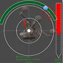
Schwalbe, M; Hansen, C; Weber, S; Lu, H
An image-guidance system for vascular malformation treatment: Concept, design and evaluation on a patient-specific phantom Journal Article
In: 2015.
@article{schwalbe_image-guidance_2015,
title = {An image-guidance system for vascular malformation treatment: Concept, design and evaluation on a patient-specific phantom},
author = {M Schwalbe and C Hansen and S Weber and H Lu},
url = {https://www.var.ovgu.de/pub/Schwalbe_2015_CARS.pdf},
year = {2015},
date = {2015-01-01},
urldate = {2015-01-01},
keywords = {},
pubstate = {published},
tppubtype = {article}
}
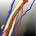
Meyer, A; Schnurr, A; Schwalbe, M; Weber, S; Hansen, C
AngioPlan: A Software Assistant to Support the Treatment of Arterio-Venous Malformations Journal Article
In: 2015.
@article{meyer_angioplan_2015,
title = {AngioPlan: A Software Assistant to Support the Treatment of Arterio-Venous Malformations},
author = {A Meyer and A Schnurr and M Schwalbe and S Weber and C Hansen},
url = {https://www.var.ovgu.de/pub/Meyer_2015_CARS.pdf},
year = {2015},
date = {2015-01-01},
urldate = {2015-01-01},
keywords = {},
pubstate = {published},
tppubtype = {article}
}
Lawonn, K; Luz, M; Preim, B; Hansen, C
Illustrative Visualization of Vascular Models for Static 2D Representations Book Section
In: Navab, N; Hornegger, J; Wells, W; Frangi, A (Ed.): Medical Image Computing and Computer-Assisted Intervention – MICCAI 2015, vol. 9350, pp. 399–406, Springer International Publishing, Cham, 2015, ISBN: 978-3-319-24570-6 978-3-319-24571-3, (Series Title: Lecture Notes in Computer Science).
@incollection{navab_illustrative_2015,
title = {Illustrative Visualization of Vascular Models for Static 2D Representations},
author = {K Lawonn and M Luz and B Preim and C Hansen},
editor = {N Navab and J Hornegger and W Wells and A Frangi},
url = {http://link.springer.com/10.1007/978-3-319-24571-3_48},
doi = {10.1007/978-3-319-24571-3_48},
isbn = {978-3-319-24570-6 978-3-319-24571-3},
year = {2015},
date = {2015-01-01},
urldate = {2015-01-01},
booktitle = {Medical Image Computing and Computer-Assisted Intervention – MICCAI 2015},
volume = {9350},
pages = {399–406},
publisher = {Springer International Publishing},
address = {Cham},
abstract = {Depth assessment of 3D vascular models visualized on 2D displays is often difficult, especially in complex workspace conditions such as in the operating room. To address these limitations, we propose a new visualization technique for 3D vascular models. Our technique is tailored to static monoscopic 2D representations, as they are often used during surgery. To improve depth assessment, we propose a combination of supporting lines, view-aligned quads, and illustrative shadows. In addition, a hatching scheme that uses different line styles depending on a distance measure is applied to encode vascular shape as well as the distance to tumors. The resulting visualization can be displayed on monoscopic 2D monitors and on 2D printouts without the requirement to use color or intensity gradients. A qualitative study with 15 participants and a quantitative study with 50 participants confirm that the proposed visualization technique significantly improves depth assessment of complex 3D vascular models.},
note = {Series Title: Lecture Notes in Computer Science},
keywords = {},
pubstate = {published},
tppubtype = {incollection}
}
Hettig, J; Mewes, A; Riabikin, O; Skalej, M; Preim, B; Hansen, C
Exploration of 3D Medical Image Data for Interventional Radiology using Myoelectric Gesture Control Journal Article
In: 2015.
@article{hettig_exploration_2015,
title = {Exploration of 3D Medical Image Data for Interventional Radiology using Myoelectric Gesture Control},
author = {J Hettig and A Mewes and O Riabikin and M Skalej and B Preim and C Hansen},
url = {https://www.var.ovgu.de/pub/2015_Hettig_MyoArmband_VCBM.pdf},
year = {2015},
date = {2015-01-01},
urldate = {2015-01-01},
abstract = {Human-computer interaction with medical images in a sterile environment is a challenging task. It is often delegated to an assistant or performed directly by the physician with an interaction device wrapped in a sterile plastic sheath. This process is time-consuming and inefficient. To address this challenge, we introduce a gesture-based interface for a medical image viewer that is completely touchlessly controlled by the Myo Gesture Control Armband (Thalmic Labs). Based on a clinical requirement analysis, we propose a minimal gesture set to support basic interaction tasks with radiological images and 3D models. We conducted two user studies and a clinical test to evaluate the interaction device and our new gesture control interface. The evaluation results prove the applicability of our approach and provide an important foundation for future research in physician-machine interaction.},
keywords = {},
pubstate = {published},
tppubtype = {article}
}
Li, M; Hansen, C; Rose, G
A robust electromagnetic tracking system for clinical applications Journal Article
In: 2015.
@article{li_robust_2015,
title = {A robust electromagnetic tracking system for clinical applications},
author = {M Li and C Hansen and G Rose},
url = {https://www.var.ovgu.de/pub/Li_2015_CURAC.pdf},
year = {2015},
date = {2015-01-01},
urldate = {2015-01-01},
abstract = {Electromagnetic tracking systems (EMTS) are utilized to track surgical instruments during navigated interventions. Compared to optical tracking technologies, the non-line-of-sight feature is the biggest advantage of EMTS. However, significant distortions due to nearby metallic objects and a small working volume are serious drawbacks preventing ETMS from being widely used. We worked on developing new methods to improve the accuracy of EMTS even in the vicinity of large metal objects while speeding up the position estimation frame rate. Therefore, we proposed a method to realize frequency deviation multiplexing (FDM) for EMTS in order to speed up the frame rate, and a quadratic-rectangular (QR) excitation method to remove the distorted voltage induced by the nearby conductive distorter. The results show that the measurement speed and robustness to nearby metal objects of our EMTS were improved by the FDM and QR method.},
keywords = {},
pubstate = {published},
tppubtype = {article}
}
2014
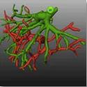
Wang, L; Hansen, C; Zidowitz, S; Hahn, H
Segmentation and Separation of Venous Vasculatures in Liver CT Images Proceedings Article
In: Proc. SPIE, pp. 90350Q–90350Q–8, San Diego, USA, 2014.
@inproceedings{wang_segmentation_2014,
title = {Segmentation and Separation of Venous Vasculatures in Liver CT Images},
author = {L Wang and C Hansen and S Zidowitz and H Hahn},
url = {https://www.var.ovgu.de/pub/Wang_SPIE2014_Manuscript.pdf},
year = {2014},
date = {2014-01-01},
urldate = {2014-01-01},
booktitle = {Proc. SPIE},
volume = {9035},
pages = {90350Q–90350Q–8},
address = {San Diego, USA},
abstract = {Computer-aided analysis of venous vasculatures including hepatic veins and portal veins is important in liver
surgery planning. The analysis normally consists of two important pre-processing tasks: segmenting both vascu-
latures and separating them from each other by assigning different labels. During the acquisition of multi-phase
CT images, both of the venous vessels are enhanced by injected contrast agent and acquired either in a common
phase or in two individual phases. The enhanced signals established by contrast agent are often not stably ac-
quired due to non-optimal acquisition time. Inadequate contrast and the presence of large lesions in oncological
patients, make the segmentation task quite challenging. To overcome these difficulties, we propose a framework
with minimal user interactions to analyze venous vasculatures in multi-phase CT images. Firstly, presented
vasculatures are automatically segmented adopting an efficient multi-scale Hessian-based vesselness filter. The
initially segmented vessel trees are then converted to a graph representation, on which a series of graph filters
are applied in post-processing steps to rule out irrelevant structures. Eventually, we develop a semi-automatic
workflow to refine the segmentation in the areas of inferior vena cava and entrance of portal veins, and to si-
multaneously separate hepatic veins from portal veins. Segmentation quality was evaluated with intensive tests
enclosing 60 CT images from both healthy liver donors and oncological patients. To quantitatively measure the
similarities between segmented and reference vessel trees, we propose three additional metrics: skeleton distance,
branch coverage, and boundary surface distance, which are dedicated to quantifying the misalignment induced
by both branching patterns and radii of two vessel trees.},
keywords = {},
pubstate = {published},
tppubtype = {inproceedings}
}
surgery planning. The analysis normally consists of two important pre-processing tasks: segmenting both vascu-
latures and separating them from each other by assigning different labels. During the acquisition of multi-phase
CT images, both of the venous vessels are enhanced by injected contrast agent and acquired either in a common
phase or in two individual phases. The enhanced signals established by contrast agent are often not stably ac-
quired due to non-optimal acquisition time. Inadequate contrast and the presence of large lesions in oncological
patients, make the segmentation task quite challenging. To overcome these difficulties, we propose a framework
with minimal user interactions to analyze venous vasculatures in multi-phase CT images. Firstly, presented
vasculatures are automatically segmented adopting an efficient multi-scale Hessian-based vesselness filter. The
initially segmented vessel trees are then converted to a graph representation, on which a series of graph filters
are applied in post-processing steps to rule out irrelevant structures. Eventually, we develop a semi-automatic
workflow to refine the segmentation in the areas of inferior vena cava and entrance of portal veins, and to si-
multaneously separate hepatic veins from portal veins. Segmentation quality was evaluated with intensive tests
enclosing 60 CT images from both healthy liver donors and oncological patients. To quantitatively measure the
similarities between segmented and reference vessel trees, we propose three additional metrics: skeleton distance,
branch coverage, and boundary surface distance, which are dedicated to quantifying the misalignment induced
by both branching patterns and radii of two vessel trees.
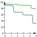
Hansen, C; Zidowitz, S; Stravrou, G; Oldhafer, K; Hahn, H
Impact of Model-based Risk Analysis for Liver Surgery Planning Journal Article
In: International Journal of Computer Assisted Radiology and Surgery, vol. 9, no. 2, pp. 473–480, 2014, ISSN: 1861-6410.
@article{hansen_impact_2014,
title = {Impact of Model-based Risk Analysis for Liver Surgery Planning},
author = {C Hansen and S Zidowitz and G Stravrou and K Oldhafer and H Hahn},
url = {https://www.var.ovgu.de/pub/Hansen_RiskAnalysis_IJCARS2013.pdf},
doi = {10.1007/s11548-013-0937-0},
issn = {1861-6410},
year = {2014},
date = {2014-01-01},
urldate = {2014-01-01},
journal = {International Journal of Computer Assisted Radiology and Surgery},
volume = {9},
number = {2},
pages = {473–480},
abstract = {Purpose A model-based risk analysis for oncologic liver surgery was described in previous work [1].
In this paper, we present an evaluation of this method.
Methods To prove whether and how the risk analysis facilitates the process of liver surgery planning,
an explorative user study with 10 liver experts was conducted. The purpose was to compare and
analyze their decision making.
Results The results of the study show that model-based risk analysis enhances the awareness of
surgical risk in the planning stage. Participants preferred smaller resection volumes and agreed more
on the safety margins width in case the risk analysis was available. In addition, time to complete the
planning task and confidence of participants was not increased when using the risk analysis.
Conclusion This work shows that the applied model-based risk analysis may influence important
planning decisions in liver surgery. It lays a basis for further clinical evaluations and points out
important fields for future research.},
keywords = {},
pubstate = {published},
tppubtype = {article}
}
In this paper, we present an evaluation of this method.
Methods To prove whether and how the risk analysis facilitates the process of liver surgery planning,
an explorative user study with 10 liver experts was conducted. The purpose was to compare and
analyze their decision making.
Results The results of the study show that model-based risk analysis enhances the awareness of
surgical risk in the planning stage. Participants preferred smaller resection volumes and agreed more
on the safety margins width in case the risk analysis was available. In addition, time to complete the
planning task and confidence of participants was not increased when using the risk analysis.
Conclusion This work shows that the applied model-based risk analysis may influence important
planning decisions in liver surgery. It lays a basis for further clinical evaluations and points out
important fields for future research.
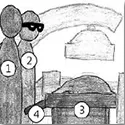
Hübler, A; Hansen, C; Beuing, O; Skalej, M; Preim, B
Workflow Analysis for Interventional Neuroradiology using Frequent Pattern Mining Journal Article
In: 2014.
@article{hubler_workflow_2014,
title = {Workflow Analysis for Interventional Neuroradiology using Frequent Pattern Mining},
author = {A Hübler and C Hansen and O Beuing and M Skalej and B Preim},
url = {https://www.var.ovgu.de/pub/Huebler_2014_CURAC.pdf},
year = {2014},
date = {2014-01-01},
urldate = {2014-01-01},
abstract = {Modern interventional imaging systems provide a wide range of functionalities and tools in order to perform complex radiological interventions. The aim of this work is to analyze and evaluate the workflow of such interventions with a focus on human-computer interaction. In our study, 25 neuroradiological CT interventions using the Siemens Artis Q imaging system were recorded on video. Based on the videos, the interaction between neuroradiologists, medical technicians and the interventional imaging system was analyzed. A frequent pattern analysis revealed that users concentrate on functionalities that are easy to reach and to understand. In addition, we observed several usability problems that were systematically collected, analyzed, and visualized using graphs and storyboards. The results of our study serve as a basis for further usability studies and workflow analyses, as well as for the design and development of future human-computer interfaces in interventional radiology.},
keywords = {},
pubstate = {published},
tppubtype = {article}
}
Hansen, C; Heckel, F; Ojdanic, D; Schenk, A; Zidowitz, S; Hahn, H
Genauigkeit und Fehlerquellen im Operationssaal am Beispiel der Leberchirurgie Book Section
In: Der digitale Operationssaal, pp. 69–87, DE GRUYTER, 2014, (Section: 6).
@incollection{hansen_genauigkeit_2014,
title = {Genauigkeit und Fehlerquellen im Operationssaal am Beispiel der Leberchirurgie},
author = {C Hansen and F Heckel and D Ojdanic and A Schenk and S Zidowitz and H Hahn},
url = {https://www.var.ovgu.de/pub/Hansen_2014_DOS.pdf},
year = {2014},
date = {2014-01-01},
urldate = {2014-01-01},
booktitle = {Der digitale Operationssaal},
pages = {69–87},
publisher = {DE GRUYTER},
note = {Section: 6},
keywords = {},
pubstate = {published},
tppubtype = {incollection}
}
Mewes, A; Adler, S; Rose, G; Hansen, C
Augmented-Reality-Mikroskop – Implementierung einer flexiblen Datenverbindung zwischen CT-Angiographieanlage und Mikroskop Proceedings Article
In: Proceedings of the Annual Meeting of the German Society of Computer- and Robot-Assisted Surgery, pp. 28–31, München, 2014.
@inproceedings{mewes_augmented-reality-mikroskop_2014,
title = {Augmented-Reality-Mikroskop - Implementierung einer flexiblen Datenverbindung zwischen CT-Angiographieanlage und Mikroskop},
author = {A Mewes and S Adler and G Rose and C Hansen},
url = {https://www.var.ovgu.de/pub/Mewes_2014_CURAC.pdf},
year = {2014},
date = {2014-01-01},
urldate = {2014-01-01},
booktitle = {Proceedings of the Annual Meeting of the German Society of Computer- and Robot-Assisted Surgery},
pages = {28–31},
address = {München},
abstract = {Während einer neurochirurgischen Intervention muss es dem Operateur möglich sein diagnostische Bilder zu aktuali-
sieren. Bei dem hier verwendeten neurochirurgischen Mikroskop können diagnostische Bilder als virtuelle Modelle und
Schnittbilder eingeblendet werden, um einen Sichtwechsel zwischen Patient und externen Bildschirmen zu vermeiden.
Um eine intraoperative Aktualisierung der Modelle zu ermöglichen, wurde im Rahmen dieser Arbeit das Mikroskop an
eine C-Arm-Angiographieanlage über einen PACS-Server angebunden. Außerdem wird ein effizienter Workflow vorge-
stellt, der u. a. die Modellgenerierung aus den DICOM-Bildern mit Hilfe der Entwicklungsumgebung MeVisLab vor-
sieht. Unter Laborbedingungen durchgeführte Experimente ergaben eine mittlere Dauer der Modellaktualisierung von
maximal 18,5 ± 2,5 min. Die Abweichung der virtuellen Überlagerung von realen Strukturen im Mikroskop liegt im We-
sentlichen im Submillimeterbereich. Das Mikroskopsystem bildet mit diesem Workflow eine solide Forschungsgrundlage
für zukünftige Projekte im Bereich der neurochirurgischen Navigation},
keywords = {},
pubstate = {published},
tppubtype = {inproceedings}
}
sieren. Bei dem hier verwendeten neurochirurgischen Mikroskop können diagnostische Bilder als virtuelle Modelle und
Schnittbilder eingeblendet werden, um einen Sichtwechsel zwischen Patient und externen Bildschirmen zu vermeiden.
Um eine intraoperative Aktualisierung der Modelle zu ermöglichen, wurde im Rahmen dieser Arbeit das Mikroskop an
eine C-Arm-Angiographieanlage über einen PACS-Server angebunden. Außerdem wird ein effizienter Workflow vorge-
stellt, der u. a. die Modellgenerierung aus den DICOM-Bildern mit Hilfe der Entwicklungsumgebung MeVisLab vor-
sieht. Unter Laborbedingungen durchgeführte Experimente ergaben eine mittlere Dauer der Modellaktualisierung von
maximal 18,5 ± 2,5 min. Die Abweichung der virtuellen Überlagerung von realen Strukturen im Mikroskop liegt im We-
sentlichen im Submillimeterbereich. Das Mikroskopsystem bildet mit diesem Workflow eine solide Forschungsgrundlage
für zukünftige Projekte im Bereich der neurochirurgischen Navigation
2013

Hansen, C; Black, D; Lange, C; Rieber, F; Lamade, W; Donati, M; Oldhafer, K; Hahn, H
Auditory Support for Resection Guidance in Navigated Liver Surgery Journal Article
In: International Journal of Medical Robotics and Computer Assisted Surgery, vol. 9, no. 1, pp. 36–43, 2013.
@article{hansen_auditory_2013,
title = {Auditory Support for Resection Guidance in Navigated Liver Surgery},
author = {C Hansen and D Black and C Lange and F Rieber and W Lamade and M Donati and K Oldhafer and H Hahn},
url = {https://onlinelibrary.wiley.com/doi/full/10.1002/rcs.1466?casa_token=9UlwLmOWMLsAAAAA%3AitwaNmk2KVOn8dleZ-MmI46wBzVHqAPOM1C4ptkiXxo4DodiHBYOWCbhGwzD9OejBcKdNEsCb2YVxkOM},
year = {2013},
date = {2013-01-01},
urldate = {2013-01-01},
journal = {International Journal of Medical Robotics and Computer Assisted Surgery},
volume = {9},
number = {1},
pages = {36–43},
abstract = {Background
An alternative mode of interaction with navigation systems for open liver surgery was requested. Surgeons who use such systems are impeded by having to constantly switch between viewing the navigation system screen and the patient during an operation.
Methods
To this end, an auditory display system for open liver surgery is introduced with support for guiding the tracked instrument towards and remaining on a predefined resection line. To evaluate the method, a clinically orientated user study with 12 surgeons was conducted.
Results
It is shown in qualitative results from the user study that the proposed auditory display is recognized as a useful addition to the current visual mode of interaction. It was revealed in a statistical analysis that participants spent less time looking on the screen (10% vs. 96%). Accuracy for resection guidance was significantly improved when using auditory display as an additional information channel (0.6 vs. 1.4 mm); however, the overall time for the resection task was shorter without auditory display (47 vs. 24 s).
Conclusions
By reducing dependence on the visual modality during resection guidance, the auditory display is well suited to become integrated in navigation systems for liver surgery. Copyright © 2012 John Wiley & Sons, Ltd.},
keywords = {},
pubstate = {published},
tppubtype = {article}
}
An alternative mode of interaction with navigation systems for open liver surgery was requested. Surgeons who use such systems are impeded by having to constantly switch between viewing the navigation system screen and the patient during an operation.
Methods
To this end, an auditory display system for open liver surgery is introduced with support for guiding the tracked instrument towards and remaining on a predefined resection line. To evaluate the method, a clinically orientated user study with 12 surgeons was conducted.
Results
It is shown in qualitative results from the user study that the proposed auditory display is recognized as a useful addition to the current visual mode of interaction. It was revealed in a statistical analysis that participants spent less time looking on the screen (10% vs. 96%). Accuracy for resection guidance was significantly improved when using auditory display as an additional information channel (0.6 vs. 1.4 mm); however, the overall time for the resection task was shorter without auditory display (47 vs. 24 s).
Conclusions
By reducing dependence on the visual modality during resection guidance, the auditory display is well suited to become integrated in navigation systems for liver surgery. Copyright © 2012 John Wiley & Sons, Ltd.
Wang, L; Hansen, C; Zidowitz, S; Hahn, H
Interactive Segmentation of Vascular Structures in CT Images for Liver Surgery Planning Proceedings Article
In: Proceedings of the Annual Meeting of the German Society of Computer- and Robot-Assisted Surgery (CURAC), pp. 98–102, Univ. Prof. Dr. Mag. Wolfgang Freysinger, Innsbruck , Österreich, 2013.
@inproceedings{wang_interactive_2013,
title = {Interactive Segmentation of Vascular Structures in CT Images for Liver Surgery Planning},
author = {L Wang and C Hansen and S Zidowitz and H Hahn},
url = {https://www.var.ovgu.de/pub/Wang_CURAC2013_Manuscript.pdf},
year = {2013},
date = {2013-01-01},
urldate = {2013-01-01},
booktitle = {Proceedings of the Annual Meeting of the German Society of Computer- and Robot-Assisted Surgery (CURAC)},
pages = {98–102},
publisher = {Univ. Prof. Dr. Mag. Wolfgang Freysinger},
address = {Innsbruck , Österreich},
abstract = {Vasculature analysis based on CT images is indispensable in liver surgical planning and diagnosis of liver disease. We
propose a novel framework and efficient workflow with minimal user interactions to analyze liver vasculature in multi-
phase CT images. To ensure segmentation quality and efficiency, a set of semi-automatic algorithms are applied to initially
segment different vascular structures in different phase. A fully automatic vessel separation procedure runs parallel to
separately connected hepatic and portal veins. In addition, an interactive editing method is integrated into the framework
to refine the segmentation of each individual structure. Quantitative evaluations of segmented vessels conducted for 60
test data sets are demonstrated.},
keywords = {},
pubstate = {published},
tppubtype = {inproceedings}
}
propose a novel framework and efficient workflow with minimal user interactions to analyze liver vasculature in multi-
phase CT images. To ensure segmentation quality and efficiency, a set of semi-automatic algorithms are applied to initially
segment different vascular structures in different phase. A fully automatic vessel separation procedure runs parallel to
separately connected hepatic and portal veins. In addition, an interactive editing method is integrated into the framework
to refine the segmentation of each individual structure. Quantitative evaluations of segmented vessels conducted for 60
test data sets are demonstrated.
Black, D; Issawi, J Al; Rieder, C; Hansen, C; Hahn, H
Auditory Support for Navigated Radiofrequency Ablation Proceedings Article
In: Proceedings of the Annual Meeting of the German Society of Computer- and Robot-Assisted Surgery (CURAC), pp. 30–33, Univ. Prof. Dr. Mag. Wolfgang Freysinger, Innsbruck , Österreich, 2013.
@inproceedings{black_auditory_2013,
title = {Auditory Support for Navigated Radiofrequency Ablation},
author = {D Black and J Al Issawi and C Rieder and C Hansen and H Hahn},
url = {https://www.var.ovgu.de/pub/curac2013_dblack_Auditory_support_for_Navigated_Radiofrequency_Ablation.pdf},
year = {2013},
date = {2013-01-01},
urldate = {2013-01-01},
booktitle = {Proceedings of the Annual Meeting of the German Society of Computer- and Robot-Assisted Surgery (CURAC)},
pages = {30–33},
publisher = {Univ. Prof. Dr. Mag. Wolfgang Freysinger},
address = {Innsbruck , Österreich},
abstract = {Radiofrequency ablation is applied to treat a lesion using a needle inserted into the patient, which delivers local radiof-
requency energy. Guided surgical methods allow surgeons to view the placement of the needle in relation to the patient
to aid in guiding the tip of the needle to the target point. Unfortunately, such methods require that surgeons remove at-
tention from the patient in order to receive guidance information from a screen. We introduce a novel method to align
and insert an ablation needle using auditory display, allowing the surgeon to retain attention on the patient. First eval-
uation results show that novice users can successfully guide a needle towards a target point using primarily auditory
display. We hypothesize that successful auditory display will lead to increased attention on the patient and reduce un-
necessary operator head and neck movements.},
keywords = {},
pubstate = {published},
tppubtype = {inproceedings}
}
requency energy. Guided surgical methods allow surgeons to view the placement of the needle in relation to the patient
to aid in guiding the tip of the needle to the target point. Unfortunately, such methods require that surgeons remove at-
tention from the patient in order to receive guidance information from a screen. We introduce a novel method to align
and insert an ablation needle using auditory display, allowing the surgeon to retain attention on the patient. First eval-
uation results show that novice users can successfully guide a needle towards a target point using primarily auditory
display. We hypothesize that successful auditory display will lead to increased attention on the patient and reduce un-
necessary operator head and neck movements.
Hansen, C; Zidowitz, S; Ritter, F; Lange, C; Oldhafer, K; Hahn, HK
Risk maps for Liver Surgery Journal Article
In: International Journal of Computer Assisted Radiology and Surgery, vol. 8, no. 3, pp. 419–428, 2013.
@article{hansen_risk_2013,
title = {Risk maps for Liver Surgery},
author = {C Hansen and S Zidowitz and F Ritter and C Lange and K Oldhafer and HK Hahn},
url = {https://link.springer.com/article/10.1007/s11548-012-0790-6},
doi = {10.1007/s11548-012-0790-6},
year = {2013},
date = {2013-01-01},
urldate = {2013-01-01},
journal = {International Journal of Computer Assisted Radiology and Surgery},
volume = {8},
number = {3},
pages = {419–428},
keywords = {},
pubstate = {published},
tppubtype = {article}
}
Mönch, J; Mühler, K; Hansen, C; Oldhafer, K; Stavrou, G; Hillert, C; Logge, C; Preim, B
The LiverSurgeryTrainer: training of Computer-based Planning in Liver Resection Surgery Journal Article
In: International Journal of Computer Assisted Radiology and Surgery, vol. 8, no. 5, pp. 809–818, 2013.
@article{monch_liversurgerytrainer_2013,
title = {The LiverSurgeryTrainer: training of Computer-based Planning in Liver Resection Surgery},
author = {J Mönch and K Mühler and C Hansen and K Oldhafer and G Stavrou and C Hillert and C Logge and B Preim},
url = {https://link.springer.com/article/10.1007/s11548-013-0812-z
https://pubmed.ncbi.nlm.nih.gov/23337995/, PubMed},
doi = {10.1007/s11548-013-0812-z},
year = {2013},
date = {2013-01-01},
urldate = {2013-01-01},
journal = {International Journal of Computer Assisted Radiology and Surgery},
volume = {8},
number = {5},
pages = {809–818},
abstract = {Purpose The training of liver surgeons depends on local conditions such as the specialization of the clinic, the spectrum of cases, and the instructing surgeons. We present the LiverSurgeryTrainer a software application to support the training of prospective surgeons in preoperative decision making.
Methods The LiverSurgeryTrainer visualizes radiological images, volumetric information, and interactive 3D models of patients’ liver anatomy. In addition, it provides special interaction techniques and tools to perform individual resections on the training data. To assess the correctness of decisions made by the learner, comments and decisions from experienced liver surgeons are provided for each case. To complete the case, additional material concerning the actual surgery (e.g., videos, reports) is presented. The application workflow is derived from a scenario-based design process and is based on an instructional design model.
Results The LiverSurgeryTrainer was evaluated in several steps. A formative usability evaluation identified workflow and user interface flaws that were resolved in further development process. A summative evaluation shows the improvement of the LiverSurgeryTrainer in nearly all analyzed aspects. First results of a learning success evaluation show that learners experience a learning effect.
Conclusion Our training system allows surgeons to train procedures and interaction techniques for computer-based planning of liver interventions. The evaluations showed acceptance and usability.},
keywords = {},
pubstate = {published},
tppubtype = {article}
}
Methods The LiverSurgeryTrainer visualizes radiological images, volumetric information, and interactive 3D models of patients’ liver anatomy. In addition, it provides special interaction techniques and tools to perform individual resections on the training data. To assess the correctness of decisions made by the learner, comments and decisions from experienced liver surgeons are provided for each case. To complete the case, additional material concerning the actual surgery (e.g., videos, reports) is presented. The application workflow is derived from a scenario-based design process and is based on an instructional design model.
Results The LiverSurgeryTrainer was evaluated in several steps. A formative usability evaluation identified workflow and user interface flaws that were resolved in further development process. A summative evaluation shows the improvement of the LiverSurgeryTrainer in nearly all analyzed aspects. First results of a learning success evaluation show that learners experience a learning effect.
Conclusion Our training system allows surgeons to train procedures and interaction techniques for computer-based planning of liver interventions. The evaluations showed acceptance and usability.
2012
Hansen, C; Zidowitz, S; Preim, B; Oldhafer, K; Hahn, H
Impact of Model-based Risk Analyses for Liver Surgery Planning Proceedings Article
In: Düsseldorf, 2012.
@inproceedings{hansen_impact_2012,
title = {Impact of Model-based Risk Analyses for Liver Surgery Planning},
author = {C Hansen and S Zidowitz and B Preim and K Oldhafer and H Hahn},
url = {https://link.springer.com/article/10.1007/s11548-013-0937-0},
year = {2012},
date = {2012-01-01},
urldate = {2012-01-01},
address = {Düsseldorf},
series = {Proceedings of the Annual Meeting of the German Society of Computer- and Robot-Assisted Surgery},
abstract = {Purpose
A model-based risk analysis for oncologic liver surgery was described in previous work (Preim et al. in Proceedings of international symposium on computer assisted radiology and surgery (CARS), Elsevier, Amsterdam, pp. 353–358, 2002; Hansen et al. Int I Comput Assist Radiol Surg 4(5):469–474, 2009). In this paper, we present an evaluation of this method.
Methods
To prove whether and how the risk analysis facilitates the process of liver surgery planning, an explorative user study with 10 liver experts was conducted. The purpose was to compare and analyze their decision-making.
Results
The results of the study show that model-based risk analysis enhances the awareness of surgical risk in the planning stage. Participants preferred smaller resection volumes and agreed more on the safety margins’ width in case the risk analysis was available. In addition, time to complete the planning task and confidence of participants were not increased when using the risk analysis.
Conclusion
This work shows that the applied model-based risk analysis may influence important planning decisions in liver surgery. It lays a basis for further clinical evaluations and points out important fields for future research.},
keywords = {},
pubstate = {published},
tppubtype = {inproceedings}
}
A model-based risk analysis for oncologic liver surgery was described in previous work (Preim et al. in Proceedings of international symposium on computer assisted radiology and surgery (CARS), Elsevier, Amsterdam, pp. 353–358, 2002; Hansen et al. Int I Comput Assist Radiol Surg 4(5):469–474, 2009). In this paper, we present an evaluation of this method.
Methods
To prove whether and how the risk analysis facilitates the process of liver surgery planning, an explorative user study with 10 liver experts was conducted. The purpose was to compare and analyze their decision-making.
Results
The results of the study show that model-based risk analysis enhances the awareness of surgical risk in the planning stage. Participants preferred smaller resection volumes and agreed more on the safety margins’ width in case the risk analysis was available. In addition, time to complete the planning task and confidence of participants were not increased when using the risk analysis.
Conclusion
This work shows that the applied model-based risk analysis may influence important planning decisions in liver surgery. It lays a basis for further clinical evaluations and points out important fields for future research.
Hansen, C; Zidowitz, S; Ritter, F; Lange, C; Oldhafer, KJ; Hahn, HK
Risk Maps for Liver Surgery Proceedings Article
In: International Journal of Computer Assisted Radiology and Surgery, pp. 144–145, Pisa, Italien, 2012.
@inproceedings{hansen_risk_2012,
title = {Risk Maps for Liver Surgery},
author = {C Hansen and S Zidowitz and F Ritter and C Lange and KJ Oldhafer and HK Hahn},
year = {2012},
date = {2012-01-01},
urldate = {2012-01-01},
booktitle = {International Journal of Computer Assisted Radiology and Surgery},
pages = {144–145},
address = {Pisa, Italien},
series = {Proceedings of International Congress for Computer-Assisted Radiology and Surgery (CARS)},
keywords = {},
pubstate = {published},
tppubtype = {inproceedings}
}
2010
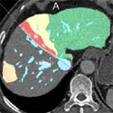
Demedts, D; Schenk, A; Hansen, C; Peitgen, H
Evaluation of Resection Proposals for Liver Surgery Planning Proceedings Article
In: Proc. of the 9th Annual Meeting of the German Society of Computer- and Robot-Assisted Surgery, pp. 13–16, 2010.
@inproceedings{demedts_evaluation_2010,
title = {Evaluation of Resection Proposals for Liver Surgery Planning},
author = {D Demedts and A Schenk and C Hansen and H Peitgen},
url = {https://ceur-ws.org/Vol-1475/Proceedings_CURAC_2010_Paper_3.pdf},
year = {2010},
date = {2010-01-01},
urldate = {2010-01-01},
booktitle = {Proc. of the 9th Annual Meeting of the German Society of Computer- and Robot-Assisted Surgery},
pages = {13–16},
abstract = {Modern software for surgery planning allows for definition of virtual resections within the liver. Thus, surgeons can si-
mulate different resection strategies and assess the associated surgical risk preoperatively. Until now, it was impossible
to measure the quality of different resection plans objectively. The choice for the optimal resection strategy was based
on subjective judgment acquired by other examinations and subsequent risk analyses. We present a fast method for
quality assessment of resection proposals with respect to surgical risk factors such as safety margin, remnant volume,
remnant perfusion, surface curvature, and resection area. Our new method has been integrated into planning software
used in the daily routine. The results from a preliminary user study confirm that the interactive quality feedback is
beneficial for precise liver surgery planning.},
keywords = {},
pubstate = {published},
tppubtype = {inproceedings}
}
mulate different resection strategies and assess the associated surgical risk preoperatively. Until now, it was impossible
to measure the quality of different resection plans objectively. The choice for the optimal resection strategy was based
on subjective judgment acquired by other examinations and subsequent risk analyses. We present a fast method for
quality assessment of resection proposals with respect to surgical risk factors such as safety margin, remnant volume,
remnant perfusion, surface curvature, and resection area. Our new method has been integrated into planning software
used in the daily routine. The results from a preliminary user study confirm that the interactive quality feedback is
beneficial for precise liver surgery planning.
Hansen, C; Zidowitz, S; Schenk, A; Oldhafer, K; Lang, H; Peitgen, H
Risk Maps for Navigation in Liver Surgery Proceedings Article
In: Proceedings of SPIE Medical Imaging, pp. 762528–1–8, San Diego, USA, 2010.
@inproceedings{hanse_risk_2010,
title = {Risk Maps for Navigation in Liver Surgery},
author = {C Hansen and S Zidowitz and A Schenk and K Oldhafer and H Lang and H Peitgen},
url = {https://www.spiedigitallibrary.org/conference-proceedings-of-spie/7625/762528/Risk-maps-for-navigation-in-liver-surgery/10.1117/12.843493.short},
year = {2010},
date = {2010-01-01},
urldate = {2010-01-01},
booktitle = {Proceedings of SPIE Medical Imaging},
pages = {762528–1–8},
address = {San Diego, USA},
abstract = {The optimal transfer of preoperative planning data and risk evaluations to the operative site is challenging. A common practice is to use preoperative 3D planning models as a printout or as a presentation on a display. One important aspect is that these models were not developed to provide information in complex workspaces like the operating room. Our aim is to reduce the visual complexity of 3D planning models by mapping surgically relevant information onto a risk map. Therefore, we present methods for the identification and classification of critical anatomical structures in the proximity of a preoperatively planned resection surface. Shadow-like distance indicators are introduced to encode the distance from the resection surface to these critical structures on the risk map. In addition, contour lines are used to accentuate shape and spatial depth. The resulting visualization is clear and intuitive, allowing for a fast mental mapping of the current resection surface to the risk map. Preliminary evaluations by liver surgeons indicate that damage to risk structures may be prevented and patient safety may be enhanced using the proposed methods.},
keywords = {},
pubstate = {published},
tppubtype = {inproceedings}
}
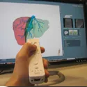
Hansen, C; Wieferich, J; Ritter, F; Rieder, C; Peitgen, H
Illustrative Visualization of 3D Planning Models for Augmented Reality in Liver Surgery Journal Article
In: International Journal of Computer Assisted Radiology and Surgery, vol. 5, no. 2, pp. 133–141, 2010.
@article{hansen_illustrative_2010,
title = {Illustrative Visualization of 3D Planning Models for Augmented Reality in Liver Surgery},
author = {C Hansen and J Wieferich and F Ritter and C Rieder and H Peitgen},
url = {https://link.springer.com/article/10.1007/s11548-009-0365-3},
doi = {10.1007/s11548-009-0365-3},
year = {2010},
date = {2010-01-01},
urldate = {2010-01-01},
journal = {International Journal of Computer Assisted Radiology and Surgery},
volume = {5},
number = {2},
pages = {133–141},
abstract = {Purpose
Augmented reality (AR) obtains increasing acceptance in the operating room. However, a meaningful augmentation of the surgical view with a 3D visualization of planning data which allows reliable comparisons of distances and spatial relations is still an open request.
Methods
We introduce methods for intraoperative visualization of 3D planning models which extend illustrative rendering and AR techniques. We aim to reduce visual complexity of 3D planning models and accentuate spatial relations between relevant objects. The main contribution of our work is an advanced silhouette algorithm for 3D planning models (distance-encoding silhouettes) combined with procedural textures (distance-encoding surfaces). In addition, we present a method for illustrative visualization of resection surfaces.
Results
The developed algorithms have been embedded into a clinical prototype that has been evaluated in the operating room. To verify the expressiveness of our illustration methods, we performed a user study under controlled conditions. The study revealed a clear advantage in distance assessment with the proposed illustrative approach in comparison to classical rendering techniques.
Conclusion
The presented illustration methods are beneficial for distance assessment in surgical AR. To increase the safety of interventions with the proposed approach, the reduction of inaccuracies in tracking and registration is a subject of our current research.},
keywords = {},
pubstate = {published},
tppubtype = {article}
}
Augmented reality (AR) obtains increasing acceptance in the operating room. However, a meaningful augmentation of the surgical view with a 3D visualization of planning data which allows reliable comparisons of distances and spatial relations is still an open request.
Methods
We introduce methods for intraoperative visualization of 3D planning models which extend illustrative rendering and AR techniques. We aim to reduce visual complexity of 3D planning models and accentuate spatial relations between relevant objects. The main contribution of our work is an advanced silhouette algorithm for 3D planning models (distance-encoding silhouettes) combined with procedural textures (distance-encoding surfaces). In addition, we present a method for illustrative visualization of resection surfaces.
Results
The developed algorithms have been embedded into a clinical prototype that has been evaluated in the operating room. To verify the expressiveness of our illustration methods, we performed a user study under controlled conditions. The study revealed a clear advantage in distance assessment with the proposed illustrative approach in comparison to classical rendering techniques.
Conclusion
The presented illustration methods are beneficial for distance assessment in surgical AR. To increase the safety of interventions with the proposed approach, the reduction of inaccuracies in tracking and registration is a subject of our current research.
Black, D; Hansen, C; Loviscach, J; Peitgen, H
Auditory Support for Image-Guided Liver Surgery Proceedings Article
In: International Journal of Computer Assisted Radiology and Surgery, pp. 197–188, Genf, Schweiz, 2010.
@inproceedings{black_auditory_2010,
title = {Auditory Support for Image-Guided Liver Surgery},
author = {D Black and C Hansen and J Loviscach and H Peitgen},
url = {https://www.researchgate.net/profile/David-Black-14/publication/313655446_Auditory_Support_for_Image-Guided_Liver_Surgery/links/58a1a699a6fdccf5e97089fc/Auditory-Support-for-Image-Guided-Liver-Surgery.pdf},
year = {2010},
date = {2010-01-01},
urldate = {2010-01-01},
booktitle = {International Journal of Computer Assisted Radiology and Surgery},
pages = {197–188},
address = {Genf, Schweiz},
series = {Proceedings of International Congress for Computer-Assisted Radiology and Surgery (CARS)},
keywords = {},
pubstate = {published},
tppubtype = {inproceedings}
}
Hansen, C; Lindow, B; Zidowitz, S; Schenk, A; Peitgen, H
Towards Automatic Generation of Resection Surfaces for Liver Surgery Planning Proceedings Article
In: International Journal of Computer Assisted Radiology and Surgery, pp. 119–120, Genf, Schweiz, 2010.
@inproceedings{hansen_towards_2010,
title = {Towards Automatic Generation of Resection Surfaces for Liver Surgery Planning},
author = {C Hansen and B Lindow and S Zidowitz and A Schenk and H Peitgen},
year = {2010},
date = {2010-01-01},
urldate = {2010-01-01},
booktitle = {International Journal of Computer Assisted Radiology and Surgery},
pages = {119–120},
address = {Genf, Schweiz},
series = {Proceedings of International Congress for Computer-Assisted Radiology and Surgery (CARS)},
keywords = {},
pubstate = {published},
tppubtype = {inproceedings}
}
2009
Ritter, F; Hansen, C; Wilkens, K; Köhn, A; Peitgen, H
User Interfaces for Direct Interaction with 3D Planning Data in the Operation Room Journal Article
In: i-com – Journal of Interactive Media, vol. 8, no. 1, pp. 24–31, 2009.
@article{ritter_user_2009,
title = {User Interfaces for Direct Interaction with 3D Planning Data in the Operation Room},
author = {F Ritter and C Hansen and K Wilkens and A Köhn and H Peitgen},
year = {2009},
date = {2009-01-01},
urldate = {2009-01-01},
journal = {i-com - Journal of Interactive Media},
volume = {8},
number = {1},
pages = {24–31},
keywords = {},
pubstate = {published},
tppubtype = {article}
}
Hansen, C; Zidowitz, S; Hindennach, M; Schenk, A; Hahn, H; Peitgen, H
Interactive Determination of Robust Safety Margins for Oncologic Liver Surgery Proceedings Article
In: International Journal of Computer Assisted Radiology and Surgery, pp. 469–474, Berlin, 2009.
@inproceedings{hansen_interactive_2009,
title = {Interactive Determination of Robust Safety Margins for Oncologic Liver Surgery},
author = {C Hansen and S Zidowitz and M Hindennach and A Schenk and H Hahn and H Peitgen},
url = {https://link.springer.com/article/10.1007/s11548-009-0359-1},
year = {2009},
date = {2009-01-01},
urldate = {2009-01-01},
booktitle = {International Journal of Computer Assisted Radiology and Surgery},
pages = {469–474},
address = {Berlin},
series = {Proceedings of International Congress for Computer-Assisted Radiology and Surgery (CARS)},
abstract = {Objective
Complex oncologic interventions in the liver require an extensive and careful preoperative analysis. Particularly the achievement of an optimal safety margin around tumors remains a difficult task for surgeons.
Methods
We present new methods for evaluating different safety margins and their effect on the associated interruption of vascular supply or drainage. The characteristic of vascular risk distributions can be evaluated in real-time by exploiting precomputed safety maps that provide a volume curve for each vascular system. By applying fast visualization methods in 3D it is possible to assist the surgeon in the determination of a tumor-free safety margin while preserving sufficient vital hepatic parenchyma. The combination of risk analysis from different vascular systems and their sensitivity is considered.
Results
We provide physicians with a novel computer-aided planning tool that allows for interactive determination of safety margins in real-time. The planning tool integrates smoothly into the preoperative workflow. Preliminary evaluations confirm that the width of safety margins can be determined more precisely, which may affect the proposed resection strategy.
Conclusion
Our new methods provide interactive feedback and support for decision making during the preoperative planning stage and thus might potentially improve the outcome of surgical interventions.},
keywords = {},
pubstate = {published},
tppubtype = {inproceedings}
}
Complex oncologic interventions in the liver require an extensive and careful preoperative analysis. Particularly the achievement of an optimal safety margin around tumors remains a difficult task for surgeons.
Methods
We present new methods for evaluating different safety margins and their effect on the associated interruption of vascular supply or drainage. The characteristic of vascular risk distributions can be evaluated in real-time by exploiting precomputed safety maps that provide a volume curve for each vascular system. By applying fast visualization methods in 3D it is possible to assist the surgeon in the determination of a tumor-free safety margin while preserving sufficient vital hepatic parenchyma. The combination of risk analysis from different vascular systems and their sensitivity is considered.
Results
We provide physicians with a novel computer-aided planning tool that allows for interactive determination of safety margins in real-time. The planning tool integrates smoothly into the preoperative workflow. Preliminary evaluations confirm that the width of safety margins can be determined more precisely, which may affect the proposed resection strategy.
Conclusion
Our new methods provide interactive feedback and support for decision making during the preoperative planning stage and thus might potentially improve the outcome of surgical interventions.
2008
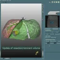
Hansen, C; Schlichting, S; Zidowitz, S; Kleemann, M; Peitgen, H
Intraoperative Adaptation of Planning Data for Oncologic Liver Surgery Proceedings Article
In: pp. 36–45, New York, USA, 2008.
@inproceedings{hansen_intraoperative_2008-1,
title = {Intraoperative Adaptation of Planning Data for Oncologic Liver Surgery},
author = {C Hansen and S Schlichting and S Zidowitz and M Kleemann and H Peitgen},
url = {https://publica.fraunhofer.de/bitstreams/c9f194d5-3de7-4302-a8e2-f6de0c73c02d/download#page=41},
year = {2008},
date = {2008-01-01},
urldate = {2008-01-01},
pages = {36–45},
address = {New York, USA},
series = {Proceedings of MICCAI Workshop on Image Guidance and Computer Assistance for Soft-Tissue Interventions},
abstract = {Recent surgical planning software provides valuable tools for
evaluating different resection strategies preoperatively. Risk analyses and
virtual resections may be carried out before surgery in order to assess the
resection feasibility with respect to tumors and risk structures. However,
in oncologic liver surgery, additional tumors that were not seen in the
preoperative images are often found during the intervention using intra-
operative ultrasound. Due to such findings, the resection strategy must
be updated or completely revised. We have developed new methods for
intraoperative adaptation of planning data using an ultrasound-based
navigation system. Fast adaptation of planning data and the communi-
cation with the navigation system is supported by our system, the In-
traoperative Planning Assistant. The system has been evaluated in the
operating room during laparoscopic liver interventions on pigs. For the
first time, surgeons are provided with a tool for intraoperative adapta-
tion of planning data that offers crucial decision support, is easy to use
and integrates smoothly into the clinical workflow.},
keywords = {},
pubstate = {published},
tppubtype = {inproceedings}
}
evaluating different resection strategies preoperatively. Risk analyses and
virtual resections may be carried out before surgery in order to assess the
resection feasibility with respect to tumors and risk structures. However,
in oncologic liver surgery, additional tumors that were not seen in the
preoperative images are often found during the intervention using intra-
operative ultrasound. Due to such findings, the resection strategy must
be updated or completely revised. We have developed new methods for
intraoperative adaptation of planning data using an ultrasound-based
navigation system. Fast adaptation of planning data and the communi-
cation with the navigation system is supported by our system, the In-
traoperative Planning Assistant. The system has been evaluated in the
operating room during laparoscopic liver interventions on pigs. For the
first time, surgeons are provided with a tool for intraoperative adapta-
tion of planning data that offers crucial decision support, is easy to use
and integrates smoothly into the clinical workflow.
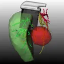
Mühler, K; Hansen, C; Neugebauer, M; Preim, B
Automatische Kamerapositionierung für Intraoperative Visualisierungen in der Onkologischen Leberchirurgie Proceedings Article
In: Bildverarbeitung für die Medizin 2008, pp. 143–147, Berlin, 2008.
@inproceedings{muhler_automatische_2008,
title = {Automatische Kamerapositionierung für Intraoperative Visualisierungen in der Onkologischen Leberchirurgie},
author = {K Mühler and C Hansen and M Neugebauer and B Preim},
url = {https://link.springer.com/chapter/10.1007/978-3-540-78640-5_29},
year = {2008},
date = {2008-01-01},
urldate = {2008-01-01},
booktitle = {Bildverarbeitung für die Medizin 2008},
pages = {143–147},
address = {Berlin},
series = {Proceedings of Bildverarbeitung für die Medizin},
abstract = {In diesem Beitrag wird ein Verfahren vorgestellt, mit
dessen Hilfe automatisch gute Blickpunkte auf dreidimensionale Pla-
nungsmodelle f¨ur die Leberchirurgie berechnet werden k¨onnen. Das Ver-
fahren passt die Position der virtuellen Kamera w¨ahrend einer Operation
dynamisch an, insbesondere im Falle einer Aktualisierung von onkologi-
schen Planungsdaten durch neue intra-operative Befunde.},
keywords = {},
pubstate = {published},
tppubtype = {inproceedings}
}
dessen Hilfe automatisch gute Blickpunkte auf dreidimensionale Pla-
nungsmodelle f¨ur die Leberchirurgie berechnet werden k¨onnen. Das Ver-
fahren passt die Position der virtuellen Kamera w¨ahrend einer Operation
dynamisch an, insbesondere im Falle einer Aktualisierung von onkologi-
schen Planungsdaten durch neue intra-operative Befunde.
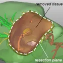
Hansen, C; Köhn, A; Schlichting, S; Weiler, F; Konrad, O; Zidowitz, S; Peitgen, H
Intraoperative Modification of Resection Plans for Liver Surgery Proceedings Article
In: International Journal of Computer Assisted Radiology and Surgery, pp. 291–297, Barcelona, Spanien, 2008.
@inproceedings{hansen_intraoperative_2008,
title = {Intraoperative Modification of Resection Plans for Liver Surgery},
author = {C Hansen and A Köhn and S Schlichting and F Weiler and O Konrad and S Zidowitz and H Peitgen},
url = {https://link.springer.com/article/10.1007/s11548-008-0161-5},
year = {2008},
date = {2008-01-01},
urldate = {2008-01-01},
booktitle = {International Journal of Computer Assisted Radiology and Surgery},
pages = {291–297},
address = {Barcelona, Spanien},
series = {Proceedings of International Congress for Computer-Assisted Radiology and Surgery (CARS)},
abstract = {Objective
Recent surgical planning software provides valuable tools for evaluating different resection strategies preoperatively. With such virtual resections, predictions and quantitative analyses may be carried out to assess the resection feasibility with respect to tumors and risk structures. In oncologic liver surgery, additional tumors that were not seen in the preoperative images are often found during the intervention using intraoperative ultrasound (IOUS). Due to such findings, the resection strategy must be updated or completely revised.
Materials and methods
Therefore, we have developed methods for the intraoperative modification of resection plans. The probe of an ultrasound-based navigation system and alternatively the pointing device Wiimote are proposed as intraoperative interaction devices. Fast adaptation of planning information and the communication with both interaction devices is supported by our system, the Intraoperative Planning Assistant (IPA). The IPA has been evaluated in the operation room (OR) during laparoscopic liver interventions on pigs.
Results
Our preliminary results confirm that intraoperative modifications of resection plans are both feasible and beneficial for liver surgery. After the intraoperative modification task, updated remaining liver volume and resection volume were displayed and quantified within 10 s.
Conclusion
For the first time, surgeons are provided with a system for intraoperative modification of resection plans that offers a crucial decision support, is easy to use and integrates smoothly into the clinical workflow. The new system provides major support for decision making in the OR and thus improves the safety of surgical interventions.},
keywords = {},
pubstate = {published},
tppubtype = {inproceedings}
}
Recent surgical planning software provides valuable tools for evaluating different resection strategies preoperatively. With such virtual resections, predictions and quantitative analyses may be carried out to assess the resection feasibility with respect to tumors and risk structures. In oncologic liver surgery, additional tumors that were not seen in the preoperative images are often found during the intervention using intraoperative ultrasound (IOUS). Due to such findings, the resection strategy must be updated or completely revised.
Materials and methods
Therefore, we have developed methods for the intraoperative modification of resection plans. The probe of an ultrasound-based navigation system and alternatively the pointing device Wiimote are proposed as intraoperative interaction devices. Fast adaptation of planning information and the communication with both interaction devices is supported by our system, the Intraoperative Planning Assistant (IPA). The IPA has been evaluated in the operation room (OR) during laparoscopic liver interventions on pigs.
Results
Our preliminary results confirm that intraoperative modifications of resection plans are both feasible and beneficial for liver surgery. After the intraoperative modification task, updated remaining liver volume and resection volume were displayed and quantified within 10 s.
Conclusion
For the first time, surgeons are provided with a system for intraoperative modification of resection plans that offers a crucial decision support, is easy to use and integrates smoothly into the clinical workflow. The new system provides major support for decision making in the OR and thus improves the safety of surgical interventions.
Hansen, C; Zidowitz, S; Schenk, A; Oldhafer, K; Lang, H; Peitgen, H
Intraoperative Adaptation and Visualization of Preoperative Risk Analyses for Oncologic Liver Surgery Proceedings Article
In: Proceedings of SPIE Medical Imaging, pp. 691809–1–10, San Diego, USA, 2008.
@inproceedings{hansen_intraoperative_2008-2,
title = {Intraoperative Adaptation and Visualization of Preoperative Risk Analyses for Oncologic Liver Surgery},
author = {C Hansen and S Zidowitz and A Schenk and K Oldhafer and H Lang and H Peitgen},
url = {https://www.spiedigitallibrary.org/conference-proceedings-of-spie/6918/691809/Intraoperative-adaptation-and-visualization-of-preoperative-risk-analyses-for-oncologic/10.1117/12.770243.short},
year = {2008},
date = {2008-01-01},
urldate = {2008-01-01},
booktitle = {Proceedings of SPIE Medical Imaging},
pages = {691809–1–10},
address = {San Diego, USA},
series = {Proceedings of SPIE Medical Imaging},
abstract = {Tumor resections from the liver are complex surgical interventions. With recent planning software, risk analyses based on individual liver anatomy can be carried out preoperatively. However, additional tumors within the liver are frequently detected during oncological interventions using intraoperative ultrasound. These tumors are not visible in preoperative data and their existence may require changes to the resection strategy. We propose a novel method that allows an intraoperative risk analysis adaptation by merging newly detected tumors with a preoperative risk analysis. To determine the exact positions and sizes of these tumors we make use of a navigated ultrasound-system. A fast communication protocol enables our application to exchange crucial data with this navigation system during an intervention. A further motivation for our work is to improve the visual presentation of a moving ultrasound plane within a complex 3D planning model including vascular systems, tumors, and organ surfaces. In case the ultrasound plane is located inside the liver, occlusion of the ultrasound plane by the planning model is an inevitable problem for the applied visualization technique. Our system allows the surgeon to focus on the ultrasound image while perceiving context-relevant planning information. To improve orientation ability and distance perception, we include additional depth cues by applying new illustrative visualization algorithms. Preliminary evaluations confirm that in case of intraoperatively detected tumors a risk analysis adaptation is beneficial for precise liver surgery. Our new GPU-based visualization approach provides the surgeon with a simultaneous visualization of planning models and navigated 2D ultrasound data while minimizing occlusion problems.},
keywords = {},
pubstate = {published},
tppubtype = {inproceedings}
}
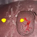
Hansen, C; Wieferich, J; Ritter, F; Rieder, C; Peitgen, H
Illustrative Visualization of 3D Planning Models for Augmented Reality in Liver Surgery Proceedings Article
In: CURAC 2008 – Tagungsband, pp. 1–4, Leipzig, 2008.
@inproceedings{hansen_illustrative_2008,
title = {Illustrative Visualization of 3D Planning Models for Augmented Reality in Liver Surgery},
author = {C Hansen and J Wieferich and F Ritter and C Rieder and H Peitgen},
url = {https://link.springer.com/article/10.1007/s11548-009-0365-3},
year = {2008},
date = {2008-01-01},
urldate = {2008-01-01},
booktitle = {CURAC 2008 - Tagungsband},
pages = {1–4},
address = {Leipzig},
series = {Proceedings of the Annual Meeting of the German Society of Computer- and Robot-Assisted Surgery},
abstract = {Purpose
Augmented reality (AR) obtains increasing acceptance in the operating room. However, a meaningful augmentation of the surgical view with a 3D visualization of planning data which allows reliable comparisons of distances and spatial relations is still an open request.
Methods
We introduce methods for intraoperative visualization of 3D planning models which extend illustrative rendering and AR techniques. We aim to reduce visual complexity of 3D planning models and accentuate spatial relations between relevant objects. The main contribution of our work is an advanced silhouette algorithm for 3D planning models (distance-encoding silhouettes) combined with procedural textures (distance-encoding surfaces). In addition, we present a method for illustrative visualization of resection surfaces.
Results
The developed algorithms have been embedded into a clinical prototype that has been evaluated in the operating room. To verify the expressiveness of our illustration methods, we performed a user study under controlled conditions. The study revealed a clear advantage in distance assessment with the proposed illustrative approach in comparison to classical rendering techniques.
Conclusion
The presented illustration methods are beneficial for distance assessment in surgical AR. To increase the safety of interventions with the proposed approach, the reduction of inaccuracies in tracking and registration is a subject of our current research.},
keywords = {},
pubstate = {published},
tppubtype = {inproceedings}
}
Augmented reality (AR) obtains increasing acceptance in the operating room. However, a meaningful augmentation of the surgical view with a 3D visualization of planning data which allows reliable comparisons of distances and spatial relations is still an open request.
Methods
We introduce methods for intraoperative visualization of 3D planning models which extend illustrative rendering and AR techniques. We aim to reduce visual complexity of 3D planning models and accentuate spatial relations between relevant objects. The main contribution of our work is an advanced silhouette algorithm for 3D planning models (distance-encoding silhouettes) combined with procedural textures (distance-encoding surfaces). In addition, we present a method for illustrative visualization of resection surfaces.
Results
The developed algorithms have been embedded into a clinical prototype that has been evaluated in the operating room. To verify the expressiveness of our illustration methods, we performed a user study under controlled conditions. The study revealed a clear advantage in distance assessment with the proposed illustrative approach in comparison to classical rendering techniques.
Conclusion
The presented illustration methods are beneficial for distance assessment in surgical AR. To increase the safety of interventions with the proposed approach, the reduction of inaccuracies in tracking and registration is a subject of our current research.
2007
Hansen, C; Koehn, A; Ritter, F; Zidowitz, S; Peitgen, H
Simultaneous Visualization of Preoperative Planning Models and Intraoperative Ultrasound for Liver Surgery Proceedings Article
In: Eurographics 2007 – Short Papers, pp. 117–120, Prag, Tschechische Republik, 2007.
@inproceedings{hansen_simultaneous_2007,
title = {Simultaneous Visualization of Preoperative Planning Models and Intraoperative Ultrasound for Liver Surgery},
author = {C Hansen and A Koehn and F Ritter and S Zidowitz and H Peitgen},
url = {http://mre-web.mevis.fraunhofer.de/~ritter/awakeideas/files/publications/EG2007.pdf},
year = {2007},
date = {2007-01-01},
urldate = {2007-01-01},
booktitle = {Eurographics 2007 - Short Papers},
pages = {117–120},
address = {Prag, Tschechische Republik},
series = {Proceedings of Eurographics},
abstract = {This paper introduces new techniques for simultaneous visualization of preoperative planning models and in-
traoperative 2D ultrasound for open liver surgery. The driving motivation for our work is to improve the visual
presentation of a moving ultrasound plane within a complex, interweaving three-dimensional model of hepatic
vascular structures. Major drawbacks of existing systems are occlusions of the ultrasound plane by the planning
model and fade-out of crucial context information. Our system allows the surgeon to focus on the ultrasound image
while perceiving context-relevant planning information without discarding important morphological information
and depth cues. The contribution of this paper is a GPU-accelerated rendering pipeline including new illustrative
visualization algorithms for focus & context rendering, distance and intersection accentuation, as well as a hybrid
technique for high quality silhouette and hatching stroke generation.},
keywords = {},
pubstate = {published},
tppubtype = {inproceedings}
}
traoperative 2D ultrasound for open liver surgery. The driving motivation for our work is to improve the visual
presentation of a moving ultrasound plane within a complex, interweaving three-dimensional model of hepatic
vascular structures. Major drawbacks of existing systems are occlusions of the ultrasound plane by the planning
model and fade-out of crucial context information. Our system allows the surgeon to focus on the ultrasound image
while perceiving context-relevant planning information without discarding important morphological information
and depth cues. The contribution of this paper is a GPU-accelerated rendering pipeline including new illustrative
visualization algorithms for focus & context rendering, distance and intersection accentuation, as well as a hybrid
technique for high quality silhouette and hatching stroke generation.
Hansen, C; Schlichting, S; Markert, M; Hindennach, M; Zidowitz, S; Peitgen, H
Intraoperative Adaptation of Preoperative Risk Analyses for Oncologic Liver Surgery using tracked 2D ultrasound Proceedings Article
In: CURAC 2007 – Tagungsband, pp. 155–158, Karlsruhe, 2007.
@inproceedings{hansen_intraoperative_2007,
title = {Intraoperative Adaptation of Preoperative Risk Analyses for Oncologic Liver Surgery using tracked 2D ultrasound},
author = {C Hansen and S Schlichting and M Markert and M Hindennach and S Zidowitz and H Peitgen},
url = {https://www.researchgate.net/profile/Stefan-Schlichting/publication/234038197_Intraoperative_Adaptation_of_Preoperative_Risk_Analyses_for_Oncologic_Liver_Surgery_using_Navigated_2D_Ultrasound/links/02bfe50e74a032f2e6000000/Intraoperative-Adaptation-of-Preoperative-Risk-Analyses-for-Oncologic-Liver-Surgery-using-Navigated-2D-Ultrasound.pdf},
year = {2007},
date = {2007-01-01},
urldate = {2007-01-01},
booktitle = {CURAC 2007 - Tagungsband},
pages = {155–158},
address = {Karlsruhe},
series = {Proceedings of the Annual Meeting of the German Society of Computer- and Robot-Assisted Surgery},
abstract = {Preoperative risk analyses for oncologic liver interventions have only a limited value when new tumour findings are
made during operation. We propose a new method that allows an intraoperative risk analysis adaptation by merging
newly detected tumors with a preoperative risk analysis. To determine the exact positions and sizes of these tumors
we make use of an ultrasound-based navigation system. For the first time, we provide surgeons with an
intraoperational tool for risk analyses adaptation that can easily be integrated into a surgical workflow.},
keywords = {},
pubstate = {published},
tppubtype = {inproceedings}
}
made during operation. We propose a new method that allows an intraoperative risk analysis adaptation by merging
newly detected tumors with a preoperative risk analysis. To determine the exact positions and sizes of these tumors
we make use of an ultrasound-based navigation system. For the first time, we provide surgeons with an
intraoperational tool for risk analyses adaptation that can easily be integrated into a surgical workflow.
2006
Ritter, F; Hansen, C; Dicken, V; Konrad, O; Preim, B; Peitgen, H
Real-time Illustration of Vascular Structures Journal Article
In: IEEE Transactions on Visualization and Computer Graphics, vol. 12, no. 5, pp. 877–884, 2006, (Place: Baltimore, Maryland, USA).
@article{ritter_real-time_2006,
title = {Real-time Illustration of Vascular Structures},
author = {F Ritter and C Hansen and V Dicken and O Konrad and B Preim and H Peitgen},
url = {https://ieeexplore.ieee.org/abstract/document/4015442},
year = {2006},
date = {2006-01-01},
urldate = {2006-01-01},
journal = {IEEE Transactions on Visualization and Computer Graphics},
volume = {12},
number = {5},
pages = {877–884},
abstract = {We present real-time vascular visualization methods, which extend on illustrative rendering techniques to particularly accentuate spatial depth and to improve the perceptive separation of important vascular properties such as branching level and supply area. The resulting visualization can and has already been used for direct projection on a patient's organ in the operation theater where the varying absorption and reflection characteristics of the surface limit the use of color. The important contributions of our work are a GPU-based hatching algorithm for complex tubular structures that emphasizes shape and depth as well as GPU-accelerated shadow-like depth indicators, which enable reliable comparisons of depth distances in a static monoscopic 3D visualization. In addition, we verify the expressiveness of our illustration methods in a large, quantitative study with 160 subjects},
note = {Place: Baltimore, Maryland, USA},
keywords = {},
pubstate = {published},
tppubtype = {article}
}